Báo cáo khoa hoc:" Diffuse idiopathic pulmonary neuroendocrine cell hyperplasia (DIPNECH) in association with an adenocarcinoma: a case report" ppsx
Bạn đang xem bản rút gọn của tài liệu. Xem và tải ngay bản đầy đủ của tài liệu tại đây (300.76 KB, 3 trang )
BioMed Central
Page 1 of 3
(page number not for citation purposes)
Journal of Medical Case Reports
Open Access
Case report
Diffuse idiopathic pulmonary neuroendocrine cell hyperplasia
(DIPNECH) in association with an adenocarcinoma: a case report
Arne Warth*
1
, Esther Herpel
1
, Astrid Schmähl
2
, Konstantina Storz
3
and
Philipp A Schnabel
1
Address:
1
Institute of Pathology, University Hospital Heidelberg, Heidelberg, Germany,
2
Department of Radiology, Thoraxklinik Heidelberg,
University of Heidelberg, Germany and
3
Department of Thoracic Surgery, Thoraxklinik Heidelberg, University of Heidelberg, Germany
Email: Arne Warth* - ; Esther Herpel - ;
Astrid Schmähl - ; Konstantina Storz - ;
Philipp A Schnabel -
* Corresponding author
Abstract
Introduction: Diffuse idiopathic pulmonary neuroendocrine cell hyperplasia (DIPNECH) is a rare
disorder and information on this disease is limited, especially with regard to its management and
prognosis. It has become generally accepted that DIPNECH is a precursor lesion to pulmonary
carcinoid tumors.
Case presentation: Here we report on a 60-year-old female patient with DIPNECH and an
associated pulmonary adenocarcinoma.
Conclusion: This case contributes to a better understanding of the disorder and its associated
pathologies.
Introduction
Diffuse idiopathic pulmonary neuroendocrine cell hyper-
plasia (DIPNECH) is an exceedingly rare disorder and
only 40 cases have been described in the literature to date
[1]. According to the current WHO classification, this dis-
order is characterized by one of the following: a general-
ized proliferation of scattered single cells, small nodules
or linear proliferations of pulmonary neuroendocrine
cells [2]; in addition, it is considered to be a precursor for
pulmonary carcinoid tumors.
Case presentation
Here we report on a patient with DIPNECH who coinci-
dently developed a pulmonary adenocarcinoma. The 60-
year-old female patient was initially referred to our hospi-
tal because computed tomography scans revealed a
tumor-like lesion measuring 2.9 cm in its widest diameter
in segment 2 (right upper lobe, posterior segment) of the
right lung. Additionally, several lesions as large as 0.6 cm
were evident in segments 4 (right middle lobe, lateral seg-
ment) and 6 (right lower lobe, superior segment) of the
right lung. These lesions were suggested to represent
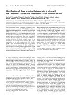
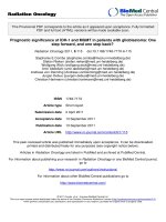
metastases of the lesion in segment 2. A CT scan of the
chest (Fig. 1) was indicated following detection of a pul-
monary nodule in the right upper field on routine chest -
x-ray. The patient had a 10 pack-year smoking history and
complained of shortness of breath upon admission; the
remaining review of symptoms was negative. Pre-opera-
tive diagnostics revealed arterial hypertension and moder-
ate left ventricular hypertrophy and pulmonary function
tests were unsuspicious (VC 143%, FEV1 128%). Since the
CT findings raised the suspicion of a malignancy, a diag-
Published: 25 January 2008
Journal of Medical Case Reports 2008, 2:21 doi:10.1186/1752-1947-2-21
Received: 14 August 2007
Accepted: 25 January 2008
This article is available from: />© 2008 Warth et al; licensee BioMed Central Ltd.
This is an Open Access article distributed under the terms of the Creative Commons Attribution License ( />),
which permits unrestricted use, distribution, and reproduction in any medium, provided the original work is properly cited.
Journal of Medical Case Reports 2008, 2:21 />Page 2 of 3
(page number not for citation purposes)
nostic thoracotomy with a concurrent sleeve lobectomy of
the right upper lobe was performed in combination with
a systematic lymphadenectomy. Pathological processing
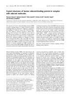
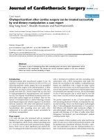
of the specimens revealed a 3.5 × 3 × 2.8 cm adenocarci-
noma of a mixed subtype with partial neuroendocrine dif-
ferentiation (Fig. 2). The tumor was strongly positive for
CK7, CK18, TTF1 and SPA and focally positive for CEA,
NSE and chromogranin A. The proliferation rate (Ki67)
was 20–30%. Besides this main tumor there were multiple
small metastases with a similar degree of differentiation in
the upper right lobe as well as in segment 4 of the middle
right lobe. Tumor infiltration of intrapulmonary and
mediastinal lymph nodes was also present. Therefore, the
TNM classification for the pulmonary adenocarcinoma
was pT4, pN2 (16/27), pM1, G3. However, further
processing of the multiple small lesions in the upper and
middle lobe revealed five foci less than 5 mm in diameter
with a different trabecular and nest-like morphology (Fig.
2). In these lesions, the cells were strongly positive for
CD56, synaptophysin, NSE and chromogranin A and
focally positive for CK7, CK18, TTF1 with a proliferation
rate (Ki67) of 1–2%. Therefore, the diagnosis of multiple
tumorlets (microcarcinoids) was made. Due to the multi-
centricity of the lesions and a size of <5 mm in diameter,
the correct diagnosis was DIPNECH. Lesions >5 mm are
classified as carcinoids according to the current WHO clas-
sification[2]. There were no clinical symptoms suggestive
of any proteins and/or hormones released, but interest-
ingly, NSE (20 ng/ml) and CEA (33 ng/ml) were slightly
elevated, whereas Cyfra was within the normal range (1.4
ng/ml). The postoperative course of the patient was une-
ventful. Six months after the operation the patient is still
alive and no tumor recurrence has been detected so far.
DIPNECH is an exceedingly rare disease involving gener-
alized proliferation of pulmonary neuroendocrine cells,
which leads to an occlusion of the bronchial lumina and
consequent clinical symptoms such as shortness of
breath. Due to its neuroendocrine origin, its similar mor-
phology to pulmonary carcinoids and particularly due to
its association with pulmonary carcinoids, the disease is
considered to be a precursor lesion for theses entities.
Only 40 cases of DIPNECH have been reported in the lit-
erature to date [1] and there are no predictive histological
or genetic data available so far. However, it has become
generally accepted that DIPNECH is a precursor to pulmo-
nary carcinoid tumors [3]. In a recent study including
1090 patients, who received resections for primary lung
tumors, Ruffini and colleagues found that the overall
prevalence of pre-invasive lesions for lung carcinomas was
6.7%. Only 3 of these 1090 cases were associated with
DIPNECH and the primary tumors were carcinoids in all
of these cases [4]. We have recently reported a similar case
in which DIPNECH was associated with a carcinoid [5].
The current report represents the first case of a patient with
DIPNECH accompanied by a pulmonary adenocarci-
noma of mixed subtype with partial neuroendocrine dif-
ferentiation. The adenocarcinoma was positive for typical
markers such as CK7, CK18, TTF1, and SPA and addition-
ally, it was positive for NSE and CEA, which were also
measured to be elevated in the patient's serum. Interest-
ingly, besides typical carcinoid markers such as CD56,
synaptophysin, and chromogranin A, the DIPNECH
lesions were also positive for NSE. However, it remains
elusive if the elevated NSE levels in the patient's serum
belong to the adenocarcinomas, the DIPNECH lesions or
a combination of both. Nevertheless, our findings raise
the hypothesis of a common pathogenic background of
pulmonary tumors with neuroendocrine differentiation,
which should further be investigated. Although it is
unlikely that DIPNECH is a precursor lesion for other
tumors of the lung with neuroendocrine differentiation
besides carcinoid tumors [6], this possibility cannot yet be
excluded considering the small number of the cases
described to date. Since it has been suggested that DIP-
NECH represents an underrecognized spectrum of disease
and since it is being increasingly diagnosed [7], we report
this case to contribute to a better understanding of the dis-
order and its associated pathologies. However, the associ-
ation of DIPNECH with a higher overall cancer incidence
should be regarded carefully, since there is evidence that
malignancies at other sites lead to an increased use of
imaging and thereby to a more frequent detection of DIP-
NECH [7]. Therefore, more data are needed to accurately
draw a conclusion on the incidence of associated malig-
nancies.
Preoperative CT scansFigure 1
Preoperative CT scans. The preoperative CT scans clearly
demonstrate the main tumor in the upper lobe of the right
lung.
Publish with BioMed Central and every
scientist can read your work free of charge
"BioMed Central will be the most significant development for
disseminating the results of biomedical research in our lifetime."
Sir Paul Nurse, Cancer Research UK
Your research papers will be:
available free of charge to the entire biomedical community
peer reviewed and published immediately upon acceptance
cited in PubMed and archived on PubMed Central
yours — you keep the copyright
Submit your manuscript here:
/>BioMedcentral
Journal of Medical Case Reports 2008, 2:21 />Page 3 of 3
(page number not for citation purposes)
Conclusion
DIPNECH is a rare disorder and it is considered to be a
precursor lesion for pulmonary carcinoid tumors. Infor-
mation on the disease is still limited, especially with
regard to management and prognosis. This case is the first
report of a patient with DIPNECH in association with a
pulmonary adenocarcinoma. Since an increasing inci-
dence of DIPNECH cases has been noted in the past few
years, we report this case to contribute to a better under-
standing of the disorder and its associated pathologies.
Abbreviations
DIPNECH = diffuse idiopathic neuroendocrine cell hyper-
plasia; CK7 = cytokeratin; CK18 = cytokeratin 18; TTF1 =
thyroid transcription factor 1; SPA = surfactant protein A;
CEA = carcinoembryonic antigen; NSE = neuron specific
enolase.
Competing interests
The author(s) declare that they have no competing inter-
ests.
Authors' contributions
AW wrote the manuscript. EH diagnosed the specimens
and collected data. AS performed and diagnosed the CT
scans. KS performed the operation, pre- and post-opera-
tive patient management. PAS diagnosed the specimens
and made final corrections of the manuscript. All authors
read and approved the final manuscript.
Consent
Written consent was obtained from the patient for the
publication of the report.
References
1. Ge Y, Eltorky MA, Ernst RD, Castro CY: Diffuse idiopathic pul-
monary neuroendocrine cell hyperplasia. Ann Diagn Pathol
2007, 11:122-126.
2. Gosney JR, Travis WD: Diffuse idiopathic pulmonary neuroen-
docrine cell hyperplasia. In Tumours of the lung, pleura, thy-
mus and heart. Edited by: Travis WD, Brambilla E, Müller-
Hermelink HK, Harris CC. IARC Press; 2004:76-77.
3. Kerr KM: Pulmonary preinvasive neoplasia. J Clin Pathol 2001,
54:257-271.
4. Ruffini E, Bongiovanni M, Cavallo A, Filosso PL, Giobbe R, Mancuso M,
Molinatti M, Oliaro A: The significance of associated pre-inva-
sive lesions in patients resected for primary lung neoplasms.
Eur J Cardiothorac Surg 2004, 26:165-172.
5. Johney EC, Pfannschmidt J, Rieker RJ, Schnabel PA, Mechtersheimer
G, Dienemann H: Diffuse idiopathic pulmonary neuroendo-
crine cell hyperplasia and a typical carcinoid tumor. J Thorac
Cardiovasc Surg 2006, 131:1207-1208.
6. Gosney JR: Diffuse idiopathic pulmonary neuroendocrine cell
hyperplasia as a precursor to pulmonary neuroendocrine
tumors. Chest 2004, 125:108.
7. Davies SJ, Gosney JR, Hansell DM, Wells AU, du Bois RM, Burke MM,
Sheppard MN, Nicholson AG: Diffuse idiopathic pulmonary neu-
roendocrine cell hyperplasia: an under-recognised spectrum
of disease. Thorax 2007, 62:248-252.
Histology of the adenocarcinoma and a representative tumorletFigure 2
Histology of the adenocarcinoma and a representative
tumorlet. Pathological processing of the resected specimens
revealed an adenocarcinoma of a mixed subtype with partial
neuroendocrine differentiation (A; primary magnification
×10) and multiple tumorlets, i.e. diffuse idiopathic pulmonary
neuroendocrine cell hyperplasia (DIPNECH; B; primary mag-
nification ×10).
Publish with BioMed Central and every
scientist can read your work free of charge
"BioMed Central will be the most significant development for
disseminating the results of biomedical research in our lifetime."
Sir Paul Nurse, Cancer Research UK
Your research papers will be:
available free of charge to the entire biomedical community
peer reviewed and published immediately upon acceptance
cited in PubMed and archived on PubMed Central
yours — you keep the copyright
Submit your manuscript here:
/>BioMedcentral