Báo cáo y học: "Rapid construction of a dendritic cell vaccine through physical perturbation and apoptotic malignant T cell loading" pot
Bạn đang xem bản rút gọn của tài liệu. Xem và tải ngay bản đầy đủ của tài liệu tại đây (2.83 MB, 16 trang )
BioMed Central
Page 1 of 16
(page number not for citation purposes)
Journal of Immune Based Therapies
and Vaccines
Open Access
Original research
Rapid construction of a dendritic cell vaccine through physical
perturbation and apoptotic malignant T cell loading
Maria Salskov-Iversen
1
, Carole L Berger*
2
and Richard L Edelson
2
Address:
1
Department of Immunology, AArhus University, Aarhus, Denmark and
2
Department of Dermatology, Yale University, School of
Medicine, New Haven, CT, USA
Email: Maria Salskov-Iversen - ; Carole L Berger* - ; Richard L Edelson -
* Corresponding author
Abstract
We have demonstrated that adherence and release of monocytes from a plastic surface drives their
differentiation into immature dendritic cells (DC,) that can mature further during overnight
incubation in the presence of apoptotic malignant T cells. Based on these results, we sought to
develop a clinically, practical, rapid means for producing DC loaded with malignant cells.
A leukapheresis harvest containing the clonal, leukemic expansion of malignant CD4
+
T cells was
obtained from the blood of patients with cutaneous T cell lymphoma (CTCL). CTCL cells were
purified with a CD3-magnetic bead column where CD3 engagement rendered the malignant T cells
apoptotic. The monocyte fraction was simultaneously activated by column passage, re-added to the
apoptotic CTCL cells and co-cultured overnight. CTCL cell apoptosis, DC differentiation and
apoptotic malignant T cell ingestion were measured by immunostaining.
The results demonstrate that as monocytes passed through the column matrix, they became
activated and differentiated into semi-mature DC expressing significantly increased levels of class
II, CD83 and CD86 (markers associated with maturing DC) and reduced expression of the
monocyte markers CD14 and CD36. Apoptotic malignant T cells were avidly engulfed by the
phagocytic transitioning DC. The addition of supportive cytokines further enhanced the number of
DC that contained apoptotic malignant T cells.
Functional studies confirmed that column passaged DC increased class II expression as shown by
significantly enhanced stimulation in mixed leukocyte culture compared to control monocytes. In
addition, DC loaded with apoptotic CTCL cells stimulated an increase in the percentage and
absolute number of CD8 T cells compared to co-cultivation with non-loaded DC. After CD8 T
cells were stimulated by DC loaded with malignant cells, they mediated increased apoptosis of
residual CTCL cells and TNF-α secretion indicating development of enhanced cytolytic function.
We report a simple one-step procedure where maturing DC containing apoptotic malignant T cells
can be prepared rapidly for potential use in vaccine immunotherapy. Ready access to both the DC
and apoptotic cells provided by this system will allow extension to other malignancies through the
addition of a variety of apoptotic tumor cells and maturation stimuli.
Published: 19 July 2005
Journal of Immune Based Therapies and Vaccines 2005, 3:4 doi:10.1186/1476-
8518-3-4
Received: 04 April 2005
Accepted: 19 July 2005
This article is available from: />© 2005 Salskov-Iversen et al; licensee BioMed Central Ltd.
This is an Open Access article distributed under the terms of the Creative Commons Attribution License ( />),
which permits unrestricted use, distribution, and reproduction in any medium, provided the original work is properly cited.
Journal of Immune Based Therapies and Vaccines 2005, 3:4 />Page 2 of 16
(page number not for citation purposes)
Background
Cutaneous T cell lymphoma (CTCL) is a malignant expan-
sion of mature, clonal CD4 T cells with an affinity for epi-
dermal localization [1]. The tumor cells proliferate in the
epidermis around a central Langerhans cell (LC) and pre-
vious studies have demonstrated that immature DC play
a crucial role in the life cycle of the malignancy [2]. The
final stages of CTCL are characterized by systemic spread,
immunosuppression and a poor prognosis. Despite the
malignancy's dependence on immature DC for prolifera-
tive support, DC immunotherapy has been of benefit in
this disease [3,4].
Two strategies for the treatment of CTCL, extracorporeal
photopheresis (ECP) and transimmunization, have been
used to successfully treat this aggressive malignancy [4,5].
The underlying principle of these treatments is extracor-
poreal establishment and re-infusion of malignant T cell-
loaded DC [6]. In both therapies, a leukapheresis product
is treated with the drug 8-methoxypsoralen (8-MOP) and
passed through a plastic ultraviolet light (UVA) exposure
plate. The 8-MOP intercalates in the DNA of nucleated
cells and is cross-linked to adjacent pyrimidine bases by
UVA light activation. The cross-link formation is a lethal
defect and replicating cells are rendered apoptotic. At the
same time, monocytes are activated by adherence and
release from the plastic exposure plate surface and begin
to transition into immature DC [6]. In the ECP treatment,
both apoptotic CTCL cells and transitioning DC are re-
infused into the patient immediately and association of
the DC and apoptotic tumor cells occurs inefficiently in
vivo.
The transimmunization procedure was devised as a more
effective modification of ECP and named to designate the
transfer of tumor antigens to competent antigen present-
ing cells (APC) that could display the full complement of
tumor antigens in the context of co-stimulatory and adhe-
sion molecules. In the transimmunization procedure, the
apoptotic malignant T cells and the transitioning DC are
co-cultured overnight enabling the up-take of the apop-
totic cells by the avidly phagocytic immature DC [6]. The
activated monocytes produce cytokines that comprise the
constituents of monocyte conditioned media thereby,
potentiating the maturation of the malignant T cell-
loaded DC [3]. The differentiating DC are re-infused the
next day into the patient where they can further mature
and have the potential to migrate to lymph nodes and
induce anti-tumor immunity.
In the current studies, we sought to explore the role of
physical perturbation in the monocyte to DC transition by
examining whether passage through a separation column
that contains a porous matrix is sufficient to induce over-
night DC differentiation from monocytes. Studies [7] sug-
gest that trans-migrating monocytes passing through the
small spaces of an endothelial cell layer become activated
and assume the phenotype of immature DC. This mono-
cyte-to-DC transition can be preserved by phagocytosis of
particulate material such as zymosan [7]. We have also
previously demonstrated that CD3-binding renders anti-
gen-experienced proliferating CTCL cells apoptotic [2].
We therefore sought to take advantage of the dual obser-
vations of the role of physical stimulation in DC matura-
tion and the rapid apoptotic cell death mediated by CD3-
binding to develop in one day a clinically practical vac-
cine. We demonstrate that a simple one-step procedure
using CD3-magnetic beads to render the malignant T cells
apoptotic and the separation column matrix to simultane-
ously activate monocytes results in overnight production
of apoptotic cell-loaded DC. These immature DC gener-
ated in the absence of cytokines could be driven to differ-
entiate further when exogenous cytokines were added.
Functional evaluation of the malignant T cell loaded DC,
developed by this methodology, demonstrated a signifi-
cantly enhanced stimulatory capacity in mixed leukocyte
culture and the ability to promote CD8 T cell expansion
and cytolytic capacity.
Therefore, this approach yields malignant cell loaded DC
in a rapid time-frame without extensive cell culture, exog-
enous factors or cell isolation and manipulation. This
method may provide a clinically practical means for the
production of immunogenic DC for cancer vaccine
therapy.
Materials and methods
Patient Population
Therapeutic leukapheresis specimens were obtained from
7 CTCL patients (in accordance with the guidelines of the
Yale human investigation committee). All patients had
advanced disease with clonal CD4
+
T cell populations
present in the peripheral circulation as determined by
immunophenotyping with antibodies to the clonotypic
variable region of family-specific T cell receptor (TCR) or
polymerase chain reaction to detect rearrangements of the
beta or gamma chain of the TCR. All patients were under-
going treatment with standard ECP.
Cell Isolation
Mononuclear cells (MNC) were isolated by centrifugation
over a ficoll-hypaque gradient followed by two washes in
RPMI 1640 (Gibco, Gaithersburg, MD) containing 10%
AB serum and 2 mM EDTA. MNC (2 × 10
7
) were incu-
bated with 40 µl Macs α-human CD3 microBeads
(Miltenyi Bioteck, Auburn CA) following the manufac-
turer's directions. The cells were separated by passage
through a Macs Separation Column (Miltenyi Bioteck)
consisting of a magnetized iron matrix. CD3 positive and
negative cells were counted, re-mixed together and
Journal of Immune Based Therapies and Vaccines 2005, 3:4 />Page 3 of 16
(page number not for citation purposes)
incubated overnight. As a control, MNC (2 × 10
7
) were
also incubated with 40 µl Macs α-human CD4
microBeads. After treatment, the cells were incubated in 3
ml RPMI 1640 containing 15% AB serum and 15% autol-
ogous plasma in one well of a 12 well tissue culture plate
(Falcon). In some experiments half of the recombined
cells obtained after CD3 column passage were incubated
overnight in RPMI containing 10% FCS (Gibco) in the
presence of the cytokines GM-CSF 800 U/ml and IL4 1000
U/ml (R & D Systems, Minneapolis, MN). Day 0 baseline
cells were immediately removed for immunostaining
while Day 1 cells were incubated overnight.
Immunophenotyping
In order to monitor DC differentiation, the cells were
stained by two-color immunofluorescence with a panel of
antibodies to monocytes, DC and apoptotic cells. Cells (1
× 10
6
) were incubated with 10–20 µl of fluorocrome con-
jugated monoclonal antibody for 30 minutes in the dark
at 4°C. The antibodies were directly conjugated to fluores-
cein (FITC) or phycoerythrin (PE) and included: CD14-
FITC (monocytes) + CD86-PE (co-stimulatory molecule
highly expressed on DC); HLA-DR-FITC (anti-class II
MHC molecule) and CD83-PE (DC maturation marker);
and their isotype matched controls (Beckman Coulter
Immuno-Tech, Hialeah, FL). Cells were washed once and
suspended in PBS and read on a XL flow cytometer (Beck-
man Coulter) within 24 hours.
Combined membrane and cytoplasmic staining was per-
formed following manufacturers instructions (Intraprep
kit, Beckman Coulter). Antibody combinations included:
membrane CD36-FITC (receptor for apoptotic cells) +
cytoplasmic CD83 PE; DR-FITC + cytoplasmic CD83-PE;
and isotype controls (Beckman Coulter). To detect apop-
totic cells, lymphocytes were stained with: membrane
HLA-DR-FITC (class II MHC) + cytoplasmic Apo2.7-PE
(apoptotic cells); and isotype controls. Data was analyzed
using the CXP software (Beckman Coulter).
Confocal Microscopy
Cells were double-stained for membrane HLA-DR-FITC +
cytoplasmic Apo2.7-PE following the manufacturer's
instructions for combined membrane and cytoplasmic
staining (see immunophenotyping). In addition, cells
were double stained for cytoplasmic LAMP-2 FITC (lyso-
somal marker, Research Diagnostics) and HLA-DR-PE.
Cells were prepared for microscopy following the instruc-
tions for Molecular Probes "Slow Fade Light" anti-fade kit
(Molecular Probes Inc, Eugene, OR). Specimens were kept
in the dark at 4° until microscopy was performed on a
Zeiss confocal microscope.
Mixed leukocyte culture assay
The mixed leukocyte culture assay was performed by iso-
lating control leukocytes from two normal donors. Con-
trol T cells were purified with CD4 magnetic beads and
the column effluent containing monocytes and B cells was
γ-irradiated to prevent differentiation and used as a source
of stimulators. Transitioning DC from CTCL patients were
obtained one day prior to the normal control cells and
cultured overnight without cytokines, γ-irradiated and
used as stimulators for the control lymphocytes. The cells
were adjusted to 4 × 10
6
/ml and 50 µl of responding cells
and 50 µl of stimulating cells co-cultured in round bot-
tom microtiter wells with the addition of 100 µl of RPMI
1640 containing 15% AB serum and 15% autologous
plasma for 6 days at 37°C under a 5% CO
2
atmosphere.
The wells were pulsed with 1 µCi/well
3
[H]-thymidine 16
hours prior to harvest (PhD harvester, Cambridge Tech.,
Cambridge, MA). The incorporation of the isotope was
evaluated in a liquid scintillation counter.
CD8 T cell purification and expansion
CD8 T cells were purified with CD8-magnetic beads
(≥96% purity) and suspended in RPMI 1640/15% autolo-
gous serum and IL2 and added to DC that had been col-
umn eluted from the same CTCL patient. The cells were
co-cultured overnight with 1.1 × 10
6
CD8 T cells/well
added to CD3-bead rendered apoptotic CTCL cells or via-
ble CTCL cells (4 × 10
6
/well). After overnight culture, the
cells were harvested, counted, and immunophenotyped
for markers of T cells (CD3, CD4, CD8) and apoptosis
(Apo2.7).
Tumor necrosis-
α
(TNF-
α
) ELISA
The production of TNF-α was measured in an ELISA assay
(R&D Systems, Minneapolis, MN) essentially as described
by the manufacturer.
Statistical evaluation
The expression of DC markers and the MLC response was
evaluated statistically by the student's t test or if the data
was not normally distributed the Mann-Whitney Rank
Sum Test using the Sigma Stat analysis program.
Results
Passage of monocytes through a separation column
induces monocyte to DC transition
Monocytes were obtained from a leukapheresis harvest
performed therapeutically on CTCL patients and were cul-
tured overnight with and without passage through a mag-
netic bead separation column. Monocyte differentiation
into semi-mature DC was monitored by 2-color immun-
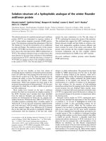
ofluorescence. In a representative experiment, (Fig. 1,
gated on the monocyte population as identified by co-
expression of CD14 and CD86), the loss of monocyte
membrane marker CD14 is revealed by a decrease in the
Journal of Immune Based Therapies and Vaccines 2005, 3:4 />Page 4 of 16
(page number not for citation purposes)
mean fluorescence intensity (MFI) of the CD14 fluoro-
chrome. CD14 expression declined as the degree of
manipulation of the cells increased from primary isola-
tion (Fig. 1a) to simple overnight culture of the leukapher-
esis product (Fig. 1b), compared to the addition to the
differentiating DC of CTCL cells that were selected by the
CD4 antibody, (Fig 1c) to the maximum reduction in
monocyte CD14 expression found when the activated
transitioning DC were cultured with CTCL cells rendered
apoptotic by CD3 antibody (Fig. 1d). In total, (Fig 1e) the
expression of CD14 was reduced by 54%, from a mean
fluorescent intensity (MFI) of 13 on primary isolation to
5.89, when the column separated monocytes were co-cul-
tured with the CD3-treated apoptotic CTCL cells. As the
differentiating DC lost the monocyte marker, a 3-fold
increase in expression of CD86, a co-stimulatory mole-
DC differentiation from monocytes induced by column activationFigure 1
DC differentiation from monocytes induced by column activation. CTCL cells and DC were isolated from a leuka-
pheresis by CD4 or CD3-antibody conjugated to magnetic beads. The cells were separated by passage through a column
placed in a magnetic field and the purified CTCL cells were re-added to the column activated monocytes and cultured over-
night. Binding of fluorochromes was analyzed using flow cytometry and 2-color quadstats were gated on the monocyte popula-
tion. The results demonstrate membrane CD14-FITC and CD86-PE co-expression on cells obtained a: Day 0, primary
isolation; and after overnight culture of b: leukapheresis cells; c: cells obtained by CD4-magnetic bead isolation and re-cultured
overnight with column activated monocytes; and d: cells obtained from CD3-magnetic bead isolation and re-cultured overnight
with column activated monocytes. e: Bar graph showing the reduction in mean fluorescent intensity (MFI) of CD14 expression
on primary isolation (Day 0) and after overnight incubation of the leukapheresis (leuk) or column passaged and recombined cell
populations using CD4 or CD3-magnetic bead isolation (negative control isotype staining is presented in the first bar). f: Bar
graph showing the increase in MFI of CD86 expression (as described in e).
Journal of Immune Based Therapies and Vaccines 2005, 3:4 />Page 5 of 16
(page number not for citation purposes)
cule, was found ranging from an MFI of 1.98 on Day 0 to
6.4 after passage through the CD3-magnetic bead column
and overnight incubation (Fig. 1f).
Transitioning DC increase their expression of the
maturation marker, CD83
In Fig. 2A &2B, the differentiation of monocytes into
semi-mature DC is demonstrated by an increase in the
percentage of cells that exhibit reduced fluorescent inten-
sity of membrane CD36 (receptor for up-take of apoptotic
cells, a marker that is lost as DC mature) and increased
expression of cytoplasmic CD83 (DC maturation
marker). Fig. 2A-a, demonstrates that only 4% of the cells
co-express membrane CD36 and cytoplasmic CD83 on
primary isolation. When the cells were cultured overnight,
the percentage of cells co-expressing CD36/CD83
increased as the level of manipulation rose from 25% in
the overnight culture of the leukapheresis (Fig. 2A-b) and
in cells separated with a CD4-magnetic bead control anti-
body and re-added to the column effluent (Fig. 2A-c) to
the maximal differentiation of 34% found when apop-
totic CD3-treated CTCL cells were re-added to the acti-
vated transitioning DC (Fig. 2A-d). In Fig 2A-e, the
reduction in CD36 MFI is shown by a decline from a MFI
of 34 on primary isolation to 7.7 (77% reduction) in the
monocyte/DC population activated by passage through
the separation column and recombined for overnight cul-
ture in the presence of CTCL cells rendered apoptotic with
CD3 antibody.
The increase in cytoplasmic CD83 expression is shown in
Fig. 2B. As expected only a small percentage of cells
express the DC differentiation marker, CD83 on primary
isolation (0.5%, Fig. 2B-a). Overnight incubation of the
leukapheresis (Fig 2B-b) increases CD83 expression to an
equivalent degree as CD83 expression detected after pas-
sage through a CD4-magnetic bead column (Fig. 2B-c).
More than one third of the monocytes transitioned into
semi-mature DC as shown by the increased expression of
cytoplasmic CD83 (Fig. 2B-d) found when CD3-separated
apoptotic CTCL cells were added to the column activated
monocytes.
Induction of simultaneous DC differentiation and CTCL
cell apoptosis and engulfment
Further confirmation of enhanced differentiation of
monocytes to DC was found when membrane class II
expression (HLA-DR) was measured and the up-take of
apoptotic CTCL cells was assessed. In figure 3A, the per-
centage of DR-positive transitioning monocytes contain-
ing apoptotic cells was determined by measurement of the
cytoplasmic expression of the early apoptotic marker
APO2-PE. On primary isolation (Fig. 3A-a), or after over-
night incubation of the leukapheresis without further
processing (Fig. 3A-b), only a small percentage of the
monocyte-DC population contained apoptotic material
in the cytoplasm. CD4-treatment and column passage
damaged enough cells to increase the number of apop-
totic CTCL cells ingested by the activated monocyte-DC
population (Fig. 3A-c). As previously reported [2], CD3-
binding to CTCL cells rendered the malignant T cells
apoptotic and material from the damaged and dying
CTCL cells could be detected inside the developing DC
population (Fig. 3A-d). While only 19% of the transition-
ing DC were reactive with DR/APO2-PE, this probably
represents only a minimal level of engulfed apoptotic cells
since processing and degradation of the apoptotic blebs
during overnight incubation could have reduced the
detectable expression of APO2-PE positive material.
Differentiation of the DC population was also demon-
strated by the increase in expression of membrane class II
MHC molecules. Physical manipulation did not increase
class II expression from the primary value obtained on ini-
tial isolation (Fig. 3B-a), when leukapheresis cells were
cultured overnight (Fig. 3B-b). No enhancement of class II
expression was noted even when the column activated
monocytes were co-cultured overnight with CD4-bead
separated CTCL cells (Fig. 3B-c). However, the overnight
addition of apoptotic CTCL cells, obtained after CD3-
binding, to transitioning DC increased class II expression
from 55% (Day 0, Fig. 3B-a) to 72% (Fig. 3B-d).
Statistical evaluation of the enhanced expression of DC
differentiation markers
We evaluated the overall increase in markers of DC differ-
entiation from monocytes in leukocytes obtained from
seven CTCL patients. While substantial variation in the
expression of several antigens precluded analysis, the
results showed that overall expression of class II MHC
antigen was significantly up-regulated in differentiating
DC obtained after column passage with (P ≤ 0.005) and
without (P ≤ 0.002) the addition of apoptotic CTCL cells
(Fig. 4a). In addition, CD86 (P ≤ 0.025) expression was
significantly increased when CTCL cells were co-cultured
with column passaged transitioning DC loaded with
apoptotic CTCL cells and CD83 (P ≤ 0.001) was enhanced
irrespective of the presence of apoptotic CTCL cells (Fig.
4b &4c). These results confirm that the physical perturba-
tion encountered after passage through the small spaces of
separation column significantly enhances the entry of
monocytes into the DC pathway.
Demonstration of DC loading with apoptotic cells by
confocal microscopy
In Fig. 5A, CTCL cells were rendered apoptotic with CD3-
magnetic bead conjugated antibody (Fig. 5A a–c ) or as a
control treated with CD4-magnetic bead conjugated
antibody (Fig. 5A d–f), run through the separation col-
umn and co-cultured with the simultaneously activated
Journal of Immune Based Therapies and Vaccines 2005, 3:4 />Page 6 of 16
(page number not for citation purposes)
differentiating DC. The activated monocyte/DC popula-
tion was double-stained for expression of membrane class
II (green) and the marker of early apoptotic cells, intracel-
lular APO-2 (red). Representative class II-positive cells
(green fluorescence) are seen in Figures 5A-a and 5A-c. In
Figure 5A-b, three cells that were rendered apoptotic after
CD3-binding, were identified (white arrows) and material
from one of these cells is contained in a class II positive
cell (merge, Fig. 5A-c). In Figure 5A-e (CD4-treatment),
only a small amount of apoptotic material is found and
none of this material is associated with the class II positive
cell (Fig. 5A-f, merge).
To confirm that class II molecules co-localized in lyso-
somal compartments in a pattern found in semi-mature
DC [8], cells were stained with a lysosomal marker LAMP2
DC maturation induced after column separation and overnight incubationFigure 2
DC maturation induced after column separation and overnight incubation. Fig. 2A: CTCL cells and monocyte/DC
isolated as described in Figure 1 were fixed and permeabilized and stained with CD36-FITC (membrane) and CD83-PE (cyto-
plasm). The results show 2-color quadstats gated on the monocyte population of cells obtained from a: Day 0, primary isola-
tion; after overnight culture of b: leukapheresis cells; c: CD4-magnetic bead isolation and re-addition to column activated
monocytes; d: CD3-magnetic bead purification and re-addition to column activated monocytes; e: Bar graph of the MFI of
membrane CD36 expression on the cell populations. Fig. 2B: Demonstration of cytoplasmic CD83 expression in the mono-
cyte/DC population gated by side-scatter (SS) on 100% of the monocyte population. Cell treatment a–d as described for Fig
2A.
Journal of Immune Based Therapies and Vaccines 2005, 3:4 />Page 7 of 16
(page number not for citation purposes)
and an antibody to class II MHC molecules (Fig. 5B). In
Fig. 5B-a, a cell that has been activated by passage through
the separation column and co-cultivated overnight with
CTCL cells rendered apoptotic by CD3-magnetic bead
binding was stained with an anti-class II antibody (red).
In Fig. 4B-b lysosomal compartments were visualized
with an antibody that binds to the lysosomal membrane
(LAMP2, green). Merging of the 2 fluorochromes (Fig. 5B-
c, yellow) demonstrates colocalization of class II MHC
molecules in lysosomal compartments. When class II
staining was monitored on column activated transitional
cells that had been co-incubated with control CTCL cells
selected by CD4-magnetic bead separation (Fig. 5B-d,
red), strong membrane staining was found. Weak lyso-
somal staining was localized beneath the plasma
membrane (Fig 5B-e, green). When the pictures were
merged, class II MHC molecules did not exhibit entry into
the lysosomal compartment (Fig. 5B-f). The presence of
class II MHC molecules in lysosomes is consistent with
differentiation into semi-mature DC [8], and suggests that
Increased class II expression on semi-mature DC after ingestion of apoptotic CTCL cellsFigure 3
Increased class II expression on semi-mature DC after ingestion of apoptotic CTCL cells. Fig. 3A: CTCL cells and
DC prepared as described in Figure 1 were fixed and permeabilized and stained with DR-FITC (anti-class II MHC antibody,
membrane) and APO2-PE (cytoplasm). The results present 2-color quadstats gated on the monocyte population of cells
obtained from a: Day 0, primary isolation; b: leukapheresis cells; c: CD4-magnetic bead isolation and re-additon to column
activated monocytes; d: CD3-magnetic bead purification and re-addition to column activated monocytes. Fig. 3B: Membrane
DR staining on the monocyte/DC population gated on the total monocyte population by SS. Cell treatment a–d as described
for Fig 3A.
Journal of Immune Based Therapies and Vaccines 2005, 3:4 />Page 8 of 16
(page number not for citation purposes)
class II molecules have migrated to lysosomal compart-
ments where they would have the opportunity for loading
with peptides derived from processed apoptotic material.
The addition of supportive cytokines enhances monocyte
to DC differentiation
We sought to maximize induction of maturing DC loaded
with apoptotic malignant T cells through the addition of
exogenous cytokines known to be important for DC
differentiation [9]. To study the effect of supportive
cytokines on the phenotype of the developing DC, we
divided the column separated cells in half and co-incu-
bated them overnight with CD3-bead rendered apoptotic
CTCL cells with and without GM-CSF and IL-4.
The addition of cytokines to the co-cultured apoptotic
CTCL cells and column activated transitioning monocytes
increased the overall maturation of the DC. In Figure 6,
the level of CD14 expression is reduced as shown by an
increase in the CD14 negative population (Gate AA1)
from 4.8% at baseline (Fig. 6a) to 10% when the
transitioning DC were incubated with apoptotic cells
Statistical analysis of DC differentiation markersFigure 4
Statistical analysis of DC differentiation markers. The expression of markers of DC differentiation were compiled from
the overnight culture of DC induced by column passage with and without apoptotic cell loading that had been obtained from 7
CTCL patients, averaged and analyzed for significance in comparison to the values obtained on primary isolation. a: Mean fluo-
rescence intensity (MFI) of class II expression on Day 0, primary isolation (Pre Tx; pre-treatment), or Day 1 column activated
cells loaded with apoptotic CTCL or co-cultivated in the presence of viable CTCL cells (Mann-Whitney Rank Sum Test). b:
Percent of monocytes expressing CD86 on primary isolation, or after column activation and overnight culture with and with-
out apoptotic cell ingestion (t test). c: Percent of monocytes expressing cytoplasmic CD83 on primary isolation or after col-
umn activation and overnight cultivation with and without apoptotic cell up-take (Mann-Whitney Rank Sum Test).
Journal of Immune Based Therapies and Vaccines 2005, 3:4 />Page 9 of 16
(page number not for citation purposes)
without cytokines (Fig. 6b). The addition of cytokines
enhanced the loss of CD14 expression resulting in 36% of
the cells becoming CD14-negative after overnight culture
(Fig. 6c). As the differentiating monocytes lost CD14
expression, a concomitant increase in CD86 expression
was noted. CD86 expression rose from a baseline level of
61% (Fig. 6d) to more than 80% CD86-positive transi-
tioning DC after column separation and co-cultivation
with CD3-rendered apoptotic cells without cytokines (Fig.
6e) or in the presence of exogenous cytokines (Fig. 6f).
Confocal microscopic demonstration of apoptotic cell ingestion and class II localization in lysosomal compartments in differen-tiating DCFigure 5
Confocal microscopic demonstration of apoptotic cell ingestion and class II localization in lysosomal compart-
ments in differentiating DC. Fig. 5A: Cell populations prepared as described in Figure 1 were evaluated by confocal
microscopy after fixation and permeabilization and staining. A representative activated monocyte/DC is shown after CD3 col-
umn passage and recombination with the apoptotic CTCL cells as detected by a: membrane class II-FITC (green); b: cytopolas-
mic APO2-PE (red, white arrows) and c: merged image demonstrating internalization of apoptotic material in a class II positive
cell. A representative activated monocyte/DC is shown after CD4 column passage and recombination with viable CTCL cells
as detected by d: membrane class II-FITC (green); e: cytopolasmic APO2-PE (red, white arrow) and f: merged image demon-
strating absence of internalization of apoptotic material in a class II positive cell. Fig. 5B: Cells prepared as described in Fig. 5A
were passed through the CD3 column and stained for a: membrane class II-PE (red); b: lysosomal membrane marker, LAMP
(green); and c: merged image showing co-localization of class II molecules in lysosomal compartments. Cells obtained after pas-
sage through the CD4 column were stained for d: membrane class II-PE (red); e: lysosomal membrane marker, LAMP (green);
and f: merged image showing an absence of co-localization of class II molecules in lysosomal compartments.
Journal of Immune Based Therapies and Vaccines 2005, 3:4 />Page 10 of 16
(page number not for citation purposes)
Cytokines enhance DC maturation
The percentage of semi-mature DC differentiated after
overnight co-culture that co-expressed membrane CD36
and intracytoplasmic CD83 was enhanced by the addition
of cytokines. In Fig. 7a, on primary isolation the
monocytes expressed intermediate levels of CD36 and did
not contain cytoplamic CD83 (Fig. 7a). Co-expression of
CD36/CD83 (Fig. 7b) rose to 50%, after overnight culture
in the absence of cytokines, on differentiating DC that had
passed through the separation column and were recom-
bined with CD3 rendered apoptotic CTCL cells. This
increased expression of a receptor for apoptotic cells may
have been driven by the presence of very high levels of
apoptotic material in the co-cultures (Fig. 8). Further mat-
uration was observed in the presence of cytokines (Fig. 7c)
leading to 53% CD36 expression on the transitioning DC
and the identification of 7% CD36-negative cells that
contained CD83 in the cytoplasm. The percentage of dif-
ferentiating DC that expressed cytoplasmic CD83 rose
from 0% at baseline (Fig. 7d) to 49% after column sepa-
ration and co-incubation with CTCL cells rendered apop-
totic by CD3-magnetic bead binding (Fig. 7e) to 59%
when cytokines were added to the cultured cells (Fig. 7f).
Class II expression and up-take of apoptotic material is
enhanced in the presence of cytokines
The baseline expression of class II MHC molecules on the
cell membrane of monocytes on primary isolation is
shown in Fig. 8a. Freshly isolated monocytes express a
reduced intensity of class II expression and contain a
Exogenous cytokines enhance DC differentiation from monocytes activated by column passageFigure 6
Exogenous cytokines enhance DC differentiation from monocytes activated by column passage. Monocyte/DC
populations isolated as described in Figure 1 were stained for membrane co-expression of CD14-FITC and CD86-PE. The
results present 2-color quadstats gated on the monocyte population of cells obtained from a: Day 0, primary isolation; b: CD3-
magnetic bead purification and re-addition to column activated monocytes; c: the same CD3 column purified and activated
recombined cell population cultured with the cytokines GM-CSF and IL4. Demonstration of membrane CD86 expression on
the monocyte/DC population gated by side-scatter on 100% of the monocyte population. d: Day 0, primary isolation; e: CD3-
magnetic bead purification and re-addition to column activated monocytes; f: the same CD3 column purified and activated
recombined cell population cultured with cytokines.
Journal of Immune Based Therapies and Vaccines 2005, 3:4 />Page 11 of 16
(page number not for citation purposes)
small percentage of cytoplasmic apoptotic material. After
column separation and co-incubation with CD3-magnetic
bead treated apoptotic cells, membrane class II expression
is enhanced (Fig. 8b) and large amounts of apoptotic
material can be detected in the cytoplasm of the transi-
tioning DC. Exogenous cytokines further increase the
percentage of class II-positive cells that contain apoptotic
material (Fig. 8c). Therefore, the addition of exogenous
cytokines enhances both the differentiation of immature
DC and the ingestion of apoptotic material improving the
overnight yield of maturing apoptotic T cell loaded DC.
Functional analysis of the differentiating DC obtained
after column passage
Transitioning DC obtained after column passage were
evaluated for their stimulatory capacity in MLC (Fig. 9).
The results demonstrate that DC induced by column pas-
sage of leukocytes from two normal controls were
significantly better stimulators (P ≤ 0.034 & P ≤ 0.036) in
MLC than autologous monocytes irrespective of apoptotic
cell loading. These results confirm that column activation
of monocytes and overnight culture enhances the mem-
brane expression of class II MHC molecules recognized by
alloresponsive CD4 T cells. Therefore, DC harvested after
physical activation and overnight culture could expand
CD4 T cells and potentially provide the help required for
licensing of anti-tumor CD8 T cell responses [10].
We have begun to investigate the capacity of the DC har-
vested after column perturbation and apoptotic malig-
nant T cell loading to induce and expand an anti-tumor
CD8 T cell response. In these initial studies (Fig. 10A), we
Exogenous cytokines increase DC maturation induced after column separation and overnight incubationFigure 7
Exogenous cytokines increase DC maturation induced after column separation and overnight incubation.
Monocyte/DC populations isolated as described in Figure 1 were fixed and permeabilized and stained for expression of mem-
brane CD36-FITC and cytoplasmic CD83-PE. The results present 2-color quadstats gated on the monocyte population of cells
obtained from a: Day 0, primary isolation; b: CD3-magnetic bead purification and re-addition to column activated monocytes;
c: the same CD3 column purified and activated recombined cell population cultured with the cytokines GM-CSF and IL4. Dem-
onstration of cytoplasmic CD83 expression in the monocyte/DC population gated by side-scatter on 100% of the monocyte
poulation. d: Day 0, primary isolation; e: CD3-magnetic bead purification and re-addition to column activated monocytes; f: the
same CD3 column purified and activated recombined cell population cultured with cytokines.
Journal of Immune Based Therapies and Vaccines 2005, 3:4 />Page 12 of 16
(page number not for citation purposes)
have found that the percentage of CD8 T cells (purified
CD8 T cells ≥96% positive, obtained from the leukapher-
esis of a patient responsive to ECP) increased by 38% in
the presence of DC fed apoptotic CTCL cells (Fig. 10A-a)
compared to the percent of CD8 T cells found after
overnight incubation with DC exposed to viable CTCL
cells (Fig. 10A-b). In addition, the absolute number of
CD8 T cells recovered from the overnight culture of differ-
entiating column passaged DC loaded with apoptotic
malignant T cells increased by 22% when compared to the
initial number of CD8 T cells while the absolute number
of CD8 T cells present in cultures of DC and viable CD4 T
cells fell by 12% (Fig. 10A-c). Therefore, exposure of CD8
T cells to malignant T cell loaded DC increases both the
percentage and absolute number of potential anti-tumor
responsive T cells.
The level of apoptosis found when CD8 T cells were cul-
tured overnight with column activated-DC loaded with
CTCL cells doubled (56%, Fig. 10B-a) in comparison to
the level of apoptosis present when CD8 T cells were
added to non-loaded DC that had been cultivated with
viable CTCL cells (27%, Fig. 10B-b). The baseline level of
apoptosis was 24% when malignant T cell loaded DC (Fig.
10B-c), or non-loaded DC (Fig. 10B-d) were cultured in
the absence of CD8 T cells. These results indicate that
residual CTCL cells may be lysed in the presence of CD8 T
cells stimulated with DC that have ingested apoptotic
malignant T cells.
Finally, further support for the contention that functional
CD8 T cells were expanded by overnight exposure to col-
umn-activated DC loaded with malignant T cells was
obtained by evaluation of the levels of TNF-α found in the
culture supernatants. In Figure 10B-e, supernatants from
CD8 T cells cultured overnight alone contained minimal
levels of TNF-α. CD8 T cells stimulated with column dif-
ferentiated DC loaded with CD3-bead rendered apoptotic
malignant T cells or not loaded both significantly (P ≤
0.001 & P ≤ 0.014) stimulated release of TNF-α. However,
DC that had engulfed apoptotic cells caused the release of
three fold more TNF-α than non-loaded DC, indicating
that CD8 T cell activation had occurred and the release of
a molecule that promotes tumor cytolysis was present.
Discussion
Development of effective DC based cancer vaccine tech-
nology has been limited by the extensive manipulation
and extended period of in vitro culture required for gener-
ation of mature DC loaded with the appropriate tumor
antigens. We have circumvented some of these limitations
through modification of a successful technology that per-
mits both DC differentiation from peripheral monocytes
and simultaneous loading of DC with apoptotic malig-
nant T cells containing the full complement of potential
tumor antigens [6]. DC are the most potent APC display-
ing when mature high levels of co-stimulatory, adhesion
and MHC molecules which can present peptides derived
from apoptotic cells to the immune system [9]. Therefore,
Exogenous cytokines enhance ingestion of apoptotic material in differentiating DCFigure 8
Exogenous cytokines enhance ingestion of apoptotic material in differentiating DC. CTCL cells and monocyte/DC
populations isolated as described in Figure 1. The cells were fixed and permeabilized and stained for expression of membrane
DR-FITC and cytoplasmic APO2-PE. The results present 2-color quadstats gated on the monocyte population of cells obtained
from a: Day 0, primary isolation; b: CD3-magnetic bead purification and re-addition to column activated monocytes; c: the
same CD3 column purified and activated recombined cell population cultured with the cytokines GM-CSF and IL4.
Journal of Immune Based Therapies and Vaccines 2005, 3:4 />Page 13 of 16
(page number not for citation purposes)
the development of a simple rapid means of generating
malignant cell-loaded DC could advance the immuno-
therapy of CTCL and perhaps other malignancies.
Immunotherapy has played a major role in the treatment
of CTCL since the introduction of ECP by Edelson and
colleagues in 1987 [5]. The mechanism underlying the
success of ECP treatment was defined by the demonstra-
tion that the simultaneous introduction of apoptotic
malignant T cells and the differentiation of monocytes
into DC resulted in patients receiving CTCL cell-loaded
DC that have the capacity to present antigen, derived from
the CTCL cells, to cytotoxic lymphocytes and initiate an
immune response towards the malignant CD4 T cells. Pre-
vious studies had demonstrated that despite the clonal
expansion of CD4
+
malignant T cells in the peripheral
blood of CTCL patients, circulating populations of CD8 T
cells that retained the capacity to lyse autologous malig-
nant T cells [11] could be identified. One antigen that
served as an immunogen recognized by cytotoxic T cells in
CTCL was determined to be peptides derived from the
beta chain of the TCR that was clonotypically displayed
on the malignant T cells [12,13]. Therefore, the potential
for development of an anti-malignant T cell immune
response exists in CTCL patients and immunotherapeutic
approaches designed to expand anti-tumor CD8 T cells
could be effective in this disease.
We sought to exploit our understanding of the mecha-
nism of ECP to develop more efficient, rapid, clinically
practical means for producing malignant T cell-loaded
DC. In the current study, we demonstrate that DC loaded
with apoptotic cells can be produced in one day without
extensive manipulation or the use of exogenous cytokines.
The use of CD3-antibody to render CTCL cells apoptotic
and passage of the treated MNC through the small pores
of the iron matrix of a separation column followed by
overnight co-incubation resulted in the generation of DC
containing material derived from apoptotic CTCL cells.
DC differentiation was demonstrated by both the reduc-
Mixed leukocyte culture response of normal T cells to monocytes or column activated loaded and non-loaded DCFigure 9
Mixed leukocyte culture response of normal T cells to monocytes or column activated loaded and non-loaded
DC. Normal CD4 T cells were magnetic bead column purified from the peripheral blood of 2 controls and stimulated with col-
umn effluent monocytes that had been γ-irradiated to prevent differentiation, or γ-irradiated CTCL cell monocytes that had
been column purified and either loaded or not with apoptotic malignant T cells and cultured overnight one day prior to the
normal T cell isolation. The results are presented as delta CPM (less background proliferation obtained by autostimulation) of
3
[H]-thymidine incorporation measured at day 6. Significance was evaluated with a student's t test.
Journal of Immune Based Therapies and Vaccines 2005, 3:4 />Page 14 of 16
(page number not for citation purposes)
tion in monocyte markers and the significant increase in
class II MHC molecules and co-stimulatory molecules, as
well as the increase in CD83, a marker of maturing DC.
The internalization of apoptotic blebs was confirmed by
localization of the apoptotic material in the cytoplasm,
indicating that processing of the apoptotic CTCL-derived
CD8 T cell response to column-activated malignant T cell loaded DCFigure 10
CD8 T cell response to column-activated malignant T cell loaded DC. Fig. 10A: CD8 T cells were magnetic-bead
enriched (≥96% CD8
+
T cells) from the leukapheresis of a CTCL patient and added to column-activated DC with and without
apoptotic malignant T cell loading. The percentage of CD8 T cells was identified by immunophenotyping and flow cytometry
and the results presented as 1-color histograms. a: Percentage of CD8 T cells found after overnight culture with column-acti-
vated DC pulsed with CD3-magnetic bead rendered apoptotic CTCL cells. b: Percentage of CD8 T cells identified after over-
night culture with DC co-cultivated with viable CTCL cells. c: Absolute number of CD8 T cells after overnight cultivation with
DC loaded with apopototic malignant T cells or DC co-incubated with viable CD4-bead isolated CTCL cells. Horizontal line
indicates the initial number of CD8 T cells added to the co-cultures on Day 0. Fig. 10B: The percentage of apoptotic cells was
determined in the co-cultures by staining for APO2.7 and flow cytometry. The quadstats are gated on the lymphocyte popula-
tion by side scatter (SS) and represent 100% of the lymphocyte population. Percent apoptotic cells found in co-cultures of a:
CD8 T cells and DC loaded with apoptotic malignant T cells; b: CD8 T cells, DC and viable CTCL cells; c: DC loaded with
apoptotic CTCL in the absence of CD8 T cells; d: DC cultured with viable CTCL without CD8 T cells. e: Culture supernatants
were obtained from CD8 T cells cultured overnight alone, or in the presence of DC loaded with apoptotic CTCL cells or DC
cultured with viable CTCL cells and the secretion of TNF-α determined in an ELISA assay. The results are presented as pg/ml
and significance determined with a student's t test.
Journal of Immune Based Therapies and Vaccines 2005, 3:4 />Page 15 of 16
(page number not for citation purposes)
material could make peptides available for MHC loading
and transport to the cell membrane [14]. The ability to
increase the number of maturing CTCL cell-loaded DC by
the addition of exogenous cytokines demonstrates that
this technique can produce cell populations that can be
manipulated to maximize the production of DC that con-
tain apoptotic material thereby providing access to a spec-
trum of CTCL cell-derived epitopes, without the
requirement for identification or isolation of individual
peptides that may be relevant for induction of an anti-
CTCL cell immune response.
Furthermore, we show that DC produced in this fashion
are effective stimulators of alloproliferation in MLC con-
firming the significant up-regulation of class II MHC
molecules. The malignant T cell loaded DC stimulated
CD8 T cell expansion and an increase in apoptotic cell
death and the significantly enhanced release of TNF-α.
These results indicate that CD8 T cells that have been
stimulated by malignant T cell loaded DC, produced by
this methodology, may develop the ability to mediate
tumor cell cytolysis.
The current studies support our previous results demon-
strating that monocyte differentiation into DC could be
driven by increasing levels of physical perturbation [6].
We confirm that leukapheresis alone generates modest
monocyte activation and conversion into immature DC
that can be enhanced by further manipulation and the
addition of apoptotic cells. We also demonstrate that
CD3-binding is a potent means of rendering CTCL cells
apoptotic [2] even when the CTCL cells are not cultured
but directly isolated from the patients. The current study
combines and extends these two previous observations
into a format for simple, rapid, clinically practical DC vac-
cine generation.
Current approaches to DC vaccine technology include
peptide pulsing [15], one week or longer of culture with
cytokines [16], cell fusion with tumor cell partners [17],
and the use of a variety of vectors designed to introduce
tumor antigens into the DC [18]. These methods are gen-
erally cumbersome, require extensive in vitro manipula-
tion, and are limited to a small set of known tumor
epitopes that may be lost from the patient's tumor, due to
immuoselective pressures. Clinical results with these tech-
niques have been variable and seldom provide long-term
responses [19]. In contradistinction, treatment of CTCL
patients with ECP has demonstrated an excellent safety
profile and in multiple studies in the literature an overall
response rate for all stages of the disease of 55.7% and a
complete response rate of 17.6% [20]. Pilot studies using
transimmunization to enhance the interaction of
apoptotic tumor cells and differentiating DC through
simple overnight incubation has demonstrated encourag-
ing results in some patients [4], that suggest that the ther-
apy retains the safety profile of ECP but may be more
potent and effective in a shorter time course.
The technology proposed in this study is likely to be as
safe as transimmunization and ECP since it retains the
same features of limited cellular manipulation and cul-
ture. The replacement of 8-MOP with CD3 antibody
should not lead to significant apoptotic cell death and
potential tumor lysis syndrome since CD3-binding
renders only 30% of the CTCL cell population apoptotic
[2]. Since the CD3 antibody is conjugated to the magnetic
beads any free antibody could be removed by a second
passage through the magnet prior to re-infusion, thereby,
limiting the induction of anti-CD3 antibodies. However,
presentation of portions of the CD3 antibody after DC
ingestion may provoke an immune response that could
prevent further therapy. These potential safety issues will
require careful monitoring in future clinical trials.
The current results demonstrate that further development
of this technology through passage over a column that
permits the one-step apoptotic cell death of CTCL cells,
sparing of normal cells and activation of monocytes into
the DC pathway may further improve the immunogenic-
ity of the reinfusate. Since only proliferating tumor cells
are rendered apoptotic by the CD3 antibody, normal rest-
ing lymphocytes will not be impacted which is in contrast
to the use of 8-MOP/UVA that targets the DNA of all
nucleated cells. This preservation of normal T cells may
serve to improve the induction of anti-CTCL immune
responses to the re-infused apoptotic cell-loaded DC by
preventing damage to by-stander normal cells and
precluding their uptake that could lead to tolerance induc-
tion [21].
Using a peristaltic pump it should be possible to rapidly
flow a leukapheresis product through a magnetic separa-
tion column. Due to the concentration of MNC obtained
with the leukapheresis procedure, high yields of mono-
cytes approaching 10
8
cells could be obtained and acti-
vated by this procedure [6]. Since CTCL patients have
large populations of circulating malignant T cells
(approaching >90% of the lymphocyte population),
CD3-treatment would provide substantial apoptotic
tumor cells for DC loading. Because both activated mono-
cytes and apoptotic malignant T cells are obtained indi-
vidually and can be re-added after treatment, the optimal
conditions for apoptotic T cell and DC co-cultivation can
be determined empirically. This access to both cell popu-
lations would permit the opportunity for loading DC with
other tumor antigens, including solid tumors rendered
apoptotic by irradiation or other methods.
Journal of Immune Based Therapies and Vaccines 2005, 3:4 />Page 16 of 16
(page number not for citation purposes)
Other studies have determined that physical separation of
DC clusters by simple pipetting [22] or cell transfer [8,23]
is among the most potent means of inducing DC matura-
tion. Furthermore, even semi-mature DC are effective at
cross-priming peptide [22] derived from exogenous
material into the class I pathway for presentation to CD8
T cells. Our simple approach to rapid DC vaccine con-
struction takes advantage of both physical stimulation
and production of apoptotic material providing access to
a broad spectrum of CTCL antigens for cross-priming into
the class I pathway.
Further studies to determine the functional ability of the
CTCL cell-loaded DC produced by this methodology will
be required to confirm the immunogenicity of the pro-
posed vaccine components. We have already demon-
strated that DC loaded with apoptotic malignant T cells
are potent immunostimulators in mixed leukocyte culture
[6], can provoke positive clinical results in treated patients
[4] and that responsive patients treated by standard ECP
develop increased levels of circulating CD8 T cells [24].
The current results indicate that the development of DC
loaded with apoptotic cells for use in immunotherapy can
be performed in a rapid, simple, clinically practical man-
ner that provides ready access to the major cell types so
that additional strategies to optimize the vaccine compo-
nents can be implemented and monitored prior to re-
infusion.
Competing interests
Drs Berger, Edelson and Yale University hold patents per-
taining to the transimmunization procedure.
Authors' contributions
Maria Salskov-Iverson has performed the majority of the
experiments presented in this manuscript and prepared
the primary draft of the paper. Drs Berger and Edelson
have defined the preliminary observations upon which
this manuscript is based and provided intellectual guid-
ance and supervision for the reported work and
manuscript.
Acknowledgements
The authors wish to acknowledge research support from: The Danish Can-
cer Society, M. S I. and the NY Cardiac Association, C.L.B. & R.L.E
References
1. Edelson RL: Cutaneous T cell lymphoma: Mycosis fungoides,
Sézary syndrome and other variants. J Am Acad Dermatol 1980,
2:89-106.
2. Berger CL, Hanlon D, Kanada D, Dhodapkar M, Lombillo V, Wang N,
Christensen I, Howe G, Crouch J, El-Fishawy P, Edelson R: The
growth of cutaneous T-cell lymphoma is stimulated by
immature dendritic cells. Blood 2002, 99:2929-2939.
3. Berger C, Hanlon D, Kanada D, Girardi M, Edelson RL: Transimmu-
nization, a novel approach for tumor immunotherapy. Trans-
fus Apheresis Sci 2002, 3:205-216.
4. Girardi M, Schechner J, Glusac E, Berger C, Edelson R: Transimmu-
nization and the evolution of extracorporeal
photochemotherapy. Transfus Apheresis Sci 2002, 3:181-190.
5. Edelson RL, Berger CL, Gasparro FP, Jegasothy B, Heald P, Wintroub
B, Vonderheid E, Knobler R, Wolff K, Plewig G, McKiernan G, Chris-
tiansen I, Oster M, Honigsmann H, Wilford H, Kokoschka E, Rehle T,
Perez M, Stingl G, Laroche L: Treatment of leukemic cutaneous
T cell lymphoma with extracorporeally photoactivated 8-
Methoxypsoralen. N Engl J Med 1987, 316:297-303.
6. Berger CL, Xu A-L, Hanlon D, Lee C, Schechner J, Glusac E, Chris-
tensen I, Snyder E, Holloway V, Tigelaar R, Edelson RL: Large-scale
induction of human tumor-loaded dendritic cells. Int J Cancer
2001, 91:4438-4447.
7. Randolph GJ, Beaulieu S, Lebecque S, Steinman RM, Muller WA: Dif-
ferentiation of monocytes into dendritic cells in a model of
transendothelial trafficking. Science 1998, 282:480-483.
8. Pierre P, Turley SJ, Gatti E, Hull M, Meltzer J, Mirza A, Inaba K, Stein-
man RM, Mellman I: Developmental regulation of MHC class II
transport in mouse dendritic cells. Nature 1997, 388:787-792.
9. Banchereau J, Briere F, Caux C, Davoust J, Lebecque S, Liu Y-H,
Pulendran B, Palucka K: Immunobiology of dendritic cells. Annu
Rev Immunol 2000, 18:767-811.
10. Lannzavecchia A: Immunology: licence to kill. Nature 1998,
393:413-414.
11. Berger CL, Wang N, Christensen I, Longley J, Heald P, Edelson RL:
The immune response to class I associated tumor-specific
cutaneous T cell lymphoma antigens. J Invest Dermatol 1996,
107:392-397.
12. Berger C, Longley BJ, Imaeda S, Christensen I, Heald P, Edelson RL:
Tumor-specific peptides in cutaneous T-cell lymphoma:
Association with class I major histocompatibility complex
and possible derivation from the clonotypic T-cell receptor.
Int J Cancer 1998, 76:304-311.
13. Winter D, Fiebiger E, Meraner P, Auer H, Brna C, Strohal R,
Trautinger F, Knobler R, Fischer GF, Stingl G, Maurer D: Definition
of TCR epitopes for CTL-mediated attack of cutaneous T
cell lymphoma. J Immunol 2003, 171:2714-2724.
14. Ackerman AL, Kyritsis C, Tampé R, Cresswell P: Access of soluble
antigens to the endoplasmic reticulum can explain cross-
presentation by dendritic cells. Nature Immunol 2004, 6:107-113.
15. Brossart P, Wirths S, Brugger W, Kanz L: Dendritic cells in cancer
vaccines. Exp Hematol 2001, 29:1247-1255.
16. Reichardt VL, Brossart P, Kanz L: Dendritic cells in vaccination
therapies of human malignant disease. Blood Rev 2004,
18:235-243.
17. Gong J, Koido S, Chen D, Tanaka Y, Huang L, Avigan D, Anderson K,
Ohno T, Kufe D: Immunization against murine multiple mye-
loma with fusions of dendritic and plasmacytoma cells is
potentiated by interleukin 12. Blood 2002, 99:2512-2517.
18. Jenne L, Schuler G, Steinkasserer A: Viral vectors for dendritic
cell-based immunotherapy. Trends Immunol 2001, 22:102-107.
19. Nencioni A, Brossart P: Cellular immunotherapy with dendritic
cells in cancer: Current status. Stem Cells 2004, 22:102-107.
20. Zic JA: The treatment of cutaneous T-cell lymphoma with
photopheresis. Dermatologic Ther 2003, 16:337-346.
21. Steinman RM, Turley S, Mellman I, Inaba K: The induction of toler-
ance by dendritic cells that have captured apoptotic cells. J
Exp Med 2000, 191:411-416.
22. Delamarre L, Holcombe J, Mellman I: Presentation of exogenous
antigens on major histocompatibility complex (MHC) class I
and MHC class II molecules is differentially regulated during
dendritic cell maturation. J Exp Med 2003, 198:111-122.
23. Gallucci S, Lolkema M, Matzinger P: Natural adjuvants endog-
enous activators of dendritic cells. Nat Med 1999, 5:1249-1255.
24. Heald P, Rook A, Perez M, Wintroub B, Knobler R, Jegasothy B,
Gasparro F, Berger C, Edelson R: Treatment of erythrodermic
cutaneous T cell lymphoma with extracorporeal
photochemotherapy. J Am Acad Dermatol 1992, 27:427-433.