Báo cáo y học: "Linear scleroderma as a rare cause of enophthalmos: a case report" pptx
Bạn đang xem bản rút gọn của tài liệu. Xem và tải ngay bản đầy đủ của tài liệu tại đây (388.82 KB, 3 trang )
BioMed Central
Page 1 of 3
(page number not for citation purposes)
Journal of Medical Case Reports
Open Access
Case report
Linear scleroderma as a rare cause of enophthalmos: a case report
Bertie S Fernando, Paul S Cannon*, Krishna Tumuluri and Anne E Cook
Address: Department of Oculoplastics, Manchester Royal Eye Hospital, Oxford Road, Manchester, M13 9WH, UK
Email: Bertie S Fernando - ; Paul S Cannon* - ; Krishna Tumuluri - ;
Anne E Cook -
* Corresponding author
Abstract
Introduction: Enophthalmos is an important physical sign which can be easily missed.
Case presentation: A 64-year old female presented with painless and progressive shrinking of
her right eye. Visual acuity was 6/6 in both eyes. The main clinical findings included
exophthalmometry readings of 14 mm in the right eye and 22 mm in the left eye and a linear scar
on her right forehead. This scar is a feature of linear scleroderma and called "en coup de sabre".
She was referred to a dermatologist for further assessment.
Conclusion: Enophthalmos is defined as the relative recession of the globe into the bony orbit
and if measuring greater than 2 mm can give a noticeable cosmetic deformity. Scleroderma is a
systemic or localised disease. Linear scleroderma has the following features-localised fibrosis of the
skin, blood vessels, subcutaneous fat, muscle and sometimes bone. Histology shows an
inflammatory and a sclerotic phase. Ophthalmic effects include enophthalmos, lash loss, lid
induration or tightening and periorbital oedema.
Introduction
Enophthalmos is a subtle, frequently missed but impor-
tant physical sign that can and should be accurately diag-
nosed. Distinction between the various causes of
enophthalmos can be difficult. The treatment and prog-
nosis differ considerably between the various causes.
Case presentation
A 64-year lady was referred to the oculoplastic clinic with
painless and progressive shrinking of her right eye. She
had no positive history for trauma or other medical prob-
lems. Her main concern was the disfiguring appearance of
her right eye (figure 1). Her visual acuity was 6/6 in both
eyes. There were no pupillary abnormalities or restriction
of extra-ocular movements. Exophthalmometry measured
14 mm in right eye and 22 mm in the left eye. Both eyes
measured an axial length of 22 mm in both eyes. There
was no periocular paraesthesia. On closer examination
she had a linear scar of 2 cm on her right forehead, which
was missed during the preliminary examination (figure
2). A CT scan of the orbit showed no orbital fractures or
any other intra orbital pathology (figure 3). The linear scar
on her forehead, which was first discarded as an innocu-
ous finding actually alludes to the early features in linear
scleroderma, called "en coup de sabre". She was referred
to the dermatologist for further assessment.
Discussion
The three basic structures that determine globe position
are the bony orbits, the ligament system and the orbital
fat. Any modification of the delicate balance between
these three parameters will result in an alteration of the
Published: 14 December 2007
Journal of Medical Case Reports 2007, 1:179 doi:10.1186/1752-1947-1-179
Received: 1 August 2007
Accepted: 14 December 2007
This article is available from: />© 2007 Fernando et al; licensee BioMed Central Ltd.
This is an Open Access article distributed under the terms of the Creative Commons Attribution License ( />),
which permits unrestricted use, distribution, and reproduction in any medium, provided the original work is properly cited.
Journal of Medical Case Reports 2007, 1:179 />Page 2 of 3
(page number not for citation purposes)
globe position. Enophthalmos is defined as the relative
recession (backward +/- downward displacement) of the
globe into the bony orbit [1]. The projection of the eye is
most commonly measured relative to the orbital rim and/
or in relation to the other eye. Enophthalmos greater than
2 mm relative to the other eye creates an observable cos-
metic deformity [2]. Depending on the aetiology other
significant morbidity may be associated [1].
Scleroderma may occur as a systemic disease or as a local-
ised form [3]. Localised scleroderma presents in three clin-
ical forms: generalised, morphoea (atrophic and sclerotic
skin lesions), and linear scleroderma [3,4]. Linear sclero-
derma is characterized by localized fibrosis of skin, blood
vessels, subcutaneous fat, muscle and sometimes bone. It
primarily affects the population during the first and sec-
ond decade [5]. Upper limbs are the most commonly
affected but the fronto-parietal area of the forehead and
scalp may also be involved initially. The skin is involved
first and appears indurated. An ivory colored, band-like
depression (en coup de sabre) of the frontoparietal region
is characteristic.
Histopathogenesis shows two phases: an inflammatory
phase and sclerotic phase [6]. Coarsened collagen bundles
in the reticular dermis with perivascular lymphocytic infil-
trates characterize the inflammatory phase. The skin
appears indurated at this time. The collagen bundles
become hyalinized, thus replacing subcutaneous fat and
muscle, characterize the late sclerotic phase. Importantly,
the elastic tissue is absent [6].
Ophthalmic manifestations may include atrophy, sclero-
sis, or inflammation of the eyelids, orbit, or globe.
Patients can present with enophthalmos, lash loss, lid
induration or tightening, periorbital edema, corneal opac-
ities and thickening, keratoconjunctivitis sicca, fornix
shortening, ocular myopathy or palsy, iritis, iris atrophy
and heterochromia, retinal hemorrhages [3]. Other con-
nective tissue disorders, lipoid dystrophies may accom-
pany linear scleroderma. But these typically affect the fat
and are bilaterally symmetrical.
Conclusion
Linear scleroderma is an unusual cause of enophthalmos,
however the presence of a linear scar on the forehead "en
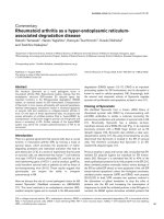
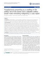
Axial CT scan demonstrating marked right enophthalmosFigure 3
Axial CT scan demonstrating marked right enophthalmos.
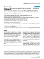
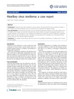
A colour photograph showing right enophthalmosFigure 1
A colour photograph showing right enophthalmos.
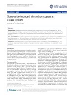
A colour photograph showing the "en coup de sabre" scar on the right forehead (marked by the arrow)Figure 2
A colour photograph showing the "en coup de sabre" scar on
the right forehead (marked by the arrow).
Publish with BioMed Central and every
scientist can read your work free of charge
"BioMed Central will be the most significant development for
disseminating the results of biomedical research in our lifetime."
Sir Paul Nurse, Cancer Research UK
Your research papers will be:
available free of charge to the entire biomedical community
peer reviewed and published immediately upon acceptance
cited in PubMed and archived on PubMed Central
yours — you keep the copyright
Submit your manuscript here:
/>BioMedcentral
Journal of Medical Case Reports 2007, 1:179 />Page 3 of 3
(page number not for citation purposes)
coup de sabre" should aid the examiner in making the
accurate diagnosis.
Competing interests
The author(s) declare that they have no competing inter-
ests. All authors declare no funding was required for the
writing and submission of the manuscript.
Authors' contributions
BSF and PSC prepared the first draft of the manuscript,
participated in the analysis and interpretation of the data.
KT and AEC designed the study. All authors contributed to
the editing and revising of the manuscript and all authors
have read and approved the final version.
Consent
Full verbal and written informed consent has been
obtained from the patient for the submission of this man-
uscript for publication and the accompanying images.
References
1. Cline RA, Rootman J: Enophthalmos: a clinical review. Ophthal-
mology 1984, 91(3):229-37.
2. Koo L, Hatton MP, Rubin PA: When is enophthalmos "signifi-
cant"? Ophthal Plast Reconstr Surg 2006, 22(4):274-7.
3. Holland KE, Steffes B, Nocton JJ, Schwabe MJ, Jacobson RD, Drolet
BA: Linear scleroderma en coup de sabre with associated
neurologic abnormalities. Pediatrics 2006, 117(1):e132-6.
4. Peterson LS, Nelson AM, Su WP: Classification of morphea
(localized scleroderma). Mayo Clin Proc 1995, 70(11):1068-76.
5. Peterson LS, Nelson AM, Su WP, Mason T, O'Fallon WM, Gabriel SE:
The epidemiology of morphea (localized scleroderma) in
Olmsted County 1960–1993. J Rheumatol 1997, 24(1):73-80.
6. Burroughs JR, Hernandez Cospin JR, Soparkar CN, Patrinely JR: Mis-
diagnosis of silent sinus syndrome. Ophthal Plast Reconstr Surg
2003, 19(6):449-54.