Báo cáo y học: "Tuberculous peritonitis in a German patient with primary biliary cirrhosis: a case report" pps
Bạn đang xem bản rút gọn của tài liệu. Xem và tải ngay bản đầy đủ của tài liệu tại đây (411.3 KB, 5 trang )
BioMed Central
Page 1 of 5
(page number not for citation purposes)
Journal of Medical Case Reports
Open Access
Case report
Tuberculous peritonitis in a German patient with primary biliary
cirrhosis: a case report
Yilin Vogel
1
, Jan C Bous
1
, Guido Winnekendonk
2
and Bernhard F Henning*
1
Address:
1
Department of Internal Medicine, Gastroenterology Unit, Marienhospital, Ruhr University, Herne, Germany and
2
Department of
Radiology, Marienhospital, Ruhr University, Herne, Germany
Email: Yilin Vogel - ; Jan C Bous - ;
Guido Winnekendonk - ; Bernhard F Henning* -
* Corresponding author
Abstract
Background: The number of cases of tuberculosis as a complication in people with
immunodeficiency, people on immunosuppressive therapy and among the immigrant population is
increasing in Germany. However, tuberculous peritonitis rarely occurs without these risks,
particularly in Germans. The incidence of tuberculous peritonitis in Germany is very low;
tuberculosis of the intestinal tract was found in approximately 0.8 % of tuberculosis cases in 2004.
The diagnosis of tuberculous peritonitis is often delayed on account of non-specific clinical
symptoms. The absence of specific biological markers, long incubation times for cultures and non-
specific radiographic or ultrasonographic signs increase the morbidity associated with this treatable
condition.
Case presentation: We report a case of tuberculous peritonitis in a 73-year-old female German
patient. Her medical history revealed primary biliary cirrhosis (PBC) since 1992. On admission, she
complained of abdominal pain, vomiting, ascites and peripheral edema. The patient has been in a
seriously reduced general condition and had fever up to 39.6°C. A few weeks earlier, the patient
was in another hospital with the same complaint. Inflammatory parameters were elevated, but the
procalcitonin level was normal. Blood culture was always negative, as was the tuberculin test.
Ultrasonography of the abdomen showed massive ascites with multiple septa. The patient
underwent a computed tomography (CT) scan of the abdomen which showed a thickened intestinal
wall in the sigmoid colon and a pronounced enhancement of the peritoneum. Computed
tomography scans of the lung showed only slight bilateral pleural effusion. Because of the
anaesthetic and bleeding risk due to thrombocytopenia, laparoscopy was not immediately
undertaken. The culture from ascites was positive for M.tuberculosis after three weeks.
Conclusion: In primary biliary cirrhosis patients with non-specific clinical symptoms, such as
vomiting, abdominal pain, ascites, weight loss, and fever, tuberculous peritonitis must be considered
in the initial differential diagnosis, although these symptoms may be attributed to cirrhosis of the
liver with spontaneous bacterial peritonitis. Ultrasonographic and CT scab findings are not specific
for tuberculous peritonitis, but an awareness of the ultrasonographic features and the features of
the CT scan may help in the diagnosis of tuberculous peritonitis and avoid clinical mismanagement.
Published: 31 January 2008
Journal of Medical Case Reports 2008, 2:32 doi:10.1186/1752-1947-2-32
Received: 29 June 2007
Accepted: 31 January 2008
This article is available from: />© 2008 Vogel et al; licensee BioMed Central Ltd.
This is an Open Access article distributed under the terms of the Creative Commons Attribution License ( />),
which permits unrestricted use, distribution, and reproduction in any medium, provided the original work is properly cited.
Journal of Medical Case Reports 2008, 2:32 />Page 2 of 5
(page number not for citation purposes)
Background
In industrialised countries, tuberculosis increasingly
occurs in the immigrant population and in patients with
acquired immune deficiency syndrome (AIDS) and those
on immunosuppressive therapy. Tuberculosis of the intes-
tinal tract ranked 8
th
of all forms of tuberculosis (0.8%) in
2004 in Germany, after pulmonary forms (79.6%),
extrathoracic lymph nodes (7%), pleura (3.6%), geni-
tourinary (3.3%), intrathoracic lymph nodes (2.4%),
osteoarticular (1%), and spine (0.9%). Tuberculous peri-
tonitis is also rare in Germany. The diagnosis of any
extrapulmonary forms of tuberculosis is quite difficult; in
the case of peritoneal tuberculosis this is because clinical
manifestations are non-specific, such as weight loss,
abdominal pain, fever, ascites, vomiting [1-3]. The diag-
nosis of tuberculous peritonitis is often delayed on
account of non-specific clinical signs or symptoms,
absence of specific biological markers, long incubation
times for cultures and non-specific radiographic or ultra-
sonographic signs. The prognosis in tuberculous peritoni-
tis was unfavorable before treatment with antituberculous
drugs became available and the mortality averaged 50 per
cent [4].
Case report
Two months before the patient visited our hospital she
had been admitted to the emergency unit of another hos-
pital with vomiting, abdominal pain and weight loss of 10
kg within three months. A diagnosis of spontaneous bac-
terial peritonitis was ruled out. Her clinical signs were ini-
tially attributed to severe gastritis and an ulcer in the
pyloric canal. She had suffered from primary biliary cir-
rhosis (PBC) since 1992 and had been treated with 750
mg of ursodeoxycholic acid daily without immunosup-
pressive therapy. She had no significant past history of
pulmonary or genital tuberculosis. She had given birth to
a son and a daughter.
Physical examination showed a blood pressure of 120/60
mmHg; regular pulse at 84/min; and a body temperature
of 39.6°C. Superficial lymph nodes were not palpable.
Chest examination revealed basal breathing. The patient's
abdomen was distended, and peristaltic sounds were not
audible. Edema of the extremities was present. The initial
laboratory data for blood (Table 1) rendered a high C-
reactive protein (CRP) level of 15.68 mg/dl. Her tubercu-
lin test was negative.
CT scan of the chest showed bilateral pleural effusions
without lymph node swellings. Abdominal ultrasonogra-
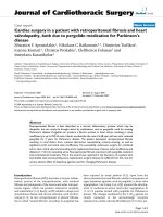
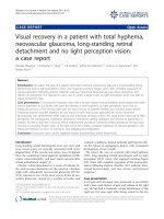
phy revealed massive ascites with multiple septa. A CT
scan of the abdomen showed a thickened intestinal wall
located in the sigmoid colon (Fig. 1) and pronounced
enhancement of the peritoneum. There were no masses or
lymph node swellings in the abdominal cavity. Esoph-
agogastroscopy and ileocoloscopy revealed no ulcer or
stenosis in the colon or ileum.
The nature of ascites was revealed by puncture and find-
ings are listed in Table 2, including a protein level of 4.6
g/dl. Microscopy was requested for malignant cells and
Mycobacterium, neither of which was discovered. The cul-
tures and polymerase chain reaction (PCR) analysis of
stool and urine as well as from bronchial lavage were neg-
ative for M. tuberculosis, but the culture of ascites returned
positive for M. tuberculosis after three weeks. The final
diagnosis was tuberculous peritonitis.
We began anti-tuberculous therapy using isoniazid,
rifampicin, ethambutol, and pyrazinamide.
In the end, the fulminating course of the disease could not
be positively influenced by this therapy and multi-organ
failure with liver failure and nephritic failure developed.
Discussion
Tuberculous peritonitis is always secondary to other
tuberculous lesions. Tuberculous peritonitis appears to be
more common in females than in males. Tuberculosis in
females commonly reaches the peritoneum through tubal
Table 1: Laboratory data for blood on admission
WBC 5.2/nl
Seg 81 %
Lymp 8 %
Mono 8 %
Eos 0 %
Baso 2 %
RBC 3.64/Pl
Hb 11.3 g/dl
Ht 34.2 %
Platelets 52/nl
Total protein 8.4 g/dl
Albumin 1.7 g/dl
GOT 41 U/l
GPT 16 U/l
LDH 301 U/l
ALP 217 U/l
Gamma-GT 90 U/l
Total bilirubin 1.7 ml/dl
CHE 2057 U/l
CRP 15.68 mg/dl
Creatinine 0.67 mg/dl
Na 135 mmol/l
K 4. 3 mmol/l
Lactate 2.47 mmol/l
AFP 1.7 ng/ml
CA 125 67.4 U/ml
CA 19-9 33 U/ml
CEA 3 ng/ml
Quick 45 %
PTT 45 Sec.
Journal of Medical Case Reports 2008, 2:32 />Page 3 of 5
(page number not for citation purposes)
infection and attacks the tubes during the sexually active
period of life. It may be due to either a local extension
from a tuberculous lymph node, Fallopian tube, tubercu-
lous intestinal ulcer, or may be caused by hematogenous
or lymphatic spread from distant sources of infection [4].
Although this patient had no previous medical history of
pulmonary or extra-pulmonary tuberculosis, and the CT
scan of the chest and abdomen showed no lymph node
swellings anywhere, we are certain that the tuberculous
infection was based on reactivation of a long-latent tuber-
culous focus in the peritoneum due to her immunocom-
promised state following a prolonged course of primary
biliary cirrhosis over 14 years.
On the basis of the history, examination and laboratory
findings a differential diagnosis of spontaneous bacterial
peritonitis, bacterial cholangitis, intra-abdominal malig-
nancy or abdominal tuberculosis was considered. The
patient was initially treated with high-dose broad-spec-
trum antibiotics. The patient's condition nevertheless
continued to deteriorate and in addition to the CT and
ultrasonographic findings, we had planned to perform a
laparoscopy. During preparation for laparoscopy the cul-
ture of ascites returned positive for M. tuberculosis after
three weeks.
Extrapulmonary manifestation of tuberculosis can be
found in about 20.4 % of cases in German population [5].
The incidence of tuberculous peritonitis in Germany has
been very low and tuberculosis of the intestinal tract was
found in approximately 0.8% of tuberculosis cases in
2004 [5]. The 'golden rule' for a rapid diagnosis of tuber-
culous peritonitis is a laparoscopy-guided biopsy. But
because of the anaesthetic and bleeding risk, laparoscopy-
guided biopsy was not an immediately available option
for our patient.
Positive cultures for M. tuberculosis have been reported
from 7.8% in a small case report [6], up to 83% [7], which
may be dependent on the fluid quantity. 1L of fluid was
recommended by Singh et al. [7].
The use of PCR to detect M. tuberculosis was diagnostically
useful in patients with ascites who were suspected of hav-
Table 2: Laboratory data of ascites on admission
Protein 4.6 mg/dl
Glucose 60 mg/dl
LDH 442 U/l
Cholesterol 43 mg/dl
Leukocytes 0.3/nl
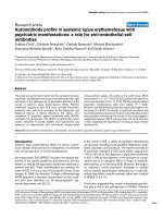
CT pelvis pronounced contrast enhancement of the peritoneum ( ); thickened wall of the sigmoid colon ( )
Figure 1
CT pelvis pronounced contrast enhancement of the peritoneum ( ); thickened wall of the sigmoid colon ( ).
Journal of Medical Case Reports 2008, 2:32 />Page 4 of 5
(page number not for citation purposes)
ing tuberculous peritonitis in order to achieve a prompter
diagnosis and treatment. The IS6110 primer was detected
in 60% of specimens [8,9]. Unfortunately the PCR analy-
sis of ascitic fluid was not performed in this case.
Adenosine deaminase (ADA) levels are used for diagnos-
ing tuberculosis in several locations and have also been
recommended in suspected tuberculous peritonitis. The
pertinent literature judges the usefulness of ADA levels in
ascitic fluid as a diagnostic test for peritoneal tuberculosis
differently. Riquelme et al. reported that ADA levels
showed a high sensitivity (100%) and specificity (97%) in
ascitic fluid using Giusti's methods [1]. Marinez-Vazquez
reported that ADA is not specific for tuberculous peritoni-
tis [10]. Lower sensitivities were reported in the context of
underlying liver cirrhosis, and false positives occurred in
malignancy and bacterial peritonitis [10,11]. ADA levels
were not measured in this instance.
The question, why the tuberculin test was negative in this
case, cannot be answered easily. New types of immuno-
logical test methods such as the Quanti FERON – TB Gold
in – tube (ELISA assay) and the T – SPOT – TB test (ELIS-
POT assay), which are based on the interferon γ (IFN – γ)
production of sensitized T lymphocytes, may yet provide
a useful additional diagnostic method. In patients with
extrapulmonary tuberculosis, a sensitivity of the IFN – γ
test of 92 % was observed, although only 13 patients were
included in the study [12]. Unfortunately, these methods
were not available in Germany at the time the patient was
admitted.
In this case, massive ascites was observed with multiple
fine delicate septa on ultrasonography, and a thickened
intestinal wall located in the sigmoid colon and pro-
nounced enhancement of peritoneum was seen on CT
scan. Case reports [13] and small case studies in the liter-
ature have already reported these findings retrospectively
and prospectively [14-17].
Although tuberculous peritonitis may be associated with
alcoholic cirrhosis of the liver, patients with PBC usually
have ascites, making the diagnosis more difficult. At the
time of diagnosis the decision to initiate anti-tuberculous
therapy turned out to be difficult due to concomitant seri-
ous liver failure and no histological or bacteriological
confirmation of infection with M. Tuberculosis. Five days
after the therapy commenced the patient died of liver and
multiple organ failure. In hindsight, an anti-tuberculous
treatment should have been started without waiting for
the culture report.
Conclusion
Tuberculous peritonitis must be considered in the initial
differential diagnosis of patients with non-specific clinical
signs and symptoms such as vomiting, abdominal pain,
ascites, weight loss and fever that mimic the picture of
spontaneous bacterial peritonitis in patients with PBC.
The sonographic findings are not specific in tuberculous
peritonitis, but can be useful in differentiating tubercu-
lous ascites. An awareness of the ultrasonographic features
may contribute valuable information, help in the diagno-
sis of tuberculous peritonitis, improve diagnostic accuracy
and avoid clinical mismanagement.
Abbreviations
ADA = adenosine deaminase activity; CT = computed
tomography; IFN – γ = Interferon γ; M = Mycobacterium;
PBC = primary biliary cirrhosis; polymerase chain reaction
= PCR.
Competing interests
The author(s) declare that they have no competing inter-
ests.
Authors' contributions
YV was responsible for the patient's management; and
manuscript design and drafting.
JB assisted with the manuscript draft and figures and pro-
vided general technical support.
GW was responsible for the radiological findings and pro-
vided the figures.
BH was responsible for the design, coordination and
supervision of the patient's management.
All authors read and approved the final manuscript.
Consent
Written informed consent was obtained from the patient's
relatives for the publication of the study.
References
1. Riquelme A, Calvo M, Salech F, Valderrama S, Pattillo A, Arellano M,
Arrese M, Soza A, Viviani P, Letelier LM: Value of adenosine
deaminase (ADA) in ascitic fluid for the diagnosis of tubercu-
lous peritonitis: a meta-analysis. J Clin Gastroenterol 2006,
40(8):705-10.
2. Bernhard JS, Bhatia G, Knauer CM: Gastrointestinal tuberculosis:
an eighteen-patient experience and review. J Clin Gastroenterol
2000, 30(4):397-402.
3. Khan R, Abid S, Jafri W, Abbas Z, Hameed K, Ahmad Z: Diagnostic
dilemma of abdominal tuberculosis in non-HIV patients: an
ongoing challenge for physicians. World J Gastroenterol
12(39):6371-5. 2006, Oct 21;
4. Sochocky S: Tuberculous peritonitis. A review of 100 cases.
Am Rev Respir Dis 1967, 95(3):398-401.
5. Brodhun B, Altmann D, Haas W: Report of epidemiology of tbc
in Germany in 2004. .
6. Demir K, Okten A, Kaymakoglu S, Dincer D, Besisik F, Cevikbas U,
Ozdil S, Bostas G, Mungan Z, Cakaloglu Y: Tuberculous peritonitis
– reports of 26 cases, detailing diagnostic and therapeutic
problems. Eur J Gastroenterol Hepatol 2001, 13(5):581-5.
Publish with BioMed Central and every
scientist can read your work free of charge
"BioMed Central will be the most significant development for
disseminating the results of biomedical research in our lifetime."
Sir Paul Nurse, Cancer Research UK
Your research papers will be:
available free of charge to the entire biomedical community
peer reviewed and published immediately upon acceptance
cited in PubMed and archived on PubMed Central
yours — you keep the copyright
Submit your manuscript here:
/>BioMedcentral
Journal of Medical Case Reports 2008, 2:32 />Page 5 of 5
(page number not for citation purposes)
7. Singh MM, Bhargava AN, Jain KP: Tuberculous peritonitis. An
evaluation of pathogenetic mechanisms, diagnostic proce-
dures and therapeutic measures. N Engl J Med 281(20):1091-4.
1969, Nov 13;
8. Tzoanopoulos D, Mimidis K, Giaglis S, Ritis K, Kartalis G: The use-
fulness of PCR amplification of the IS6110 insertion element
of M. tuberculosis complex in ascitic fluid of patients with
peritoneal tuberculosis. Eur J Intern Med 2003, 14(6):367-371.
9. Uzunkoy A, Harma M, Harma M: Diagnosis of abdominal tuber-
culosis: Experience from 11 cases and review of the litera-
ture. World J Gastroenterol 10(24):3647-3649. 2004 December 15;
10. Martinez-Vazquez JM, Ocana I, Ribera E, Segura RM, Pascual C: Ade-
nosine deaminase activity in the diagnosis of tuberculous
peritonitis. Gut 1986, 27(9):1049-53.
11. Hillebrand DJ, Runyon BA, Yasmineh WG, Rynders GP: Ascitic fluid
adenosine deaminase insensitivity in detecting tuberculous
peritonitis in the United States. Hepatology 1996,
24(6):1408-12.
12. Ravn P, Munk ME, Andersen AB, Lundgren B, Lundgren JD, Nielsen
LN, Kok-Jensen A, Andersen P, Weldingh K: Prospective evalua-
tion of a whole-blood test using Mycobacterium tuberculo-
sis-specific antigens ESAT-6 and CFP-10 for diagnosis of
active tuberculosis. Clin Diagn Lab Immonol 2005, 12(4):491-6.
13. Makiyama A, Okuyama Y, Okajima T, Fujimoto S: Tuberculous
peritonitis. J Gastroenterol 2003, 38(12):1167-70.
14. Yilmaz T, Sever A, Gur S, Killi RM, Elmas N: CT findings of abdom-
inal tuberculosis in 12 patients. Comput Med Imaging Graph 2002,
26(5):321-5.
15. Rodriguez E: Pom. Peritoneal tuberculosis versus peritoneal
carcinomatosis: distinction based on CT findings. J Comput
Assist Tomogr 1996, 20(2):269-72.
16. Lee DH, Lim JH, Ko YT, Yoon Y: Sonographic findings in Tuber-
culous peritonitis of wet-ascitic type. Clinical Radiology 1991,
44:306-310.
17. Akhan O, Demirkazik FB, Demirkazik F, Gulekon N, Eryilmaz M,
Unsal M, Besim A: Tuberculous peritonitis: ultrasonic diagno-
sis. J Clin Ultrasound 1990, 18:711-714.