báo cáo khoa học: " Identification of differentially expressed genes associated with semigamy in Pima cotton (Gossypium barbadense L.) through comparative microarray analysis" docx
Bạn đang xem bản rút gọn của tài liệu. Xem và tải ngay bản đầy đủ của tài liệu tại đây (344.88 KB, 9 trang )
RESEARCH ARTICLE Open Access
Identification of differentially expressed genes
associated with semigamy in Pima cotton
(Gossypium barbadense L.) through comparative
microarray analysis
Jessica Curtiss
1
, Laura Rodriguez-Uribe
1
, J McD Stewart
2
, Jinfa Zhang
1*
Abstract
Background: Semigamy in cotton is a type of facultative apomixis controlled by an incompletely dominant
autosomal gene (Se). During semigamy, the sperm and egg cells undergo cellular fusion, but the sperm and egg
nucleus fail to fuse in the embryo sac, giving rise to diploid, haploid, or chimeric embryos composed of sectors of
paternal and maternal origin. In this study we sought to identify differentially expressed genes related to the
semigamy genotype by implementing a comparative microarray analysis of anthers and ovules between a non-
semigametic Pima S-1 cotton and its doubled haploid natural isogenic mutant semigametic 57-4. Selected
differentially expressed genes identified by the microarray results were then confirmed using quantitative reverse
transcription PCR (qRT-PCR).
Results: The comparative analysis between isogenic 57-4 and Pima S-1 identified 284 genes in anthers and 1,864
genes in ovules as being differentially expressed in the semigametic genotype 57-4. Based on gene functions, 127
differentially expressed genes were common to both semigametic anthers and ovules, with 115 being consistently
differentially expressed in both tissues. Nine of those genes were selected for qRT-PCR analysis, seven of which
were confirmed. Furthermore, several well characterized metabolic pat hways including glycolysis/gluconeogenesis,
carbon fixation in photosynthetic organisms, sesquiterpenoid biosynthesis, and the biosynthesis of and response to
plant hormones were shown to be affected by differentially expressed genes in the semigametic tissues.
Conclusion: As the first report using microarray analysis, several important metabolic pathways affected by
differentially expressed genes in the semigametic cotton genotype have been identified and described in detail.
While these genes are unlikely to be the semigamy gene itself, the effects associated with expression changes in
those genes do mimic phenotypic traits observed in semigametic plants. A more in-depth analysis of semigamy is
necessary to understand its expression and regulation at the genetic and molecular level.
Background
Semigamy is a naturally occurring mutation that condi-
tions atypical reproductive behavior in plants. It has been
described in 13 plant species including Rudbeckia spp.,
Zephyranthes spp., Cooperia pedunculata, Coix aquatica,
Gossypium barbadense, and most recently Theobroma
cacao [1-6]. During semigamy, the sperm and egg cells
undergo syngamy or cellular fusion, but forgo karyogamy,
the fusion of the sperm and egg nuclei. In most semiga-
metic plant species, the male nucleus and its derivatives
are sequestered following syngamy and do not contribute
to the genetic makeup of the zygote [3,4]. However, in
G. barbadense and T. cacao, both of which are members
of the plant family Malvaceae, the mode of semigamy is
unique in that the male nucleus is not sequestered and
does contribute its genetic material to the embryo [5,6].
Consequently, the maternal and paternal nuclei p roceed
to divide independently resulting in several possible pro-
genies including normal tetraploids, diploids, haploids, or
chimeric embryos.
* Correspondence:
1
Department of Plant and Environmental Sciences, New Mexico State
University, Las Cruces, NM 88003, USA
Full list of author information is available at the end of the article
Curtiss et al. BMC Plant Biology 2011, 11:49
/>© 2011 Curtiss et al; licensee BioMed Central Ltd. This is an Open Acces s article distributed und er the terms of the Creative Com mons
Attribution License ( which permits unrestricted use, distribution, and reproduction in
any medium, provided the original work is properly cited.
In cotton, semigamy was first observed by Turcotte
and Feaster [5] through recovery of a doubled haploid
mutant 57-4 from a commercial non-semigametic Pima
S-1, which produced haploids at a high frequency, ran-
ging from 25 to 61% when self pollinated. Subsequent
breeding and genetic experiments revealed that semi-
gamy was an inheritable trait and controlled by a single
incompletely dominant gene, denoted Se [7,8]. A unique
feature of semigamy in cotton is that expression of the
trait in terms of haploid production is controlled by the
genotype of both male and female gametes [8]. Zhang
and Stewart [8] reported that the semigametic line 57-4
produced 44% haploids when both gametes carried the
semigametic gene Se by self pollination, but produced
only 11% haploids when crossed as female to its nonse-
migametic isoline Pima S-1. However, no haploids were
detected when 57-4 was crossed as male to Pima S-1.
This indicates that a special microenvironment in the
embryo sac provided by the semigametic genotype is
essential for haploid production. Also, a similar condi-
tion in male gametes with the semigametic genotype
can substantially facilitate semigamy expression, indicat-
ing that the semigametic ge ne is expressed in both male
and female gametes for a maximum haploid production.
This also lays the foundation for searching for the
expressed Se gene and associat ed gene expression using
both male and female tissues in the present study.
While there have been attempts at molecular analysis
related to semi gamy in cotton [9], there is currently little
known about the molecular genetics and gene expression
of semigamy. Therefore, the objective of this study was to
identify differentially expressed genes associated with the
semi gametic genotype using microarray analysi s in order
to gain insight into the underlying molecular mechanism
of semigamy in cotton. To our knowledge, this is the first
report of microarray and quantitative reverse transcrip-
tion PCR (qRT-PCR) usage associated with semigamy
and will hopefully lay the groundwork towards under-
standing its genetic mechanism, regulation and control.
Results
Microarray and data analysis
In this study, RNA from anthers and ovules of flowers at
the 0 day post-anthesis (DPA) were extracted from both
semigametic mutant 57-4 and its nonsemigametic natural
isoline Pima S-1 and compared for transcriptome analysis
using Affymetrix Gen eChip Cotton Gen ome Array. The
data were submitted to the GEO repository with the ser-
ies entry number GSE27242 .
gov/geo/query/acc.cgi?acc=GSE27242. 284 genes in
anthers and 1,864 genes in ovules were found to be dif-
ferentially expressed in the semig ametic genotype 57-4
compared to its non-semigametic isogenic line Pima S-1
(Additional file 1 and 2). Of the 284 differentially
expressed genes identified in the semigametic anther
tissue, 52 were up-regulated and 232 were down-
regulated, while in semigametic ovule tissues 149 genes
were up-regulated and 1,678 genes were down-regulated.
Since it is known that fewer genes are expressed in male
gametes of plants [10], it is not surprising to see much
few differentially e xpressed (DE) genes were identified
when anthers were used. Because the Se gene appears to
be expressed in both male and female gametophytes for
maximum haploid production [8], both ovules and
anthers were harvested for identifying genes that were
consistently up- or down- regulated in both tissues. Out
of the 2,067 total differentially expressed genes identified,
127 genes were found to be differentially expressed in
both tissues, 115 of which were consistently differentially
expressed, i.e., either up- or down- regulated, in both tis-
sues (Additional file 3), which accounted for more than
40% of the DE genes identified in the anthers. For exam-
ple, among 81 genes with the same GeneBank accession
numbers in both tissues, most genes (77) were consis-
tently down-regulated in both anther and ovule tissues of
57-4 and two genes were consistently up-regulated, while
only two differentially expressed genes were inconsistent
(i.e., up-regulated in one tissue, but down-regulated in
another, or vice versa). The correlation of the log2 ratios
between the two tissues based on the 81 genes was found
to be highly significant (r = 0.51, P < 0.01). The common
differentially expressed genes identified in both tissues
indicates common gene regulation mechanism in differ-
ent tissues by the semigamy gene in cotton. It also
demonstrated the reliability of the microarray technology
used in the current study and also provided a greater
confidence in our research results.
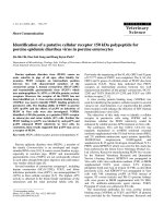
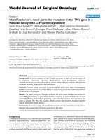
The 127 common differentially expres sed genes identi-
fied in semigametic anthers and ovules were then cate-
gorized based on their cellular function (Figure 1) and
literature pertaining to their corresponding metabolic or
biological pathways was analyzed. Several well character-
ized pathways, such as glycolysis/gluconeogenesis, carbon
fixation in photosynthetic organisms and the tricar-
boxylic acid (TCA) cycle, were found to be affected in
semigametic t issues (Table 1). Additionally, there were
several differentially expressed genes related to hormo ne
biosynthesis and response. Both 12-oxophytodienoate
reductase [GeneBank: DT466538], which is involved i n
the biosynthesis of jasmonates, and the gibberellin
response protein DELLA-GAI [GeneBank: DT468888]
were found to be up-regulated in semigametic tissues.
Conversely, an ethylene-responsive transcription factor
[GeneBank: DT047349, AW186839], allene oxide
synthase [GeneBank: DT047194] which also participates
in jasmonate synthesis, and an auxin/indole acetic
acid protein [GeneBank: DW517716, CA992726] were
found to be down-regulated in semigametic tissues.
Curtiss et al. BMC Plant Biology 2011, 11:49
/>Page 2 of 9
In addition, (+)-δ-cadinene synthase [GeneBank: U23206,
CO107110], which catalyzes the first step in gossypol
synthesis in cotton, was found to be up-regulated in
semigametic anthers and ovules. Another common find-
ing was the down-regulation of cytoskeletal proteins,
such as a-tubulin [GeneBank: DT052122] and b-tubulin
[GeneBank: CO124756, DW516614, DT507015] in semi-
gametic tissues. However, genes homologous to actin
were found to be up-regulated in semigametic anthers
but down-regulated in semigametic ovules. There were
also several genes related to oxidative stress, such as iron
superoxide dismutase (SOD) [GeneBank: DQ088821] and
Cu/Zn SOD [GeneBank: DQ088818, DQ120514], identi-
fied as down-regulated in semigametic tissues.
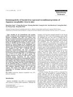
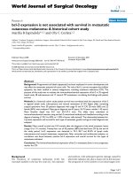
Quantitative reverse transcription PCR
Initially, the six most up-regulated and down-regulated
genes identified in semigametic tissues by microarray
were chosen for confirmation using qRT-PCR (Table 2
and 3). Of the twelve total reactions, seven including
transcription initiation factor TFIID (SeRT 05), 60S
acidic ribosomal protein P1 (SeRT 11) and b-Tubulin 8
(SeRT 19) in anthers as well as histone H1-III (SeRT
04) and high MW heat shock protein (SeRT 14) in both
anthers and ovules, produced significantly different
results between the two isogenic genotypes (Figure 2).
The statistically significant qRT-PCR results are listed in
Table 2.
Previous studies have shown that the rate of photo-
synthesis, specifically carbon dioxide (CO
2
) fixation, is
markedly decreased in semigametic 5 7-4 cotton plants
in comparison to its non-semigametic isoline Pima S-1
[8]. In plants and photosynthetic bacteria, the enzyme
Ribulose-1,5-bisphosphate carboxylase/oxygenase
(Rubisco) catalyzes the first step in photosynthetic CO
2
assimilation and is the overall rate limiting step of
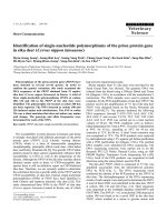
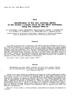
photosynthesis [11]. As a preliminary probe into any
effects of semigamy on the photosynthetic pathways,
three differentially expressed Rubisco genes identified
via microarray analysis, Rubisco activase 1 [GeneBank:
AF329934], Rubisco a ctivase 2 [GeneBank: DQ233255],
and a Rubisco small subunit precursor [GeneBank:
DN780767], were used to perform six qRT-PCR reac-
tions to study the expression of R ubisco i n semigametic
versus non-semigametic anther and ovule tissues. The
results of the reactions are presented in Figure 3. Of the
six total reactions, five were found to be statistically sig-
nificant (Table 2). Rubisco activase 1 was f ound to be
up-regulated in both semigametic anthers and ovules,
mirroring the expression found during microarray analy-
sis. However, expression of Rubisco activase 2 was
found to be down-regulated in both semigametic tissues,
contrary to what was found in the microarray results,
while there was consistent down-regulation of the
Rubisco small subunit precursor in semigametic ovules
in both the qRT-PCR and microarray results.
Discussion
While there are a few microarray platforms for cotton
available, we decided to use Affymetrix GeneChip
Cotton Genome Array for our studies due to its techni-
cal robustness and use of multiple probes for a single
gene (a total of 239,777 probe sets representing 21,854
cotton transcripts). Since 57-4 was a natural doubled
haploid mutant isolated from Pima S-1, both are natural
isogenic lines. A comparison be tween the two genotypes
allows for the identification of genetic and molecular
differences that may be further traced to the semiga-
metic gene itself. For example, Zhang and Stewart
(2005) reported that 57-4 had significantly reduced
photosynthetic rate and chlorophyll content, shorter
fiber length a nd higher microna ire (i.e., courser fiber),
comp ared with Pima S-1 [8]. In this study, 284 genes in
anthers and 1,864 genes in ovules were identified as
being differentially expressed in the semigametic geno-
type 57-4 relative to Pima S-1. Although the list of com-
mon differentially expr essed genes in semigametic
tissues is too large to analyze individually and one of
them may be the semigamy gene itself the limitation of
the current microarray analysis did not allow pinpoint-
ing of the semigamy gene. However, there were several
interesting genes in the group that deserve a closer
examination. It should also be pointed out that 17 of
the differentially expressed genes identified i n our pre-
vious d ifferential display study [12] were also identified
in our current microarray analysis, further confirming
the corroboration between the two gene expression
Figure 1 Distribution of commonly differentially expressed
genes in semigametic anthers and ovules based on cellular
function.
Curtiss et al. BMC Plant Biology 2011, 11:49
/>Page 3 of 9
technologies. Once again, it is currently unclear whether
one o f the genes is the semigamy gene without a com-
pletion of genetic and physical map-based cloning of the
Se gene.
Choline production and response to environmental stress
In plants, the metabolite choline is of vital importance
because it is used to synthesize phosphatidylcholine, a
major membrane lipid. Additionally, in some plant spe-
cies choline is oxidized to glycine betaine, which acts as
a potent osmoprotectant that confers tolerance to high
salinity, drought and other environmental stresses [13].
Phosphoethanolamine N-methyltransferase is a key
enzyme which catalyzes the steps necessary to convert
phosphoethanolamine to phosphocholine. Recent studies
have shown that silencing of phosphoethanolamine
Table 1 Noteworthy differentially expressed genes identified in semigametic tissues
Category Gene Log
2
Signal Anthers Log
2
Signal Ovules GeneBank ID
Glycolysis and TCA Fructose-bisphosphate aldolase 2.3 -1.5 CA993106/AI054483
Succinate dehydrogenase -1.0 -2.3 DT570098/CO122837
Phosphoglycerate kinase -1.3 -2.2 DW481615/DW498822
Glucose-6-isomerase - -1.1 DT456471
Pyruvate dehydrogenase subunit E1 - -2.1 DT570955
Photosynthesis Oxygen-evolving enhancer protein -3.6 -1.3 DT458079/CO093680
Rubisco small subunit precursor 3.5 -1.1 DN780767/CO496683
Rubisco activase 1 1.8 - AF329934
Rubisco activase 2 1.6 - DQ233255
Chlorophyll A/B binding protein 1.1 -1.5 CA992778
Cytochrome b5 -1.3 -1.3 CO085819/DT047754
Cytochrome c oxidase - 3.2 CA993773
Metabolism (+)-δ-cadinene synthase 1.4 1.4 U23205/CO107110
Phosphoethanolamine N-methyltransferase -1.1 -1.3 DW225135
Cytoskeleton a-Tubulin - -1.5 DT052122
Actin 1.0 -1.4 DN759693/CO084889
b-Tubulin 1 -1.0 -1.5 CO124765/DW516614
b-Tubulin 3 - -2.1 DT557030
b-Tubulin 8 -1.1 - CO124872
Tubulin -1.4 -2.4 DW507015
Hormone-related 12-oxophytodienoate reductase 1.1 1.2 DT466538
Allene oxide synthase -1.2 - DT047194
DELLA protein GAI 1.3 1.1 DT468888
Ethylene-responsive transcription factor 5 -2.5 -2.2 DT047349/AW186839
Ethylene-responsive transcription factor ERF017 -5.0 -2.2 DT049130
Auxin/Indole acetic acid protein -2.0 -2.0 DW517716/CA992726
Auxin repressed protein - -1.1 CO127792
ACC synthase -1.2 - DQ122174
ACC oxidase 1.0 - DQ116442
SOD-related FeSOD -1.9 -1.9 DQ088821
Cytosol Cu/Zn SOD - -1.4 DQ088818
Chloroplast Cu/Zn SOD - -1.1 DQ120514
Dashes designate that the gene was not found to be differentially expressed through microarray analysis.
Curtiss et al. BMC Plant Biology 2011, 11:49
/>Page 4 of 9
N-methyltransferas e in Arabidopsis thaliana resulted in
abnormal growth and temperature-sensitive male steri-
lity, which was attributed to failure to produce func-
tional pollen [13,14]. This f inding bodes well with a
previous differential display study comparing gene expres-
sion between semigametic 57-4 and non-semigametic
Pima S-1, which also identified phosphoethanolamine
N-methyltransfer ase as being down-regulated in semiga-
metic tissues [12]. While down-regulation
of phosphoethanolamine N-methyltransferase is likely
to result in decreased choline and phosphotidylcho-
line levels, it may also result in lower levels of glycine
betaine, which would render semigametic plants
more susceptible to h igh soil salinity and other
environmental stress ors, such as reactive oxygen species .
According to a previous study, some phosphoethanola-
mine N-methyltransferase mutants exhibited pale green
leaf color when subjected to high salinity [14], which may
indicate a decrease in leaf chlorophyll levels. A more
recent study into the effects of s alt stress on cotton
revealed that the rate of photosynthesis and the activity of
Rubisco decreased as salinity increased [15]. In cotton,
Zhang and Stewart [8] noted that the chlorophyll content
as well as the rate of photosynthesis is markedly reduc ed
in semigametic cotton plants. Furthermore, the rate of
photosynthesis, especially CO
2
fixation, can be severely
affected by reactive oxygen species, such as the superoxide
radical, hydrogen peroxide, and the hydroxyl radical [16].
Table 2 Statistically significant qRT-PCR results
Target Gene Tissue qRT-PCR Result Microarray Result
Histone H1-III Anthers 2.0-fold increase 6.5-fold increase
Histone H1-III Ovules 1.6-fold increase -
b-Tubulin Anthers 1.8-fold decrease 2.6-fold decrease
High MW heat shock protein Anthers 1.8-fold decrease 2.8-fold decrease
High MW heat shock protein Ovules 5.0-fold decrease 12.1-fold decrease
Transcription initiation factor TFIID Anthers 1.4-fold increase 6.5-fold increase
Rubisco activase 1 Anthers 1.7-fold increase 3.5-fold increase
Rubisco activase 1 Ovules 5.7-fold increase -
Rubisco activase 2 Anthers 2.3-fold decrease 3.0-fold increase
Rubisco activase 2 Ovules 1.1-fold decrease -
Rubisco small subunit precursor Ovules 1.1-fold decrease 2.1-fold decrease
The dashes designate that the gene was not found to be differentially expressed via microarray analysis.
Table 3 Results for each gene analyzed using qRT-PCR
Primer Name Target Gene Tissue PS-1 Expression 57-4 Expression
SeRT 04 Histone H1-III Anthers 1.000 ± 0.119 2.222 ± 0.194
Ovules 1.000 ± 0.081 1.632 ± 0.187
SeRT 05 Transcription initiation factor TFIID Anthers 1.000 ± 0.158 1.374 ± 0.100
Ovules 1.000 ± 0.149 1.076 ± 0.170
SeRT 11 60S acidic ribosomal protein P1 Anthers 1.000 ± 0.066 1.632 ± 0.073
Ovules 1.000 ± 0.108 0.960 ± 0.061
SeRT 13 E3 ubiquitin-protein ligase Anthers 1.000 ± 0.143 1.076 ± 0.151
Ovules 1.000 ± 0.077 0.954 ± 0.103
SeRT 14 High MW heat shock protein Anthers 1.000 ± 0.048 0.552 ± 0.199
Ovules 1.000 ± 0.049 0.201 ± 0.017
SeRT 19 b-Tubulin 8 Anthers 1.000 ± 0.094 0.542 ± 0.077
Ovules 1.000 ± 0.061 0.978 ± 0.099
RBC 01 Rubisco activase 1 Anthers 1.000 ± 0.089 1.674 ± 0.247
Ovules 1.000 ± 0.138 5.745 ± 0.601
RBC 02 Rubisco activase 2 Anthers 1.000 ± 0.027 0.434 ± 0.018
Ovules 1.000 ± 0.017 0.950 ± 0.013
RBC SmSub Rubisco small subunit precursor Anthers 1.000 ± 0.074 0.861 ± 0.077
Ovules 1.000 ± 0.026 0.885 ± 0.056
Curtiss et al. BMC Plant Biology 2011, 11:49
/>Page 5 of 9
Production of and response to plant hormones
Ethylene is a potent plant hormone that regulates many
aspects of plant growth and development, such as fruit
and flower maturation as well as other physiological
effects associated with aging [17]. In cotton, production
of ethylene has been shown to be one of the most
significantly up-regulated biochemical pathways during
fiber cell elongation and it was found that exogenously
applied ethylene promot ed robust fiber cell elongation,
whereas its biosynthetic inhibitor L-(2-aminoethoxyvi-
nyl)-glycine reduced fiber length [18]. The down-regula-
tion of an ethylene responsive transcription factor
identified in the semigametic tissues may have an adverse
effect on ethylene production and a decrease in ethylene
production in turn could result in the production of
shorter, coarser fibers previously observed in the semiga-
metic cotton 57-4 in comparison to Pima S-1 [8]. How-
ever, their relationship with respect to semigamy is
currently unknown.
The hormone gibberellin has an important role in plant
development and growth as well a s signal transduction
pathways which influence gene expression and plant mor-
phology [19]. Gibberellic acid signaling has been shown to
be a de-repressible system controlled by DELLA proteins
[20]. DELLA proteins act as transcriptional modulators
which repress response to gibberellins. In semigametic tis-
sues, a gibberellic acid insensitive DELLA (DELLA-GAI)
protein was found to be up-regulated in both anthers and
ovules. Previously, genetically engineered apple trees con-
taining an Arabidopsis gai gene exhibited a dwarf ed phe-
notype [21] similar to the shorter statue observed in
semigametic 57-4 cotton plants in comparison to Pima
S-1 [8]. Gibberellic acid was also shown to induce expres-
sion of xyloglucan endotransglycosylase and expansin gene
during fiber cell elongation in cotton [22]. Both xyloglucan
endotransglycosylase and several expansins were found
to be down-regulated in semigametic tissues, signifying
that gibberellins may play some part in the semigamy
phenotype.
Jasmonates are a class of plant hormone that play a key
role in the regulation o f reproduction, metabolism,
response to abiotic stress, and defense responses against
pathogens and insects [23]. Biosynthesis of jasmonates has
also been shown to be of critical importance in pollen
maturation and dehiscence. Previous studies have shown
that knock-out mutants of allene oxide synthase, the first
committed step in jasmonate synthesis result in male steri-
lity [23,24]. Additionally, a mutant of 12-oxophytodienoate
reductase was also shown to be male-sterile due to lack of
jasmonic acid synthesis [25]. In semigametic anthers,
allene oxide synthase was identified as down-regulated
while 12-oxophytodienoate reductase was found to be up-
regulated in bo th semigametic anthers and ovules. While
both of these genes are interesting due to the fact that
they can result in male sterility, the role of jasmonates in
semigamy is currently unknown.
Cytoskeletal components
Cytoskeleton plays an important critical role in plant
growth and development through regulating an array of
Figure 2 qRT-PCR results. SeRT04-Histone H1-III, SeRT05-
Transcription initiation factor TFIID, SeRT11-60S acidic ribosomal
protein, SeRT13-E3 ubiquitin-protein ligase, SeRT14-High MW heat
shock protein, SeRT19-Tubulin beta-8. The dashed line represents
gene expression in non-semigametic Pima S-1 (PS-1) tissues.
Asterisks (*) indicate that the result was statistically significant
between the two genotypes.
Figure 3 qRT-PCR results for Rubisco-related target genes.
RBC01-Rubisco activase 1, RBC02-Rubisco activase 2, RBCSmSub-
Rubisco smallchain chloroplast precursor. The dashed line represents
gene expression in non-semigametic Pima S-1 (PS-1) tissues.
Asterisks (*) indicate that the result was statistically significant
between the two genotypes.
Curtiss et al. BMC Plant Biology 2011, 11:49
/>Page 6 of 9
fundamental cellular processes such as cell division, cell
expansion, organelle motility and vesicle trafficking.
While the mechanism of movement of the sperm cells to
the egg and central cell during double fertilization
remains largely unknown, previous studies have shown
that reorganization of the cytoskeleton may play a key
role in the transport process. In studying the process of
double fertilization in Nicotiana tabacum,Huangand
Russell [26] noted dramatic changes in cytoskeletal reor-
ganization. It has been postulated that abundant actin in
the embryo sac, also called actin coronas, plays a key role
in aligning the male gametes to their target cells and
facilitating gametic fusion [26-28]. In our microarray ana-
lyses, several genes homologous to tubulins were found
to be down-regulated in semigametic tissues and actin
was found to be down-regulated in semigametic ovules
but up-regulated in semigametic anthers (Table 1). The
down-regulation of actin in semigametic ovules may
cause the misalignment of the sperm cell and inhibition
of sperm movement. Even though the function of micro-
tubules in double fertilization is minor, their involvement
in the process of semigamy in cotton is currently
unknown. In addition, the mechanism by which the
sperm nucleus migrates to the egg nucleus once it has
penetrated the egg cell still remains enigmatic.
Biosynthesis of gossypol
This study revealed that delta-cadinene synthase was
up-regulated in both anther and ovule tissues of 57-4 as
compared to these of Pima S-1. Delta-cadinene synthase
is the first committed step in a multi-enzyme process
leading to the production of gossypol, a polyphenolic
yellow pigment produced by most cotton species that
acts as a natural insecticide [29]. Gossypol is a chiral
compound due to restricted rotation between the
naphthalene ring systems, with the (-)-enantiomer being
more biologically active than the (+)-enantiomer. Pre-
vious studies have shown that Pima cotton (G. barba-
dense) produces more of the biologically active
(-)-enantiomer than the majority of other cotton species;
these of the species produce more of the biolo gically
inert (+)-enantiomer than G. barbadense [30,31]. The
compound has great pharmacological interest due to its
potential as an anti-cancer agent and for its male con-
traceptive abilities [29]. In human spermatozoa, gossypol
was shown to inhibit the motility of sperm cells through
a dos e dependent mechanism [32]. Upon a closer exam-
ination, it was found that gossypol can inhibit enzymes
of glycolysis and the TCA cycle, severely crippling
energy metabolism and ATP production. Additional stu-
dies have shown that g ossypol binds tubulin monomers
non-covalently such that they cannot participate in
microtubule polymerization [33]. As previously men-
tioned, microtubules may play a key role in transporting
the sperm nucleus to the egg nucleus during karyogamy.
Thus inability to form complete microtubules may inhi-
bit karyogamy from occurring during fertilization. Dur-
ing our microarray analyses, several genes homologous
to actin and tubulins were found to be down-regulated
in semigametic tissues (Table 1). I n yet another study
into t he effects of gossypol on a photosynt hetic protist
Dunaliella bioculata, it wa s noted t hat the motility o f
the flagellated protist dropped as expected, however the
authors also noted a significant decline in cellular
respiration and the rate of photosynthesis [34]. This
finding correlates well with the observatio ns of Zhang
and Stew art [8] in semigametic cotton. Lastly, Kennedy
et al. [35] observed that addition of gossypol to sperma-
tozoa prevented the sperm from penetrating denuded
hamster oocytes. Upon further analysis, they discerned
that gossypol ’s inhibition of the autoproteolytic conver-
sion of proacrosin to acrosin results in its contraceptive
ability. This observation is particularly interesting when
considering semigamy in cotton where the egg does not
fuse with the sperm during fertilization. Although repro-
ductive mechanisms in plants and animals are distinc-
tive in many ways, there are also many common
molecular processes [36]. If a system homologous to the
proacrosin-acrosin system in animals were to exist in
plants, the effect of gossypol may very well explain the
lack of nuclear fusion between sperm and egg nuclei
in semigamy. While the increased expression of delta-
cadinene synthase (as it correlates with gossypol concen-
tration) may explain many of the observed phenotypic
traits associated with semigamy, a more focused study
of the two active gland loci, Gl
2
and Gl
3
,orother
genes/alleles and their relationship t o semigamy s hould
be performed through gene expression studies and
molecular marker analysis. Furthermore, the actual
levels of gossypol, as well as the ratio of the two enantio-
mers, should be temporally and spatially measured in
semigametic ovules and seeds relative to non-semigametic
cotton.
Conclusion
To our knowledge this is the first report using microarray
technology and qRT-PCR associated with semigamy in
cotton. In this study, over 2,000 diff erentially expressed
gene s associated with semigamy were identified with 127
of those genes being commonly differentially expressed
in both semigametic anthers and ovules. Several impor-
tant metabolic pathways affected by differentially
expressed genes in the semigametic genotype have been
identified and described in detail. And while these genes
are not likely to be the semigamy gene itself, the effects
associated with over-expressing or under-expressing their
gene products do mimic phenotypic traits observed in
semigametic plants. As a result, a more in-depth future
Curtiss et al. BMC Plant Biology 2011, 11:49
/>Page 7 of 9
analysis of their expression and regulation with respect to
semigamy is necessary.
Methods
Plant materials and RNA isolation
Anther and ovule tissues from Pima S-1 (also designated
PS-1), a normal, non-semigametic yet obsolete G. barba-
densecultivar,andPima57-4, its naturally occurring
semigametic mutant were used. Both genotypes w ere
grown in a greenhouse in peat pots and transplanted to
the field a month later. The experimental design was a
paired comparison with three replicates and the plot
size was single row × 40 ft long. Seeding rate was 3
seed/ft and crop production wa s managed as re com-
mended locally. Anther and ovule tissues from 10 flow-
ers were collected for each replicate of each genotype at
zero days postanthesis (0 DPA) and placed in liquid
nitrogen immediately and stored at -80°C. Total RNA
from collected anthers and ovules was isolated using a
previously described hot borate method [37]. RNA yield
and quality were determined by absorbance spectra at
260 and 280 nm using a DU 530 UV/VIS spectrophot-
ometer (Beckman Coulter, Brea, CA). After quantifica-
tion, the RNA was cleaned using an RNeasy MinElute
Cleanup kit (Qiagen, Valencia, CA). RNA was stored at
-80°C until used.
Microarrays and data analysis
For the microarray experiments, RNA was pooled in an
equal molar ratio from the three biol ogical replicates
based on tissue and genotype. 2 mg cleaned to tal RNA
from each of the four samples, semigametic anthers and
ovules as well as non-semigametic anthers and ovules, and
Affymetrix GeneChips
©
Cotton Genome Array (Santa
Clara, CA) were sent to Genome Explorations (Memphis,
TN) for hybridization and preliminary data analysis.
A pair-wise comparison between semigametic 57-4 and
non-semigametic Pima S-1 tissues was conducted for both
anther and ovule samples in order to identify differentially
expressed genes. Using the Affymetrix GeneChip Operat-
ing Software the relative mean signal, detection calls, sig-
nal log ratios and change calls are independently
calculated using four different algorithms for each probe
set [38]. Excel files with statistically relevant up-regulated
anddown-regulatedgenesandtheirsignalLog
2
ratios
were provided by Genome Explorations.
The sequences of differentially expressed genes identi-
fied by the microarray experiments were collected from
NCBI GeneBank [39] and compared them to known
sequences from C otton Gene In dex [40] using the Basic
Local Alignment Search Tool (BLAST) to determine if
the re was any significant homology to kn own gene pro-
ducts. The results of the BLAST search were then
sorted based on gene function to identify common
differentially expressed genes in both semigametic
anther and ovule tissue.
Quantitative reverse transcription PCR
Nine differentially expressed genes were sele cted based
on the microarray results (i.e., 2-12 fold changes) and
putat ive gene functions were selected and analyzed using
real-time quantitative RT-PCR. Initially, the total RNA
for each sample was quantified using a DU 530 UV/VIS
spectrophotometer (Beckman Coulter, Brea, CA). The
total RNA was then diluted 5-fold with sterile molecular
biology grade water (Promega, Madison, WI) to concen-
trations of 20 ng/μL, 4 ng/μL, and 800 pg/μL. Real-time
PCR assays for each target gene were performed in tripli-
cate for each of the aforementioned concentrations of
total RNA, no reverse transcriptase and no template con-
trols on a Bio-Rad iQ5 Thermal Cycler (Hercules, CA).
One-step RT-PCR reactions of 20 μLvolumecontaining
10 μL EXPRESS SYBR GreenER qPCR SuperMix Univer-
sal (Invitrogen, Carlsbad, CA), 20 nM Fluorescein refer-
ence dye (Invitrogen, Carlsbad, CA), 0.5 μLEXPRESS
SuperScript Reverse Transcriptase (Invitrogen, Carlsbad,
CA), 0.2 μM forward and reverse primers, 1.5 μLRNA
template and 3.2 μL sterile water (Promega, Madison,
WI). Reactions were run using the pre-set one-step RT-
PCR with melt curve program, the cycling parameters of
which were 50°C for 10 min., 95°C for 5 min., followed
by 45 cycles of 95°C for 10 sec. and 60°C for 30 sec., and
ending with the melt curve program. Gene expression
and statistical analysis (Table 3) was performed using the
Bio-Rad iQ5 optical system software utilizing relative
quantification as described in the iQ5 system software
instruction manual (Bio-Rad, Hercules, CA).
Additional material
Additional file 1: Raw microarray data for semigametic anthers.
Additional file 2: Raw microarray data for semigametic ovules.
Additional file 3: BLAST results for all differentially expressed genes
in semigametic anthers and ovules.
Acknowledgements
We thank Mrs. Yingzhi Lu for her help in tissue sampling and Drs. Champa
Sengupta-Gopalan and Suman Bagga for their help in using the iQ5 real-
time thermal cycler. This research was funded by USDA-ARS, Cotton
Incorporated, and the New Mexico Agricultural Experiment Station.
Author details
1
Department of Plant and Environmental Sciences, New Mexico State
University, Las Cruces, NM 88003, USA.
2
Department of Crop, Soil, and
Environmental Sciences, University of Arkansas, Fayetteville, AR 72701, USA.
Authors’ contributions
JZ and JMcDS conceived the study, and JZ supervised the project, revised
the manuscript and finalized the paper. LRU conducted RNA isolation for
microarray analysis. JC conducted the analyses and qRT-PCR, and drafted the
Curtiss et al. BMC Plant Biology 2011, 11:49
/>Page 8 of 9
manuscript. All authors contributed to the manuscript preparation, and read
and approved the final manuscript.
Received: 12 September 2010 Accepted: 16 March 2011
Published: 16 March 2011
References
1. Battaglia E: New cytological phenomenon in embryogenesis (semigamy)
and in microsporogenesis (restitution of double nuclei). Nuovo Giornale
Botanico Italiano 1945, 52:34-38.
2. Solntseva M, Vorsobina D: Semigamy in Zephyranthes carinata Herb.
Doklady Akademii Nauk SSSR 1972, 206:1006-1009.
3. Coe G: Cytology of reproduction in Cooperia pedunculata. American
Journal of Botany 1953, 40:335-343.
4. Rao P, Narayana D: Occurrence and identification of semigamy in Coix
aquatica. Journal of Heredity 1980, 71:117-120.
5. Turcotte EL, Feaster CV: Haploids: High frequency production from single-
embryo seeds in a line of Pima cotton. Science (New York, NY) 1963,
140:1407-1408.
6. Lanaud C: Origins of haploids and semigamy in Theobroma cacao L.
Euphytica 1988, 38:221-228.
7. Turcotte EL, Feaster CV: Semigametic production of haploids in Pima
cotton. Crop Science 1969, 9:653-655.
8. Zhang JF, Stewart JMcD: Semigamy gene is associated with chlorophyll
reduction in cotton. Crop Science 2004, 44:2054-2062.
9. Zhang JF, Nepomuceno A, Stewart JMcD: Gene expression related to the
semigamy genotype in cotton (Gossypium barbadense). Proceedings of the
Beltwide Cotton Conference 1998, 2:1457-1462.
10. Borg M, Brownfield L, Twell D: Male gametophyte development: a
molecular perspective. Journal of Experimental Botany 2009,
60:1465-1478.
11. Spreitzer R, Salvucci M: Rubisco: Structure, regulatory interactions, and
possibilities for a better enzyme. Annual Reviews of Plant Biology 2002,
53:449-475.
12. Curtiss JL: Genetic and molecular analysis of semigamy in cotton
(Gossypium barbadense L.). M.S thesis New Mexico State University, Las
Cruces, NM, USA; 2010, 102.
13. Bolognese CP, McGraw P: The isolation and characterization in yeast of a
gene for Arabidopsis S-adenosylmethionine:phosphoethanolamine N-
methyltransferase. Plant Physiology 2000, 124
:1800-1813.
14. Mou Z, Wang X, Fu Z, Dai Y, Han C, Ouyang J, Bao F, Hu Y, Li J: Silencing
of phosphoethanolamine N-methyltransferase results in temperature-
sensitive male sterility and salt hypersensitivity in Arabidopsis. The Plant
Cell 2002, 14:2031-2043.
15. Desingh R, Kanagaraj G: Influence of salinity stress on photosynthesis and
antioxidative systems in two cotton varieties. General Applications in Plant
Physiology 2007, 33:221-234.
16. Scandalios JG: Oxygen stress and superoxide dismutases. Plant Physiology
1993, 101:7-12.
17. Lieberman M: Biosynthesis and action of ethylene. Annual Reviews in Plant
Physiology 1979, 30:533-591.
18. Shi Y, Zhu S, Mao X, Feng J, Qin Y, Zhang L, Cheng L, Wang Z, Zhu Y:
Transcriptome profiling, molecular biological, and physiological studies
reveal a major role for ethylene in cotton fiber cell elongation. The Plant
Cell 2006, 18:651-664.
19. Sun TP, Gubler F: Molecular mechanism of gibberellin signaling in plants.
Annual Review of Plant Biology 2004, 55:197-223.
20. Fleet CM, Sun TP: A DELLAcate balance: the role of gibberellin in plant
morphogenesis. Current Opinion in Plant Biology 2005, 8:77-85.
21. Zhu LH, Li XY, Welander M: Overexpression of the Arabidopsis gai gene in
apple significantly reduces plant size. Plant Cell Reports 2008, 27:289-296.
22. Aleman L, Kitamura J, Abdel-Mageed H, Lee J, Sun Y, Nakajima M, Ueguchi-
Tanaka M, Matsuoka M, Allen RD: Functional analysis of cotton orthologs
of GA signal transduction factors GID1 and SLR1. Plant Molecular Biology
2008, 68:1-16.
23. Devoto A, Turner JG: Regulation of jasmonate-mediated plant responses
in Arabidopsis. Annals of Botany 2003, 92:329-337.
24. Park JH, Halitschke R, Kim HB, Baldwin IT, Feldmann KA, Feyereisen R: A
knock-out mutant in allene oxide synthase results in male sterility and
defective wound signal transduction in Arabidopsis due to a block in
jasmonic acid biosynthesis. The Plant Journal 2002, 31:1-12.
25. Stintzi A, Browse J: The Arabidopsis male-sterile mutant, opr3, lacks the
12-oxophytodienoic acid reductase required for jasmonate synthesis.
Proceedings of the National Academy of Science 2000, 97:10625-10630.
26. Huang BQ, Russell SD: Fertilization in Nicotiana tabacum: Cytoskeletal
modifications in the embryo sac during synergid degeneration. Planta
1994, 194
:200-214.
27. Fu Y, Yuan M, Huang BQ, Yang HY, Zee SY, O’Brien TP: Changes in actin
organization in the living egg apparatus of Torenia fournieri during
fertilization. Sexual Plant Reproduction 2000, 12:315-322.
28. Ye XL, Yeung EC, Zee SY: Sperm movement during double fertilization of
a flowering plant, Phaius tankervilliae. Planta 2002, 215:60-66.
29. Dodou K: Investigations on gossypol: past and present developments.
Expert Opinion Investigative Drugs 2005, 14:1419-1434.
30. Hron RJ, Kim HL, Calhoun MC, Fisher GS: Determination of (+), (-), and
total gossypol in cottonseed by HPLC. Journal of American Oil Chemists
1999, 76:1351-1355.
31. Cass QB, Oliveira RV, De Pietro AC: Determination of gossypol
enantiomers ratio in cotton plants by chiral higher-performance liquid
chromatography. Journal of Agricultural and Food Chemistry 2004,
52:5822-5827.
32. Wichmann K, Käpyaho K, Sinervirta R, Jänne J: Effect of gossypol on the
motility of human spermatozoa. Journal of Reproduction and Fertility 1983,
69:259-264.
33. Medrano FJ, Andreu JM: Binding of gossypol to purified tubulin and
inhibition of its assembly into microtubules. European Journal of
Biochemistry 1986, 158:63-69.
34. Druez D, Marano F, Calvayrac B, Volochine B, Soufir JC: Effect of gossypol
on the morphology, motility, and metabolism of a flagellated protist,
Dunaliella bioculata. Journal of Submicroscopic Cytology and Pathology
1989, 21:367-374.
35. Kennedy WP, Van der Ven HH, Straus JW, Polakoski KL: Gossypol inhibition
of acrosin and proacrosin, and oocyte penetration by human
spermatozoa. Biology of Reproduction 1983, 29:999-1009.
36. Márton ML, Dresselhaus T: A comparison of early molecular fertilization
mechanisms in animals and flowering plants. Sexual Plant Reproduction
2008, 21:37-52.
37. Wan C, Wilkins TA: A modified hot borate method significantly enhances
the yield of high-quality RNA from cotton (Gossypium hirsutum L.).
Analytical Biochemistry 1994, 223:7-12.
38. Genome Explorations: [].
39. NCBI GeneBank: [].
40. Cotton Gene Index: [ />cgi].
doi:10.1186/1471-2229-11-49
Cite this article as: Curtiss et al.: Identification of differentially expressed
genes associated with semigamy in Pima cotton (Gossypium barbadense
L.) through comparative microarray analysis. BMC Plant Biology 2011
11:49.
Submit your next manuscript to BioMed Central
and take full advantage of:
• Convenient online submission
• Thorough peer review
• No space constraints or color figure charges
• Immediate publication on acceptance
• Inclusion in PubMed, CAS, Scopus and Google Scholar
• Research which is freely available for redistribution
Submit your manuscript at
www.biomedcentral.com/submit
Curtiss et al. BMC Plant Biology 2011, 11:49
/>Page 9 of 9