Practical Pediatric Gastrointestinal Endoscopy - part 3 docx
Bạn đang xem bản rút gọn của tài liệu. Xem và tải ngay bản đầy đủ của tài liệu tại đây (233.61 KB, 22 trang )
36 CHAPTER 3
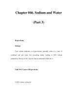
Feature RGB sequential video Color-chip video
endoscopes endoscopes
Image resolution Have a theoretical advantage
because each pixel has unique
image intensity information. The
advantage is primarily seen only
in the very smallest of
endoscopes.
Have a slight
disadvantage because
information from multiple
pixels must be combined
together.
Color accuracy Have a theoretical advantage
because each pixel has unique,
directly measured, full-color
information. Ideal for research
based on spectroscopy and
color-analysis algorithms.
Have a slight
disadvantage because
color is calculated from
values of surrounding
pixels.
Reproduction of motion Stroboscopic illumination creates
problems with rapid motion.
Motion produces color slip and
brightly colored artifacts. Newer
generation systems have
advanced image capture
algorithms to reduce the
color-slip problem.
Smooth, natural
reproduction of motion.
No stroboscopic effect. No
color artifacts. A fast
shutter mode reduces
blurring of quickly moving
subjects.
Abdominal transillumination Strobed illumination produces
very weak transillumination.
‘‘Transillumination Mode’’ results
in good transillumination but
normal imaging is impossible.
The system’s bright,
continuous, white light
illumination is ideal for
transillumination.
Light source compatibility Requires a special strobing light
source.
Videoscopes are
compatible with light
sources for fiberoptic
endoscopes.
Compatibility with laser therapy The red He–Ne aiming beam
appears white and may mask the
tissue-blanching effect. Built-in
filters enable the endoscope to be
used with Nd:YAG lasers.
Built-in filters enable the
endoscope to be used
with Nd:YAG lasers.
Table 3.1 The advantages and disadvantages of the two basic
endoscope imaging systems.
precautions, (ii) Occupational Safety and Health Administration
rules on exposure to blood-borne pathogens, (iii) procedures for
the safe handling of chemicals, (iv) professional society guide-
lines (e.g., those promulgated by ASGE, SGNA, APIC, etc.), and
(v) the manufacturer’s specific procedures for reprocessing the
equipment. Reprocessing personnel must also be adequately
outfitted with appropriate personal protective equipment
for protection against splattering of microorganisms, organic
Problem Troubleshooting
Poor air or water feeding (i) Check that the air pump is turned on and set at the proper setting.
(ii) Check that the water bottle contains sufficient water, that the lid is
screwed on tightly, and that the water bottle tube is connected to the
endoscope. NOTE: If the nozzle on the tip of the instrument is
obstructed by debris, air and water feeding will be compromised.
Thoroughly clean all instrument channels, openings, and nozzles
each time the instrument is reprocessed. Some manufacturers supply
special adapters for bedside precleaning of the air/water system.
Image is not clear (i) Feed water and then air to wash debris off distal objective lens. (ii)
If permanently obscured, clean the objective lens by carefully
rubbing with gauze moistened with alcohol. (iii) Repair the
endoscope if the distal lens is damaged or has moisture trapped
behind it. NOTE: A cracked or badly scratched lens cannot produce
sharp images. Never let the tip of the endoscope contact the floor or
other hard surfaces. Protect the distal tip of the endoscope from
damage. Moisture trapped behind the lens will cloud the image.
Have the endoscope repaired.
Image color is not correct (i) ‘‘White balance’’ the image while pointing the endoscope at a
manufacturer-supplied test fixture or a piece of white gauze. (ii) Make
sure all color adjustment controls on both the video processor and
the video monitor are set in a neutral position. (iii) Check for loose or
broken video cables. NOTE: If the endoscope is ‘‘white-balanced’’
while pointing at a nonwhite surface, distorted color will result. Many
video systems use separate wires for transmitting the red, green, and
blue component images. If one of these wires is disconnected or
broken, the color of the image on the monitor will be severely
distorted.
Image is permanently frozen
or completely absent
(i) Turn off and on again both the light source and the video
processor. This may correct the problem if it is microprocessor
related. (ii) Check all wires for accidental disconnection. (iii) Check
the input selector on the video monitor to ensure that it is set to
display the input with the endoscopic image. (iv) Press the ‘‘Reset’’
button on the video processor, if one is available. This will return the
videoprocessor settings back to their factory defaults. (e.g., if the
video processor was accidentally set to display an image from the
image management software or a VCR, rather than from the
endoscope, pressing Reset will restore the live endoscopic image.)
The image cannot be restored
and the endoscope must be
withdrawn from the patient
(i) Close and remove all accessories from the endoscope channel. (ii)
If using a colonoscope with adjustable stiffness, set the stiffness
control to the ‘‘most flexible’’ setting. (iii) Make sure that the
angulation locks are off. (iv) Return both angulation knobs to their
neutral position in order to straighten the distal tip. (v) Carefully
withdraw the endoscope. NOTE: If the endoscope cannot be
withdrawn easily, stop and contact the endoscope manufacturer’s
service center for additional instructions.
The endoscope is damaged If the endoscope insertion tube is damaged by a patient bite, by
accidental closure in the carrying case hinge, or by other means, do
not continue to use the endoscope. Futher use of the endoscope
could cause additional damage to internal components of the
instrument, adding to the repair cost.
Table 3.2 General troubleshooting information for selective problems.
38 CHAPTER 3
matter, and reprocessing chemicals. Adequate personal protec-
tive equipment includes (i) long-sleeved gowns that are imper-
vious to fluid, (ii) gloves that are long enough to extend up the
arms to protect theforearms, and (iii) eye and/or face protection.
Cleaning
Following patient use, the endoscope should be immediately
precleaned at the bedside by flushing the internal channels and
wiping down the insertion tube. Following bedside precleaning,
the endoscope is brought to the reprocessing room for manual
cleaning. Thorough manual cleaning is often described as be-
ing “the most important step’’ of the entire reprocessing proce-
dure. Cleaning removes gross debris and organic matter that can
dry on the instrumentation and hinder future performance (e.g.,
flow through the air/water nozzle). Studies have shown that
cleaning alone can reduce the number of microorganisms and
organic load on the instrument by 4 logs, or 99.99%. This signif-
icantly reduces the organic and microbial challenge to the high-
level disinfectant or sterilant. Furthermore, residual debris may
inhibit germicide penetration and shield microorganisms from
contact with the germicide. The recommended channel-cleaning
brushes and any special brushes (e.g., channel-opening-cleaning
brush) supplied by the manufacturer must be used to mechan-
ically abrade all lumens while they are wetted with detergent.
After manual cleaning is complete, there should be no visible
debris left on the instrument.
When cleaning and disinfecting the endoscope, the cleaning
tubes and attachments recommended by the endoscope manu-
facturer for flushing the internal lumens of the endoscope must
be used. This ensures that the required volume of fluid for clean-
ing, disinfection/sterilization, and rinsing passes through the in-
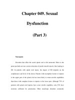
ternal channels. Figure 3.19 illustrates one such manufacturer’s
range of cleaning attachments. The Food and Drug Administra-
tion (FDA) requires that the endoscope manufacturer validate
the steps listed in each instrument’s instruction manual. These
instructions must be followed explicitly. Shortcutting the pre-
scribed procedure may result in an inadequately reprocessed
instrument that presents an infection control risk to medical per-
sonnel and the next patient.
Leak testing
Periodically performing a leak test is an essential part of the re-
processing procedure. Leak testing the endoscope ensures that
the seals, lumens, and external surface of the endoscope are fluid
tight and will not allow reprocessing fluids to enter the inte-
rior of the endoscope. If a leak if detected, have the endoscope
EQUIPMENT 39
Fig. 3.19 The cleaning attachments required to flush reprocessing chemicals through the lumens of a typical
Olympus 160/180-series video endoscope.
40 CHAPTER 3
repaired immediately. Fluid invasion of the endoscope can cause
extensive and expensive damage. Furthermore, a breach in the
surface integrity of the endoscope can allow microorganisms
to enter the endoscope body, where they can reside and later
emerge, creating an infection control risk. For all these reasons,
every endoscope should be leak tested on a regular basis. Dur-
ing leak testing, and any time the endoscope is submerged in
fluid, it is important that all sensitive components be protected
from fluid contact. Most endoscopes require the attachment of a
water-resistant cap to the electrical connector of the endoscope.
This cap must remain attached during the entire reprocessing
procedure.
High-level disinfection
In 1968, Dr Earle H. Spaulding devised a classification system
that divided medical devices into three categories (critical, semi-
critical, and noncritical) based on the risk of infection involved
with their use. Based on the Spaulding classification system, GI
endoscopes are considered by the FDA to be “semicritical med-
ical devices’’. Semicritical medical devices are instruments that
do not enter sterile areas of the body and are generally in contact
with intact mucous membranes. As such, both high-level disin-
fection and sterilization are acceptable methods for reprocessing
GI endoscopes.
High-level disinfection via an approved liquid chemical ger-
micide is the most commonly used method for reprocessing GI
endoscopes. High-level disinfection destroys all vegetative or-
ganisms, but not necessarily all bacterial endospores. The ger-
micide must be cleared by the FDA explicitly as a high-level dis-
infectant. The FDA has approved several high-level disinfectants
for use on medical devices, including 2.0–3.4% glutaraldehydes,
7.5% hydrogen peroxide, 0.2% peracetic acid, 0.08% peracetic
acid/1% hydrogen peroxide, and 0.55% ortho-phthalaldehyde.
Each of these germicides has advantages and disadvantages in
terms of cost, contact time, temperature, and fume control re-
quirements. However, it is important to note that not all of these
products are compatible with all endoscopes. Always check with
the endoscope manufacturer regarding chemical compatibility.
Endoscopes are composed of a variety of rubbers, plastics,
metals, glasses, adhesives, coatings, etc., which may be either
immediately damaged or gradually deteriorated following long-
term exposure to certain chemicals. Reported damage from in-
compatible chemical germicides includes the loss of exterior
body surface color and/or luster, loss of insertion tube stiffness,
peeling of the insertion tube coating material, pitting and cor-
rosion of anodized aluminum parts, chipping and peeling of
painted and coated parts, crazing of plastic parts, deterioration
EQUIPMENT 41
of adhesives, and insertion tubes that become sticky to the touch.
The damage produced by some chemicals is quickly apparent.
However, other chemicals may initially appear to be compatible,
with cumulative effects that only become apparent following ex-
tended use of the germicide.The compatibility ofall reprocessing
chemicals should be determined by contacting the endoscope
manufacturer. If a third-party repair organization services the
endoscope, check with the service provider regarding the chem-
ical compatibility of replacement parts.
Some germicides are suitable for manual reprocessing at room
temperature. Others require heating and are only approved
for use in automated reprocessors. Glutaraldehydes have been
used for 30 years and are available in both room temperature
and heated formulations. They are relatively inexpensive, but
may require fume control in accordance with local and state
regulations. Glutaraldehyde is an irritant and some individu-
als develop acute sensitivities resulting in irritation to the skin,
eyes, and nasal membranes, headaches, coughing, sneezing, and
asthma-like symptoms. The safe use of glutaraldehydes requires
adequate ventilation (e.g., exhaust hoods, ductless absorbent fil-
ter systems) or enclosed automated reprocessing systems.
The efficacy of any chemical germicide is dependent upon the
manufacturer’s instructions for use. The label instructions re-
garding activation (if required), reuse life, and shelf life must
be followed explicitly. All reusable germicides should be tested
regularly, as recommended by the manufacturer, to ensure that
they exceed the minimum effective concentration of the active in-
gredient. The addition of significant quantities of microbes and
organic matter, dilution by rinse water, and aging of the chemical
solution, will all result in a gradual reduction in the effectiveness
of reusable high-level disinfectants/sterilants.
Alcohol flush
Although many automated reprocessors use 0.2-µm microbial
retention filters to produce “sterile’’ water for the final rinse
following disinfection, other endoscopy units rinse their endo-
scopes in tap water. Irrespective of the quality of the final water
rinse (“tap’’ water, “bacteria-free’’water, “sterile’’water), the en-
tire endoscope should be dried and each of its channels flushed
with 70% alcohol, followed by an air purge prior to reuse or stor-
age. Alcohol aids in the drying process and inhibits the recon-
tamination of the internal channels with water-borne organisms.
Special channels
Some endoscopes have special channels, such as an auxil-
iary water or water-jet channel. These channels must be fully
42 CHAPTER 3
reprocessed after each patient use, regardless of whether the
channel was used during the preceding patient examination. Pa-
tient debris and microorganisms can enter these channels even
if they are not used during the endoscopy exam. These channels
often require additional steps and special attachments to access
and flush the channel with detergent, disinfectant, rinse water,
and alcohol (see Fig. 3.19).
Automated reprocessors
Automated reprocessors standardize the disinfection pro-
cess and decrease personnel exposure to high-level disinfec-
tants/sterilants. No currently available reprocessor is approved
for automating the entire cleaning procedure. As a result, all of
the prescribed steps for manually cleaning the endoscope must
be performed prior to placing it in the automated reprocessor.
If an automated endoscope reprocessor is used, the endoscope
must be connected to the reprocessor using the correct set of con-
necting tubes. Some endoscope models, particularly those with
special channels, may require a different set of connecting tubes
from those used on standard instruments. Failing to connect a
specific channel opening or port to the reprocessor may result in
patient debris and infectious material remaining in the channel.
Failure to reprocess any part of the endoscope poses an infection
control risk to both medical personnel and patients.
Rinsing and disposal
Whether reprocessingmanually orusing anautomated machine,
all disinfectant must be flushed from the endoscope’s internal lu-
mens during the rinse process. There have been several reports
in the medical literature of patients enduring chemical burns
and/or chemical colitis when residual disinfectant solution was
expelled from the endoscope’s channels when used on the fol-
lowing patient.
Some germicides require deactivation or dilution prior to dis-
posal. State or local ordinances may prohibit the dumping or
disposal of certain germicides into the city waste water system.
Check with the germicide manufacturer and with state and local
authorities regarding disposal requirements.
Accessories
Many endoscopic accessories are deemed to be “critical’’medical
devices by the Spaulding classification system, since they either
penetrate mucous membranes (e.g., endoscopic cutting devices)
or enter normally sterile areas of the body (e.g., biliary ducts). As
such, they should be sterilized prior to reuse. Steam sterilization
EQUIPMENT 43
is the preferred method of sterilizing any reusable endoscopic
accessory that is autoclavable. After sterilization, store sterile
accessories in an organized and protected storage system that
prevents damage to the sterile packaging.
Storage
Store reprocessed endoscopes in a well-ventilated storage area
where they are protected against damage and contamination.
Endoscopes should be stored with all valves and removable
parts removed, to facilitate drying. The endoscope-carrying case
should never be used for storage of patient-ready endoscopes.
Carrying cases are not ventilated, easily contaminated, cannot
be reprocessed, and intended for shipping and long-term stor-
age only. Never put an endoscope that has not been completely
reprocessed into its carrying case. In addition, reprocess any en-
doscope that is removed from a carrying case prior to subsequent
patient use.
SUMMARY
During the 1990s, video image endoscopes supplanted fiberop-
tic endoscopes as the preferred instrument for examining the GI
tract. The availability of two distinct technologies for generating
color images (color-chip versus RGB sequential) provides the
endoscopist with a choice of basic systems, each with its own
advantages and disadvantages. Although the basic shape and
function of the instrument have remained unchanged, recent
advancements – including the development of smaller diame-
ter insertion tubes, the incorporation of standard features into
pediatric instruments, improvements in image resolution, and
advanced video processor features – have continued the evolu-
tion of the GI endoscope. Proper reprocessing equipment and
procedures and a trained reprocessing staff are essential to en-
suring the health and safety of health care workers and patients
alike.
FURTHER READING
Alvarado CJ, Mark R. APIC guidelines for infection prevention and con-
trol in flexible endoscopy. Am J Infect Control 2000;28:138–55.
Barlow DE. Flexible endoscope technology: the video image endoscope.
In: Sivak MV Jr (ed), Gastroenterologic Endoscopy, Vol 1, 2nd edn.
Philadelphia, PA: WB Saunders; 2000:29–49.
Barlow DE. How endoscopes work. In: Ginsberg GG, Kochman ML,
Norton I, Gostout CJ (eds), Clinical Gastrointestinal Endoscopy.
Philadelphia, PA: Elsevier Saunders; 2005:29–47.
Food and Drug Administration. FDA-cleared sterilants and high level
disinfectants with general claims for processing reusable medical and
44 CHAPTER 3
dental devices – May 13,2005.FDA-CDRH,Rockville, MD; 2005. Avail-
able at: www.fda.gov/cdrh/ode/germlab.html. Accessed July 7, 2005.
Kawahara I, Ichikawa H. Flexible endoscope technology: the fiberoptic
endoscope. In: Sivak MV Jr (ed), Gastroenterologic Endoscopy, Vol 1,
2nd edn. Philadelphia, PA: WB Saunders; 2000:16–28.
Knyrim K, Seidlitz H, Vakil N, et al. Optical performance of electronic
imaging systems for the colon. Gastroenterology 1989;96:776–82.
Moriyama H. Engineering characteristics and improvement of colono-
scope for insertion. Early Colorectal Cancer 2000;4(1):57–62.
Moriyama H. Variable stiffness colonoscope – structure and handling.
Clin Gastroenterol 2001;16(2):167–72.
Nelson DB, Barkun AN, Block KP, et al. Transmission of infection by
gastrointestinal endoscopy. Gastrointest Endosc 2001;54:824–8.
Nelson DB, Jarvis WR, Rutala WA, et al. Multi-society guideline for re-
processing flexible gastrointestinal endoscopes. Gastrointest Endosc
2003;58:1–8.
Recommended practice for cleaning and processing endoscopes and en-
doscopic accessories. AORN J 2003;77:434–42.
Rutala WA. APIC guideline for selection and use of disinfectants. Am J
Infect Control 1996;24:313–42.
Schapiro M. Electronic video endoscopy. A comprehensive review of the
newest technology and techniques. Pract Gastroenterol 1986;10:8–18.
SGNA Guidelines. Guidelines for the use of high-level disinfectants and
sterilants for reprocessing of flexible gastrointestinal endoscopes. Gas-
troenterol Nurs July/August 2004;27(4):198–206.
Sivak MV Jr, Fleischer DE. Colonoscopy with a video endoscope. Pre-
liminary experience. Gastrointest Endosc 1984;30:1–5.
Video colonoscope systems. Health Devices 1994;23:151–205.
45
4
Patient Preparation
The patient preparation before pediatric gastrointestinal endo-
scopies targets anxiety reduction, reassurance of child well-
being, and creation of the optimal conditions for safe sedation
and monitoring during procedure and recovery. A high level of
anxiety is typical for children and parents before endoscopy. This
has to be reduced as much as possible to minimize a child’s stress
and parental frustration and to create a suitable condition for the
patient–nurse interaction during placement of the intravenous
access, obtaining baseline vital signs, and placement of the mon-
itoring sensors. It is extremely important to find a delicate bal-
ance between the full disclosure of the invasive nature of the
procedure and related complications and anticipated parental
and patient responses to the disclosed information. This is one
of the moments when a pediatric gastroenterologist should act
as a well-trained psychologist for the parents and the patient be-
cause parental cooperation and support is an important element
of patient preparation. The preprocedure conversation empha-
sizes the fact that the routine endoscopic procedure is safe and
is going to be performed by a very well trained physician and
assistants in the medical facility, which is fully equipped for any
supportive care necessary.
The assessment of the patient is focused on the child well-
being and recognition of any risk factors, such as recent meals,
allergies, recent respiratory illness, or chronic conditions, such
as asthma, gastroesophageal reflux disease, seizure disorder, or
other diseases, which may complicate a sedation or the patient’s
recovery. Children with the American Society of Anesthesiol-
ogists (ASA) physical status 3 and 4 and patients who are go-
ing to have procedures such as achalasia dilation, foreign body
removal, and percutaneous endoscopic gastrostomy placement
are typically selected for general anesthesia and should be as-
sessed by an anesthesiologist. Children with congenital heart
diseases or a compromised immune system are candidates for
endocorditis prophylaxis. The intravenous access should be es-
tablished and secured by well-trainedmedical personnel.Special
attention has to be paid to the right size and placement of the
pulse oximeter sensors because detachment from the skin, dis-
placement of the two diodes more than 2–3 mm, or exposure to
ambient light may lead to an optical shunt and false high or low
readings. Parental cooperation and support is also an important
element of the patient’s preparation.
Practical Pediatric Gastrointestinal Endoscopy
George Gershman, Marvin Ament
Copyright © 2007 by Blackwell Publishing Ltd
46 CHAPTER 4
PEDIATRIC-MONITORED SEDATION AND
ANESTHESIA FOR DIAGNOSTIC AND
THERAPEUTIC PROCEDURES IN ENDOSCOPY
Monitored sedation and analgesia for diagnostic and therapeu-
tic procedures performed by pediatric gastroenterologists and
anesthesiologists outside of the operating room has dramatically
increased. In the recent past there has been a great understand-
ing on how to perform sedation and analgesia in infants and
children within and outside the operating room in ways that
minimize their potential fear or pain.
Endoscopic procedures performed outside of the operating
room require the same attention to anxiolysis, analgesia, and
sedation as procedures performed in the operating room.
Painful procedures suchas endoscopy andliver biopsy require
analgesia and often monitored sedation. The sedation which is
usually provided may reach levels that are quite deep or be sim-
ilar to general anesthesia.
Young children as well as those that are developmentally and
mentally handicapped are often unable to remain motionless
for even short periods of time. This is not all that different in
children without these disabilities. The fear and anxiety, which is
often associated with the contemplationof procedures, is at times
difficult to control and may be worsened by parental anxiety,
separation from the parents, and the anticipation of potential
pain from the procedure.
One of the major reasons for the increase in procedures out-
side of the operating room that require monitored sedation and
analgesia has been the emphasis by managed care providers on
lowering cost.
Third care providers and managed care providers have the
belief that performing these procedures outside of the operating
room will lower cost. It is assumed that monitored sedation and
analgesia for diagnostic and therapeutic procedures can be pro-
vided more cheaply, conveniently, and efficiently outside of the
operating room.
In many instances endoscopic procedures, which in the past
may have been performed in the operating or recovery room, are
now performed in offices or special procedure rooms, at times
without the direct supervision of an anesthesiologist.
Anesthesiologists have helped to develop institution guide-
lines for monitored sedation as well as anesthesia outside of the
operating room as required by the Joint Commission on Accred-
itation of Healthcare Organizations.
Definition of levels of sedation
Sedation and analgesia for diagnostic procedures such as up-
per intestinal endoscopy and colonoscopy represents a contin-
uum of consciousness to unconsciousness,with three levels more
PATIENT PREPARATION 47
commonly described: conscious sedation, deep sedation, and
general anesthesia. Sedation and analgesia for procedures is a
continuum; a patient may easily pass from a light level of seda-
tion to general anesthesia. The American Academy of Pediatrics
(AAP) formalized and defined the concepts of conscious seda-
tion, deep sedation, and general anesthesia as follows: conscious
sedation is defined as a medically controlled state of depressed
consciousness that allows protected reflexes to be maintained,
retains the ability to maintain a patent airway independently
and continuously, and permits appropriate responses by the pa-
tient to physical stimulation or verbal commands; for example,
“open your eyes.’’
Deep sedation is defined as a medically controlled state of de-
pressed consciousness or unconsciousness from which the pa-
tient is not easily aroused. It may be accompanied by a partial or
complete loss of protective reflexes, and includes the inability to
maintain a patent airway independently and respond purpose-
fully to physical stimulation of verbal command.
General anesthesia is defined as a medically controlled state of
unconsciousness accompanied by a loss of protective reflexes,
including the inability to maintain an airway independently
and respond purposefully to physical stimulation of verbal
command.
A common problem with assessing a sedated child is the dif-
ficulty of interpreting any movement in response to pain as “ap-
propriate’’ and therefore a sign of “conscious sedation.’’ An in-
fant or a child who is consciously sedated should respond to
pain by saying “ouch,’’pushing your hand away and/or pulling
the covers over himself or herself. Reflex withdrawal from pain
is considered a sign of deep sedation and not conscious seda-
tion and should lead to escalation of care of the patient since
respiratory depression may occur.
Most procedures in children requiring sedation can be done
only during deep sedation. The ability to achieve a state of anxi-
olysis and immobility during a painful or frightening procedure
in small children using conscious sedation is extremely difficult.
Small children can very easily move from conscious to deep se-
dation with loss of airway reflexes. It should be assumed that
children younger than 6 years will require a greater level of
vigilance than that required for deep sedation. We believe that
it is virtually impossible to do conscious sedation in children
younger than 6 years.
Goals of sedation
What are the goals of pediatric sedation? They can be summa-
rized as follows:
1 Guard the patient’s safety and welfare
2 Minimize physical discomfort or pain
48 CHAPTER 4
3 Minimize negative psychological responses to treatment by
providing analgesia and anxiolysis and maximize the potential
for amnesia
4 Control behavior
5 Return the patient to a state in which safe discharge is possible
Risks and complications associated
with monitored sedation
The goal is always to optimize patient safety by minimizing
complications. There are many case reports describing pediatric
sedation complications but limited hard data of the frequency
of adverse effects compared to the total number of sedations.
Risk equals the number of adverse events over the number
of patients sedated. The number of reported cases of adverse
outcome we believe is only a small number of those that oc-
cur. There are no really good studies that look at true risk fac-
tors, considering age of the patient, underlying disease, level
of sedation, type of drug, monitors, personnel, guidelines used
for sedation, severity of the event, and experience and type of
practitioner.
In a very large dental study involving 3000 dentists, 74% of
adverse reactions did not require hospitalizations, whereas 26%
required intubation and life support. The overall incidence of
adverse effects was 1 in 5000 where narcotics were administered
and 1 in 20,000 sedated with nonnarcotics. Death and morbidity
was said to be 1 in 10,000 where narcotics were administered
and less when nonnarcotics were administered.
Limited numbers of pediatric studies look at the risk involved
in doing endoscopy. Carefully collected data on adverse pedi-
atric outcomes in pediatric upper intestinal and lower intesti-
nal endoscopy are not available. In a large study reported in
Pediatrics in 2000, Cote and others reported on the adverse se-
dation events in pediatrics. This was a critical incident anal-
ysis of contributory factors. The primary event in both the
hospital-based and non-hospital-based patients was respiratory,
the secondary event was cardiac arrest, and the third was in-
adequate resuscitation. The outcome in these adverse events
included 37% who died in the hospital-based series and 92%
in the non-hospital-based series. Some of the other causes of
adverse sedation events included drug–drug interactions, in-
adequate monitoring, inadequate medical evaluation, lack of
an independent observer, and inadequate management of re-
suscitation. Successful outcome was related to the use of pulse
oximetry in patients compared to those without any monitoring.
Seventy-eight percent of adverse outcomes in patients who were
not monitored resulted in death or neurologic injury, whereas
24% of patients who were monitored with pulse oximetry
PATIENT PREPARATION 49
died or had neurologic injury. All patients monitored with
pulse oximetry in hospital-based venue were rescued without
injury.
The following conclusions were determined from this study:
1 All classes of drugs (sedatives, barbituates, benzodiazepans,
and narcotics) have been associated with problems even when
administered in “recommended doses.’’
2 All areas using sedation have reported adverse events.
3 Children 1–6 years of age are at greatest risk. Most had no
severe underlying disease.
4 Respiratory depression, airway obstruction, desaturation,
and apnea are the most frequently encountered adverse effects.
5 Adverse events involved multiple drugs, drug errors or over-
dose, inadequate medical evaluation, inadequate monitoring, in-
adequate practitioner skills, and premature discharge.
6 Most complications from sedation were avoidable.
7 Uniform guidelines for both in hospital and out of hospital
sedation must include appropriate personnel skilled in airway
management and resuscitation.
8 Health care personnel who sedate children for procedures
must have advanced airway and resuscitation skills so as to suc-
cessfully manage complications and rescue the patient.
The AAP guidelines are divided into “before sedation,’’ “dur-
ing sedation,’’and “postsedation’’categories. Pediatric gastroen-
terologists and anesthesiologists should use these guidelines as
a template for their own institutional guidelines.
Before sedation
Facilities, personnel, and equipment must be immediately avail-
able to treat emergency situations arising from sedation. These
complications include vomiting, aspiration, seizures, anaphy-
laxis, respiratory depression, airway obstruction, apnea, and car-
diac arrest. A protocol for backup emergency services shall be
clearly identified. In nonhospital environments, ambulance ser-
vices must be assured. On-site equipment of appropriate sizes
must be immediately available and must include the following:
(i) positive pressure O
2
delivery system (90% O
2
for greater than
or equal to 60 min; check before each sedation); (ii) suction and
catheters; (iii) noninvasive blood pressure measurement equip-
ment; (iv) pulse oximetry; and (v) emergency cart with age- and
size-appropriate drugs and equipment.
Sedatives should not be administered at home or in a facil-
ity unsupervised by medically trained personnel, since unrec-
ognized complications may lead to disaster. Sedatives should
be administered only by appropriately trained health care
providers and only in a facility where appropriate monitoring
and personnel are available.
50 CHAPTER 4
Documentation before sedation must include the following:
1 Informed consent in accordance with local, state, and institu-
tional guidelines.
2 Verbal and written instructions to the responsible person.
These shall include the objectives of sedation, anticipated
changes in behavior, discharge instructions, and a 24-hour
telephone number for follow-up.
3 Dietary precautions must be clearly stated and documented
for elective sedation. Elective patients at risk for aspiration, for
example, uncontrolled gastroesophageal reflux and obesity, may
benefit from drugs to decrease gastric volume and/or acidity. In
emergency situations in which appropriate NPO (nil per os) sta-
tus cannot be established, the lightest effective level of sedation
should be used. An emergency patient may require intubation
to protect the airway before sedation.
4 A health evaluation must be performed either by the patient’s
practitioner or by the gastroenterologist before the procedure.
The evaluation must include the following: (i) age and weight
of patient; (ii) health history: allergies, drug usage, relevant dis-
eases, physical abnormalities,history of sedationor general anes-
thesia, relevant family history; (iii) reviews of systems: especially
note airwayproblems, forexample, loudsnoring and obstructive
sleep apnea, recent colds, croup, poorly controlled asthma, un-
explained cyanosis, central nervous system’s abnormalities, and
seizure history; (iv) physical examination, especially a focused
airway examination, looking for anatomic airway abnormali-
ties, such as large tonsils, hypoplastic mandible, midfacial hy-
poplasia, and cervical spine abnormalities; (v) vital signs, heart
rate, blood pressure, respiratory rate, and temperature; (vi) ASA
physical status classification.
A detailed health evaluation is critical in identifying children
whose underlying medical conditions may place them at in-
creased risk for sedation complications. Patients such as these
should be referred to an anesthesiologist or other qualified spe-
cialist for sedation. We are particularly concerned if a patient
has upper airway obstruction that would likely become worse
with the administration of sedatives. Tonsils and adenoid hy-
pertrophy, which are common in children, are often associated
with loud snoring or obstructive sleep apnea. Parents will fre-
quently tell the practitioner if their child snores loudly and then
“stops breathing.’’ These children are at increased risk for air-
way obstruction and should be referred to an airway specialist
for procedures requiring any sedation.
Problems for which consultation with an anesthesiologist is
suggested are as follows:
1 Medical problems:
(a) ASA class 3 or 4 status
PATIENT PREPARATION 51
(b) Pulmonary airway obstruction (tonsils/adenoids): loud
snoring, obstructive sleep apnea, poorly controlled asthma
(c) Morbid obesity greater than or equal to two times ideal
body weight
(d) Cardiovascular conditionsuch as cyanosisand congestive
heart failure
2 Prematurity less than 60 weeksofpostconceptual age: residual pul-
monary, cardiovascular, gastrointestinal, and neurologic prob-
lems
3 Neurologic conditions: poorly controlled seizures, central apnea
4 Gastrointestinal conditions: uncontrolled gastroesophageal re-
flux, procedures required during sedation in patients with a full
stomach, management problems (severe developmental delay,
patients who are difficult to control, history of failed sedation,
oversedation, hyperactive [perodoxical response to sedatives])
The physician must review the child’s presedation medica-
tions to determine if any are being used that will affect the se-
dation. Patients who are on protease inhibitors for treatment
of human immunodeficiency virus should recognize that they
are potent inhibitors of the cytochrome P450 metabolic path-
way. This pathway is responsible for the metabolism of many
sedatives, including menazolam, and may markedly prolong its
duration of action and may lead to life-threatening respiratory
depression.
DURING SEDATION
The AAP guidelines require a minimum of two persons during
sedation. We believe no less than three should be present for any
endoscopic procedure in a pediatric patient. There must be a
nurse to assist the physician in doing the endoscopic procedure
and toassist intaking biopsyspecimens and mounting them. The
third individual should be paying attention and recording vital
signs of the patient and should provide additional medication
as may be necessary as dictated by the endoscopist.
The gastroenterologist must be competent in using and ad-
ministering sedatives, providing appropriate monitoring, and
managing complications. Training in pediatric basic life support
is required. Pediatric advanced life support is strongly recom-
mended.
The third individual we feel should be responsible for doing
the monitoring and assisting in supportive care and resuscitation
if this becomes necessary. We believe this assistance should have
pediatric basiclife support training. If the infant or child becomes
deeply sedated, one person must have as his or her responsibility
the role of constantly observing the patient’s vital signs, airway
patency, and adequacy of ventilation.
52 CHAPTER 4
Documentation should occur on a time-based “sedation flow
chart’’similar to an “anesthesia record.’’ The sedation flow chart
should be uniform throughout the institution and designed to
be easy to use, complete, and comprehensive such that while fill-
ing out the form all aspects of the AAP guidelines are followed.
This includes presedation and postsedation sections. The flow
chart should have guideline instructions on the back, which an-
swer questions that are commonly asked during the sedation
process.
Baseline vital signs shall be documented on the sedation flow
chart. The name, route, time of administration, and dosage of all
drugs administered must be recorded. There must be continu-
ous quantitative monitoring of oxygen saturation and heart rate,
such as by pulse oximetry. The time-based sedation flow chart
must contain intermittent recording of respiratory rate, heart
rate, oxygen saturation, and blood pressure as well as the level
of consciousness and responsiveness.
The typical time interval for recording data during the seda-
tion is every 15 minutes unless this interferes with the procedure.
If the child becomes deeply sedated then vital signs must be doc-
umented every 5 minutes.
POSTSEDATION CARE
The child must recover in a facility with adequate cardiores-
piratory monitering, oxygen delivery system and a functioning
suction apparatus. The patient’s vital signs should be recorded at
specific intervals. Recording usually occurs every 15 minutes un-
less the child is deeply sedated, in which case vital signs should
be recorded every 5 minutes. Recommended discharge criteria
include the following:
1 Cardiovascular function and airway patency are stable and
satisfactory.
2 The child is easily aroused and protective reflexes are intact.
3 Patient can speak if age-appropriate.
4 Patient can sit up if age-appropriate or walk with assistance.
5 Presedation level of consciousness is achieved or is as close as
possible to normal level for very young or handicapped children.
6 Adequate state of hydration exists.
SPECIFIC SEDATION TECHNIQUES
Sedation treatment plan should be considered before the proce-
dures to be undertaken. The physician needs to think about the
requirements for analgesics, anxiolytics, or both. Depending on
the procedure and the anxiety of the patient and the family, the
needs may vary from child to child. Psychological techniques are
sometimes useful to put their anxiety to rest. These can include
PATIENT PREPARATION 53
cuddling of the patient, parental support, warm blankets, a gen-
tle reassuring voice, and rarely hypnosis.
The main classes of drugs used for sedation analgesia for di-
agnostic and therapeutic procedures are as follows:
1 Local anesthetics
2 Anxiolytics and sedatives
3 Barbituates
4 Opiod analgesics
5 Systemic anesthetics
Topical administration of local anesthetics is useful in the se-
dated patient. Before an intravenous line is to be placed, the
EMLA cream should be applied on the skin where the intra-
venous infusion device is to be placed about 60 minutes before-
hand. Usually two sites should be chosen in case the physician
has difficulty with accessing the intravenous site. Ideally the pa-
tient should arrive 1–1.5 hours before the procedure to check in
and to have the EMLA cream applied on the skin. This is ex-
tremely useful in reducing the pain of venipuncture.
EMLA cream is a mixture of lidocaine 2.5% and prilocaine
2.5%. The effect of the EMLA cream lasts 1–2 hours after it is
removed. Adverse effects of EMLA cream include skin blanch-
ing, erythema, itching, rashes, and rarely methemoglobinemia.
It is been contraindicated in children younger than 1 month or
those who are known to have congenital or idiopathic methe-
moglobinemia. It has also been proscribed in those receiving
phenytoin, phenobarbital, acetaminophen, and sulfonamides.
The benzodiazepines (diazepam and midazolam) are among
the most commonly used sedatives in pediatric practice. They
exert their effects by interacting with γ -aminobutyric acid re-
ceptors in the central nervous system. The sedated child usually
becomes compliant but does not lose consciousness with these
agents. Children frequently move and another agent such as a
narcotic is necessary if the patient must not move for the pro-
cedure to be successfully accomplished. Many children initially
act disinhibited following small doses of benzodiazepan; some
patients may have a paradoxical response and become more agi-
tated with higher doses. It is wise to change to a differentsedative
drug in such patients since increasing the dose of benzodiazepan
may lead to severe agitation followed by unconsciousness and
respiratory compromise. The benzodiazepans have the advan-
tage of antigrade amnesia in a significant number of patients.
Benzodiazepans produce mild respiratory depression and up-
per airway obstruction. This depression may become severe in
compromised patients or in children with tonsillar hypertrophy.
The combination of benzodiazepans and narcotics can produce a
superadditive effect on respiratory depression in which the total
depressant effect from the combination of drugs is much greater
than the sum of their anticipated individual effects. Diazepam is
54 CHAPTER 4
more fat-soluble than midazolam and has twice the duration of
sedative effect, but intravenous administration can be painful.
The onset of intravenous diazepam is approximately three times
faster than that of intravenous midazolam, whereas the onset
of oral midazolam is faster than that of oral diazepam. The
markedly prolongedand variable elimination half-life and active
metabolite of diazepam make midazolam a superior sedative
drug in children, particularly in infants. Midazolam is the only
drug in this class approved for neonates. It can be given intra-
venously, intranasally, sublingually, orally, or rectally. It can be
given through many routes but in many instances the oral route
is the preferred one. An oral cherry-flavored form is now avail-
able. Nasal burning occurs when it is administered transnasally.
Rectal administration has been used but absorption may be ir-
regular owing to many factors.
Flumazenil is a specific benzodiazepan antagonist and will
rapidly reverse the sedative and respiratory effects of benzodi-
azepans.
In patients who are taking benzodiazepans for seizures or
drug dependency, seizures may recur if flumazenil is given. The
recommended dose of flumazenil is 10 µg/kg up to 1 mg in-
travenously. Antagonism begins within 1–2 minutes and lasts
approximately 1 hour. Since resedation after 1 hour may occur,
the patient must be carefully monitored for at least 2 hours. Re-
peat flumazenil may be necessary. It should be observed that
flumazenil will not antagonize the respiratory depression sec-
ondary to opiodes. In that situation the opiate antagonist is also
required. Flumazenil should not be administered for the routine
reversal of the sedative effects of benzodiazepan, but reserved
for reversal of respiratory depression.
Until relatively recently, meperidine (Demerol) was a useful
agent in longer procedures since its clinical duration of action
is 2–4 hours. However, meperidine was never recommended in
neonates because its elimination half-life is 3–59 hours. It may
be given intravenously in dosage of 0.5–1.0 mg/kg, a maximum
being 4 mg/kg. The rectal route is not recommended for endo-
scopic procedures, nor is the intramuscular one. The time of peak
effect for meperidine is 30–90 minutes for oral and intramuscu-
lar administration and 1–3 minutes for intravenous administra-
tion. In addition to respiratory depression, the active metabolite
meperidine (normeperidine) may cause seizures. Meperidine
should not be used long-term or in patients with poor renal
clearance. The other adverse reactions against meperidine in-
clude delirium, nausea, vomiting, urinary retention, pruritis,
smooth muscle spasm, and hypotension. Special consideration
includes avoidance in patients taking monoamine oxidase in-
hibitors and in patients with cardiovascular instability. Central
nervous system toxicity may occur in patients taking tricyclic
PATIENT PREPARATION 55
antidepressants and phenothiazines. Patients taking phenytoin
or dilantin may have a lesser analgesic effect.
Naloxone reversal of meperidine due to respiratory depres-
sion may precipitate seizures caused by normeperidine.
Meperidine in the past was commonly used mixed with
promethazine and chlorpromazine as a so-called lytic cocktail.
The mixture DPT (Demerol, Phenergan, and Thorazine) is still,
on occasion, used by some but it has very long sedation dura-
tion, anywhere from 7 to 19 hours. It can also be associated with
hypotension seizures, extra pyramidal reactions, and severe pro-
longed life-threatening respiratory depression. We have not used
DPT in more than two decades and no longer see a reason for
using it.
Fentanyl is the narcotic that should replace morphine and
meperidine as one of the choice for analgesia and sedation for
endoscopic procedures in children. Fentanyl is available in a par-
enteral form or an oral transmucosal delivery form and a trans-
dermal patch delivery form (Duragesic). Duragesic is never to
be used for sedation analgesia during procedures in children.
Fentanyl is the only narcotic currently approved by the Food
and Drug Administration of the United States for sedation anal-
gesia during procedures in children. Approximately 30% of the
Fentanyl dose is absorbed via the oral mucosa as the child sucks
on a lozenge. The swallowed part of the lozenge is poorly ab-
sorbed in the stomach and intestine. Administration usually
takes 10–15 minutes. Fentanyl has been used for mildly painful
and anxiety-producing situations such as burn dressing changes
and skin laceration repair. The 2–3-hour duration of analgesia
with Fentanyl also helps with postprocedure pain relief. The in-
cidence of nausea and vomiting is similar to that for other opiate
antagonists.
Intravenous Fentanyl in doses of 0.25–0.50 µg/kg has near im-
mediate onset. Doses may be given in small aliquots and care-
fully titrated to avoid chest wall and glottic rigidity. The duration
of action is 30–45 minutes. Close postprocedure observation is
required since respiratory depression can outlast analgesia. Ad-
verse effects of both oral and intravenous forms are similar.
Opiod antagonists specifically reverse the respiratory and
analgesic effects of narcotics and should be readily available
when narcotics are used. Naloxone (Narcan) is the most com-
monly used antagonist. Opiod antagonist should not be used
for routine reversal of sedative effects of narcotics, but reserved
for reversal of respiratory depression or respiratory arrest.
Naloxone may be given intravenously, intramuscularly, or
subcutaneously. The initial dose for respiratory depression is
1–2 µg/kg titrated to effect every 2–3 minutes. A dose of 10–
100 µg/kg up to 2 mg may be required for respiratory arrest.
Adverse reactions from reverse of analgesia include nausea,
56 CHAPTER 4
vomiting, tachycardia, hypertension, delirium, and pulmonary
edema.
Patients on long-term narcotics should be given narcotic re-
versal agents in low doses and with extreme caution, since with-
drawal seizures anddelirium may occur. Patientsgiven naloxone
may narcotize after 1 hour. If naloxone is used, the patient should
be observed for a minimum of 2 hours.
Systemic anesthetics, ketamine and propofol, have been tra-
ditionally used in the operating room by anesthesiologists to
produce a state of deep sedation. With appropriate monitoring
and personnel, these agents can be safely used outside of the op-
erating room for diagnostic and therapeutic procedures. These
drugs are extremely difficult to titrate in children, and so only a
state of “conscious sedation’’is produced. The child may quickly
become deeply sedated and develop airway compromise. These
drugs should only be used by anesthesiologists or other prac-
titioners who have specific training in the use of these drugs
and have advanced airway management skills, since airway ob-
struction, apnea, and cardiovascular instability may quickly and
unpredictably occur.
Ketamine in low doses can cause intense analgesia with min-
imal respiratory and cardiovascular depression. Typical doses
are 1–2 mg/kg intramuscular or 0.25–0.50 mg/kg intravenous.
The intramuscular onset is 2–5 minutes, with a peak of 20 min-
utes. Duration can be 30–120 minutes. The intravenous onset
occurs in less than 1 minute, with a peak effect in several min-
utes and duration of action in approximately 15 minutes. Higher
doses or supplementation with other sedatives or narcotics may
produce deep sedation or general anesthesia. Ketamine should
always be administered with an antisialagogue (0.02 mg/kg) or
glycopyrrolate (0.01 mg/kg) since copious secretions from ke-
tamine alone may induce laryngospasm.
Although it was initially thought that use of ketamine would
allow for maintaining airway reflexes, this is not the case.
Ketamine will not protect against aspiration. Cardiovascular
stability and blood pressure are usually maintained. Typically,
ketamine has been associated with dysphoric reactions and hal-
lucinations during emergence up to 12%. It may be reduced by
administration of benzodiazepan. Ketamine is also associated
with nonpurposeful motion, which limits its usefulness when
mobility is necessary. It is contraindicated to use it in patients
with head injury, openglobe injury,hypertension, and psychosis.
It is recognized that ketamine can induce apnea in neonates as
well as a decrease response to hypocarbia, laryngospasm, and
coughing. There is no antagonist available.
Propofol is a short-acting sedative hypnotic in an Intralipid
formulation. It has no analgesic properties, but it does have
antiemetic and antipruritic properties. Although small doses of
PATIENT PREPARATION 57
propofol (25–50 µg/(kg min) can provide “conscious sedation’’
in adults, deep sedation airway obstruction quickly occurs in
pediatric patients. It is generally best administered by titration
with an infusion pump by individuals with advanced airway
skills. There has been much interest in using this agent outside
of the operating room, especially in pediatric intensive care units
and in endoscopy. However, cases of fatal metabolic acidosis,
mild cardiac failure, and lipemic serum have been reported in
children who received this for prolonged periods of time. Short-
term sedation with propofol has not been associated with such
problems. Propofol may cause pain on injection. This may be cir-
cumvented by using large veins or by common administration
of an opiod.
Respiratory depression and apnea are very much related to
the dose and rate in which propofol is administered. These oc-
cur when other central nervous system depressants are used.
Hypotension may occur from using the medication, especially
when it is given rapidly. Anaphylactic reactions and bacterial
contaminations have been described and have been attributed
to the lipid emulsion in which it comes. It has also been associ-
ated with metabolic acidosis and mild chronic movements.
As indicated, it can cause a concomitant depression if used
with other respiratory depressants.
The drug should be decreased in dosage when used in high-
risk or debilitated patients. The dosage of this drug should also
be lowered if the patients are hemodynamically unstable. Strict
aseptic technique must be used when one uses propofol because
it may support the growth of microorganisms.
There is no antagonist available for this agent.
FURTHER READING
Alexander CM, Gross JB. Sedative doses of midazolam depress hypoxic
ventilatory responses in humans. Anesth Analg 1988;67:377–82.
American Academy of Pediatrics Dentistry. Guidelines for the elective
use of conscious sedation, deep sedation, and general anesthesia in
pediatric patients. ASDC J Dent Child 1986;53:21–22.
American Academy of Pediatric Dentistry.Guidelines for the elective use
of pharmacologic conscious sedation and deep sedation in pediatric
dental patients. Pediatr Dent 1997;19:48–52.
American College of Emergency Physicians. The use of pediatric seda-
tion and analgesia. Ann Emerg Med 1997;29:834–5.
Bloomfield EL, Masaryk TJ, Caplin A, et al. Intravenous sedation for
MR imaging of the brain and spine in children: pentobarbitol versus
propofol. Radiology 1993;186:93–7.
Committee on Drugs, American Academy of Pediatrics. Guidelines for
monitoring and management of pediatric patients during and af-
ter sedation for diagnostic and therapeutic procedures. Pediatrics
1992;89:1110–15.