Paediatrics & Child Health - part 6 potx
Bạn đang xem bản rút gọn của tài liệu. Xem và tải ngay bản đầy đủ của tài liệu tại đây (129.87 KB, 23 trang )
Intussusception
D: Invagination of part of the intestine into itself.
A: The cause is unknown in most cases. May complicate viral infections such as
otitis media, gastroenteritis, or URTIs; swollen Peyer’s patches in the ileum
may stimulate peristalsis causing an intussusception. In 2–10%, a lead point
for the intussusception is found, i.e. inverted appendiceal stump, Meckel’s
diverticulum, an intestinal polyp, or adhesions from recent surgery.
A/R: HSP, CF patients are at risk if they become dehydrated.
E: 1–4/1000 births. M : F ¼ 4 : 1. It is the most common cause of intestinal obstruc-
tion between 3 months and 6 years of age. 60% are < 1 year, 80% occur
before 2 years. Peak age 6–9 months. Rare in neonates.
H: Classically present with a triad of symptoms:
(1) Vomiting is the most common symptom; initially non-bilious and reflexive
then becomes bilious.
(2) Colicky, severe, and intermittent pain associated with screaming and often
pallor. The child is often reported to draw legs up to the abdomen; may
appear well or lethargic in between episodes.
(3) Red currant jelly stools occur in only one-third of cases.
Note: may present with only lethargy and poor feeding.
E: Abdomen: soft and non-tender in early stages, but eventually abdomen be-
comes distended and tender. May find sausage-shaped mass in the right side
of the abdomen or blood on rectal examination.
P: The most common site of invagination is the terminal ileum into the caecum.
Constriction of the mesentery obstructs venous return, which ! engorgement
and oedema with subsequent bleeding. If the obstruction is not relieved, en-
gorgement will prevent arterial perfusion that results in intestinal infarction
and perforation.
I: Bloods: "WCC (late sign), "CRP, U&Es.
AXR: look for dilated small bowel and absence of gas in the region of the
caecum.
Barium//air//water-soluble contrast enema: diagno stic and curative in 75%
of children.
M: Reduction is an emergency.
Therapeutic enema: using barium, air, or water-soluble contrast, pressure
can be gradually " to force back the intussuscepting bowel. Therapeutic
enemas, should only be used if the history is < 24 h and if there is no evidence
of peritonism or severe dehydration.
Surgical reduction: if therapeutic enema fails or is contraindicated.
C: Prolonged intussusception can ! shock, peritonitis, and intestinal perforation.
Overzealous reduction may rarely ! perforation.
P: Untreated intussusception is almost always fatal within 2–5 days. Recovery is
best if reduction occurs within 24 h of onset. The risk or recurrence is 10%
with barium reduction and 2–5% with surgery.
97
CONDITIONS
Brough / Rapid Paediatrics and Child Health Final Proof 9.7.2004 2:45pm page 97
Klinefelter syndrome
D: Genetic defect in the sex chromosomes in males which ! a karyotype of
47XXY (80–90% of cases) or more rarely a mosaic of 46XY/47XXY.
A: Common causes:
(1) Extra X chromosome.
(2) Non-disjunction during meiosis and mitosis (50–60% due to maternal non-
disjunction, and 75% due to meiosis I errors).
(3) Anaphase lag during meiosis and mitosis.
Rare causes: structural chromosomal abnormalities.
A/R: "Maternal age, paternal non-disjunction errors.
E: 1–2/1000 live-born males. It is the commonest cause of hypogonadism in
males.
H&
E:
Antenatal: condition may be detected on chromos omal analysis.
Childhood: speech, language, and reading problem may be noted; however,
most XXY individuals have normal intelligence.
Puberty: patients may lack 28 sexual characteristics because of a decrease in
androgen production. This results in sparse facial/body/sexual hair, a high-
pitched voice, a female type of fat distribution and #muscle mass. Small testes
(< 2 cm) and gynaecomastia may also be noted due to elevated oestrogen levels
and "oestrogen/testosterone ratio.
Adulthood: usually present with infertility or gynaecomastia. Also suffer from
erectile dysfunction and low libido. Adults are usually tall, with disproportio-
nately long arms and legs.
P: Testicular biopsy: seminiferous tubular hyalinisation, sclerosis, and atrophy
with focal hyperplasia of mostly degenerated Leydig cells. Germ cells are
markedly deficient or absent.
I: Bloods: elevated LH/FSH, # testosterone.
Chromosomal studies: confirm diagnosis on karyotype analysis once sus-
pected.
Semen analysis: performed to assess whether conception is possible using
ICSI.
Testicular biopsy: for evaluation of infertility.
Bone mass density: osteoporosis may develop in 25% due to lack of sex
hormone regulation on osteoblast/osteoclast activity in the bone.
Echo: assess for MVP.
M: Hormonal treatment: testosterone replacemen t via IM injections or skin
patches; this should commence as patients enter puberty. This helps in de-
veloping 28 sexual characteristics; however, does not "fertility.
Surgical intervention: persistent gynaecomastia should be treated by mast-
ectomy as this reduces the "risk of breast carcinoma.
Regular follow-up: needed to assess bone mass density via DEXA scans and
monitor effect of treatment.
Psychosocial: support for patient in dealing with this lifelong condition.
C: (1) "Risk of breast cancer with gynaecomastia ( 20 population risk).
(2) Osteoporosis and related fractures.
(3) Infertility is seen in practically all individuals with a 47XXY karyotype who
require ICSI to reproduce. Patients with Klinefelter syndrome mosaicism
(46XY/47XXY) may be fertile.
(4) MVP (50–60% of patients).
P: Individuals have a normal lifespan, and can lead relatively normal lives.
98
CONDITIONS
Brough / Rapid Paediatrics and Child Health Final Proof 9.7.2004 2:45pm page 98
Legg–Calve
´
–Perthes disease (LCPD)
D: Avascular necrosis of the capital femoral epiphysis of the femoral head.
A: Idiopathic or 28 to DDH, sickle-cell crisis, SUFE, steroid use, and trauma.
A/R: DDH, achondroplasia, mucopolysaccharidosis, rickets.
E: 1/2000 children < 15 years. Peak age: 7 years; range: 2–12 years.
Sex: M:F¼ 5 : 1. 10–15% of cases are bilateral.
H: Wide range of presentation:
(1) Hip or groin pain that may be referred to the knee or lateral thigh.
(2) Painless limp particularly after exertion.
(3) Pain in the anterior part of the thigh with an antalgic gait.
(4) Usually no history of trauma.
E: Look: atrophy of the quadriceps muscles 28 to disuse on the affected side, leg
length inequality.
Feel: not so useful in examining the hip (hidden joint).
Move: restricted range of movement, particularly internal rotation and abduc-
tion.
Roll test: patient lies in a supine position. The examiner rolls the foot of the
affected hip into internal and external rotation. This test invokes guarding or
spasm, especially with internal rotation.
P: Rapid growth of the epiphysis of the femur during childhood may not be
matched by equal growth in blood supply. This can ! avascular necrosis of
the proximal head of the femur. Revascularisation occurs, and new bone ossi-
fication starts. At this point, some children develop LCPD, while others con-
tinue to have normal bone growth and development. LCPD occurs when there
is a subchondral fracture (smooth bone tissue beneath cartilage). This usually
results from normal physical activity, not from traumatic injuries.
I: Bloods: FBC, CRP if suspicion of septic arthritis (fever, pain at rest).
Hip X-ray (frog leg views): 5 stages of disease:
(1) Cessation of growth at the capital femoral epiphysis; smaller femoral head
epiphysis and widening of articular space on affected side.
(2) Subchondral fracture; linear radiolucency within the femoral head epiphysis.
(3) Resorption of bone.
(4) Re-ossification of new bone.
(5) Healed stage.
M: Paediatric orthopaedic referral:
(1) Rest the hip in the early irritable phase.
(2) Subsequently encourage movement of hip to overcome any stiffness.
(3) External splints or surgical osteotomy may be required to maintain hip in a
position of internal rotation and abduction so as to reduce weight bearing.
C: May result in permanent femoral head deformity and early osteoarthritis.
P: Later onset (> 10 years) and degree of radiological involvement are associated
with early osteoarthritis. Most children, however, have a favourable outcome.
99
CONDITIONS
Brough / Rapid Paediatrics and Child Health Final Proof 9.7.2004 2:45pm page 99
Leukaemia, acute lymphoblastic (ALL)
D: Uncontrolled proliferation of a clone of immature lymphoid cells from a single
abnormal cell. ALL is divided into B-cell (>80%), T-cell (15%) or mixed (2%)
depending on what cell line the clone lies.
A: Lymphoblasts (arrested at an early stage of development) with varying cyto-
genetic abnormalities (gene mutations and chromosome translocations)
undergo malignant transformation and proliferation with subsequent replace-
ment of normal marrow elements, bone marrow failure, and infiltration into
other tissues.
A/R: Risk factors: environmental (radiation, viruses).
Genetic associations: Down syndrome, Fanconi anaemia, achondroplasia,
ataxia telangiectasia, xeroderma pigmentosum, X-linked agammaglobulinae-
mia, "risk in siblings.
E: Commonest malignancy of childhood (30% of all malignancies). 85% of child-
hood leukaemias are ALL. Peak age: 3–7 years, 2nd peak in old age.
H&
E:
Symptoms of bone marrow failure: anaemia (fatigue, dyspnoea), bleed-
ing (spontaneous bruising, bleeding gums, and menorrhagia if adolescent),
infections (bacterial/viral/fungal).
Symptoms of organ infiltration: meningeal involvement (headache, visual
disturbance and nausea), cranial nerve palsies, retinal haemorrhage or papil-
loedema on fundoscopy, lymphadenopathy, tender bones, hepatosplenome-
galy, testicular swelling, mediastinal compression in T-ce ll ALL (dyspnoea).
P: Malignant lymphoblasts are subclassified using FAB classification based on
morphology (L
1
L
3
).
I: Bloods: #Hb (normochromic, normocytic) þ=#platelets, "WCC, "uric acid,
"LDH, clotting screen.
Bone marrow aspirate//trephine biopsy: hypercellular (> 30% lympho -
blasts).
Immunophenotyping: uses antibodies to identify cell antigens in order to
classify ALL cells into early B, common, pre-B, B cell, and T cell.
Cytogenetics: karyotype abnormalities such as chromosomal loss/gain, trans-
location, e.g. t(9, 22).
Cytochemical stains: B-lineage ALL stains positive with PAS stain, T-lineage
ALL stains with acid phosphatase.
LP: CSF analysis for meningeal involvement.
CXR: mediastinal lymphadenopathy, thymic enlargement, lytic lesions.
Bone X-rays: ‘punched-out’ lesions of the bones, e.g. skull due to leukaemic
infiltration.
M: Combination cytotoxic chemotherapy:
Remission induction: oral prednisolone, IV vincristine, IM/SC L-asparaginase
(3–4 weeks).
Intensification or consolidation: addition of cytosine arabinoside, daunor-
ubicin.
Prophylaxis of CNS involvement: intrathecal/IV methotrexate, cranial ir-
radiation at 1 year in high-risk cases (WCC >5010
9
, age > 9 years, T cell, pre-B
cell with t(1, 19) ).
Maintenance chemotherapy: (2–3 years) 6-mercaptopurine (/day), metho-
trexate (/week), vincristine/prednisolone (/month), co-trimoxazole (Pneumo-
cystis carinii prophylaxis).
Stem cell transplantation: only in children with t(9, 12), WCC > 20010
9
,
B cell with t(8, 14).
Supportive care: antiemetics , central venous access (Portacath/Hickman line)
blood products/growth factors, infection treatment and prophylaxis, mouth
care, counselling.
100
CONDITIONS
Brough / Rapid Paediatrics and Child Health Final Proof 9.7.2004 2:45pm page 100
Leukaemia, acute lymphoblastic continued
C: 288 to treatment: tumour lysis syndrome (rapid cell death with initiation of
chemotherapy may precipitate renal failure).
Long-term sequelae of chemotherapy: cardiotoxicity, fertility problems, 28
malignancy, (intracranial tumours, NHL), and relapse.
P: 70% 5-year survival.
Poor prognostic features: Philadelphia translocation t(9, 22); 0–15% dis-
ease-free at 5 years, age < 2 and > 10 years, males, WCC > 10010
9
=L, t(4, 11),
T-cell ALL, CNS involvement at presentation, lack of response to treatment.
Good prognostic features: t(12, 21).
101
CONDITIONS
Brough / Rapid Paediatrics and Child Health Final Proof 9.7.2004 2:45pm page 101
Leukaemia, acute myeloblastic (AML)
D: Malignant clonal disease characterised by proliferation of myeloblasts in the
bone marrow or blood.
A: Myeloblasts (arrested at an early stage of development) with varying cytogen-
etic abnormalities (gene mutations and chromosome translocations) undergo
malignant transformation and proliferation with subsequent replacement of
normal marrow elements, bone marrow failure, and infiltration into other
tissues.
A/R: AML may be 18 or arise on a background of myeloproliferative or myelodys-
plastic disease or previous chemotherapy.
Occasionally associated with Sweet syndrome (acute febrile neutrophilic
dermatitis), which also occurs in other malignant diseases.
Individuals with Down syndrome are at higher risk (some forms of AML are
linked to chromosome 21).
E: Accounts for 15% of all childhood leukaemias.
H: Symptoms of bone marrow failure: anaemia (lethargy, dyspnoea), bleed-
ing (DIC or thrombocytopenia in case of M
3
promyelocytic leukaemia), infec-
tions (bacterial/viral/fungal).
Symptoms of tissue infiltration: gum swelling/bleeding, CNS involvement
(headaches, nausea, diplopia), bone pain.
Systemic symptoms: malaise, weakness, fever.
E: Signs of bone marrow failure: pallor, cardiac flow murmur, ecchymoses,
and infections of the m outh (Candida, herpes simplex), skin (pseudomonas),
respiratory system, perianal þ perineal (E. coli, streptococcus faecalis) area.
Signs of tissue infiltration: skin rashes, gum hypertrophy, deposit of leu-
kaemic blasts may rarely be seen in the eye (‘chloroma’), hepatosplenomegaly,
and lymphadenopathy.
P: FAB classification into 8 morphological variants (M
0
M
7
Þ:
I: Bloods: #Hb, #Plt, "#WCC, "uric acid, "LDH, fibrinogen/D-dimers (if DIC is
suspected in M
3
).
Blood film: AML blasts show cytoplasmic granules or Auer rods.
Bone marrow aspirate//biopsy: hypercellular with > 30% blasts.
Special: immunophenotyping and cytogenetics for FAB classification.
M: More intensive than ALL as remission is more difficult to achieve and
maintain.
Emergency: DIC with M
3
; requires FFP and platelet transfusions.
Chemotherapy: combination cytotoxic chemotherapy weekly, e.g. cytosine
arabinoside, daunorubicin, etoposide (topoisomerase inhibitor) þ all-trans reti-
noic acid with therapy for M
3
to induce differentiation.
Stem cell transplantation.
Supportive care: central venous access, (Portacath/Hickman line) blood prod-
ucts/growth factors, infection treatment and prophylaxis, mouth care, counsel-
ling.
C: Leucostasis: WBC thrombi, causing pulmonary or CNS infarcts, may occur if
""WCC.
Sequelae of chemotherapy: infertility, cardiotoxicity, malignancy, tumour
lysis syndrome (rapid cell death with initiation of chemotherapy, may precipi-
tate renal failure).
Sequelae of BMT: GVHD, relapse, rejection.
P: 5-year survival > 50%
Poor prognostic features: WCC > 100 000, < 2 years at presentation.
102
CONDITIONS
Brough / Rapid Paediatrics and Child Health Final Proof 9.7.2004 2:45pm page 102
Liver disease, chronic
D: Chronic disease of the hepatic cells ! decrease in overall liver function.
A: (1) Chronic active hepatitis: autoimmune disease of parenchyma (auto-
immune hepatitis) or biliary tree (primary sclerosing cholangitis), HBV,
HCV, drugs (NSAIDs, antibiotics (nitrofurantoin), anticonvulsants, paraceta-
mol (indirect)).
(2) Wilson disease: genetic disease of copper metabolism which !
deposition of copper in the liver, brain, kidneys, and cornea.
(3) CF (see chapter).
A/R: Family history, developing countries (HBV/HCV).
E: HBV: 5% of the world’s population has chronic HBV infection. Age at infec-
tion determines the rate of progression from acute infection to chronic infec-
tion; 90% in the perinatal period, 20–50% in children aged 1–5 years,
and < 5% in adults.
HCV: 1% worldwide chronic infection, with development of cirrhosis and
hepatocellular carcinoma in a number of cases, after an interval of 10–15
years.
Autoimmune hepatitis: 0.1–1.2/100 000. M: F ¼ 1:4. Peak ages: 10–20
years, 45–70 years.
Wilson disease: 1/30 000 live births.
H&
E:
Presentation may be acute or insidious:
General: failure to thrive, lethargy, loss of fat and muscle bulk.
GI: distended abdomen (ascites /hepatosplenomegaly), scrotal swelling, di-
lated abdominal veins (portal hypertension).
Autoimmune hepatitis: skin rash, lupus erythematosus, arthritis, haemolytic
anaemia, or nephritis.
Wilson disease: Kayser–Fleischer rings in the corneas at > 7 years, neuro-
logical features >12 years such as speech changes, tremor, difficulty with fine
motor tasks and gait.
P: HBV/HCV: death of hepatocytes at an interface between parenchyma and
connective tissue, with infiltration of plasma cells and lymphocytes.
Autoimmune hepatitis: inflammatory cell infiltrate with ‘piecemeal’ hepa-
tocellular necrosis.
Wilson disease: autosomal recessive disorder with multiple mutations on
chromosome 13 ! a reduced synthesis of caeruloplasmin (copper-binding pro-
tein) and defective excretion of copp er in bile.
I: HBV/HCV: serology and antigen screen.
Autoimmune hepatitis: hypergammaglobulinaemia (IgG > 20g/L), positive
autoantibodies (smooth muscle cell antibodies), antinuclear antibodies, liver/
kidney microsomal antibodies.
Wilson disease: #serum caeruloplasmin, #serum copper, "
urinary copper,
"hepatic copper.
M: HBV: immunisation in at-risk infants. Supportive management þ a-interferon.
HCV: a-interferon. No vaccine is available.
Autoimmune hepatitis: 90% of children respond to prednisolone and
azathioprine.
Wilson disease: penicillamine reduces hepatic and CNS copper deposition,
zinc reduces copper absorption, pyridoxine prevents peripheral neuropathy.
All may require liver transplantation in end-stage liver disease.
103
CONDITIONS
Brough / Rapid Paediatrics and Child Health Final Proof 9.7.2004 2:45pm page 103
Liver disease, chronic continued
C: Coagulation defects, electrolyte disturbances, hypoglycaemia.
P: Depends on the underlying pathology. The prognosis is greatly improved with
adequate and prompt treatment of the underlying condition. Autoimmune
hepatitis has the best prognosis due to its high response to therapy.
104
CONDITIONS
Brough / Rapid Paediatrics and Child Health Final Proof 9.7.2004 2:45pm page 104
Liver failure, acute
D: Acute failure of the hepatic cells to maintain normal function, also called
fulminant hepatitis.
A: Acute liver failure is caused by damage to the hepatic cells by:
(1) Infection: acute viral hepatitis (A, B), EBV may precipitate infectious
mononucleosis hepatitis.
(2) Drugs//inadvertent poisoning: paracetamol, isoniazid, halothane, and
Amanita phalloides (poisonous mushrooms).
(3) Reye’s syndrome: there is convincing evidence that aspirin given in pa-
tients < 14 years of age is associated with an acute non-inflammatory en-
cephalopathy with associated liver damage.
A/R: See A.
E: Uncommon in children. EBV is common in adolescents (age 15–20) as it is
transmitted through exchange of bodily fluids of close contacts.
H&E: General: may present within hours with jaundice, encephalopathy, coagulo-
pathy, hypoglycaemia or other electrolyte disturbances.
Encephalopathy:
(1) Young children: there may be a history of alternating periods of irritability
and confusion with drowsiness.
(2) Older children may have a history of aggression and being unusually diffi-
cult.
P: Reye’s syndrome: microvesicular fatty infiltration of the liver.
I: Bloods: "bilirubin (although may be normal in the early stages), deranged
clotting, ""transaminases, "ALP, "plasma ammonia, and #glucose.
ABG sampling: for frequently associated acid–base imbalance.
Viral serology: to detect hepatitis strain.
Radiology: CT brain in encephalopathy; may show cerebral oedema.
Other: EEG may show acute hepatic encephalopathy.
M: Treatment of complications:
Hypoglycaemia: dextrose infusion.
Sepsis: IV broad-spectrum antibiotics.
Coagulation defect: FFP and H
2
-blockers/proton pump inhibitors to prevent
gastric bleed. Vitamin K is avoided unless necessary as may mask deterioration
in clotting factors, which is used as an indication for liver transplantation.
Cerebral oedema: fluid restriction and diuresis with mannitol.
Liver transplantation: with worsening clinical, biochemical, and clotting pro-
file; prothrombin time is the best marker of liver failure.
C: Cerebral oedema, haemorrhage from gastritis or coagulopathy, sepsis, and
pancreatitis.
P: Although acute liver failure is uncommon , it has a high mortality.
Poor prognostic signs:
(1) Liver starting to shrink in size.
(2) Rising bilirubin with falling transaminases.
(3) "Coagulation defect.
(4) Progression to encephalopathy and coma.
A patient who progresses to coma and does not receive a liver transplant has a
70% mortality.
105
CONDITIONS
Brough / Rapid Paediatrics and Child Health Final Proof 9.7.2004 2:45pm page 105
Lymphoma, Hodgkin’s
D: Lymphomas are neoplasms of lymphoid cells, originating in lymph nodes or
other lymphoid tissues. Hodgkin’s lymphoma is characterised histopathologi-
cally by the presence of the Reed–Sternberg cell, and T-cell dysfunction.
A: Likely to be due to environmental triggers in a genetically susceptible individ-
ual (may be due to defect in cell-mediated immunity). EBV genome has been
detected in 50% of Hodgkin’s lymphomas, but its role in its pathogenesis is
unclear.
A/R: Higher socio-economic groups. Past history of infectious mononucleosis.
E: Incidence: 1/100 000/year. Non-Hodgkin’s (85%) > Hodgkin’s (15%).
Age of presentation: late childhood/adolescence. M : F ¼ 2:1.
H: Enlarged lymph nodes: painless enlarging mass, often in neck, occasionally
axilla or groin.
Constitutional B symptoms: fevers > 388 C, night sweats, weight loss > 10%
body weight in 6 months.
Others: pruritus, cough or dyspnoea with intrathoracic disease, SVC obstruc-
tion (blackouts, dyspnoea, feeling of fullness in the head).
E: (1) Non-tender firm lymphadenopathy (cervical, axillary, or inguinal).
(2) Splenomegaly, occasionally hepatomegaly.
(3) Skin excoriations.
(4) Signs of intrathoracic disease: SVC obstruction (facial oedema, "JVP).
P: Histological subtypes:
(1) Nodular sclerosing (70%).
(2) Mixed cellularity (20%).
(3) Lymphocyte-predominant (5%).
(4) Lymphocyte-depleted (5%).
Reed–Sternberg cell: large cell with abundant pale cytoplasm and 2 or more
oval lobulated nuclei containing prominent ‘owl-eye’ eosinophilic nucleoli.
I: Bloods: #Hb (normochromic, normocytic) leucocytosis, eosinophilia, lympho-
penia (with advanced disease), "ESR, "CRP, "LDH, "AST/ALT (with liver involve-
ment).
Lymph node biopsy: immunophenotyping, cytogenetics.
Bone marrow aspirate and trephine biopsy: involvement seen only in very
advanced disease.
Imaging: CXR, CT (thorax, abdomen, pelvis), gallium scan, PET scan.
Staging (Ann Arbor):
I: single lymph node region.
II: 2 or more lymph node regions on one side of the diaphragm.
III: lymph node regions involved on both sides of the diaphragm.
IV: extranodal involvement (liver or bone marrow).
A: without B symptoms; B: with B symptoms; E: localised extranodal extension;
S: spleen involved.
M: Stage I, IIA:
radiotherapy; mantle for above the diaphragm, inverted Y for
below the diaphragm (para-aortic lymph nodes and groin) þ= chemotherapy.
Stage III, IV: cyclical (6) chemotherapy (ABVD) þ= radiotherapy.
Stem cell transplantation: for relapsed disease.
C: Late malignancy 288 to chemotherapy: AML (1% at 10 years), NHL, or solid
tumours.
Inverted Y irradiation: infertility, early menopause, skin cancer.
Mantle irradiation: thyroid disease, accelerated CAD, pulmonary fibrosis.
P: Stage I, II: 80–90% cured. Stage III, IV: 50–70% cured.
Poor prognostic factors: B symptoms or if lymphocyte-depleted.
106
CONDITIONS
Brough / Rapid Paediatrics and Child Health Final Proof 9.7.2004 2:45pm page 106
Lymphoma, non-Hodgkin’s (NHL)
D: Lymphomas are neoplasms of lymphoid cells originating from lymph nodes or
other lymphoid tissues. NHLs are a diverse group, 85% B-cell, 15% T-cell, and
NK-cell neoplasms, ranging from indolent to aggressive disease, which can be
referred to as low, intermediate, and high grade NHL.
A: Environmental triggers in a genetically susceptible individual results in DNA
mutations or translocations ! uncontrolled proliferation of lymphoid cells.
A/R: Inherited or acquired immunodeficiency syndromes:
(1) HIV and high-grade B-cell lymphomas.
(2) EBV and post-transplant lymphoproliferative disease.
(3) Prior treatment with chemo- or radiotherapy.
Infective causes:
(1) HTLV-1 ! adult T-cell leukaemia/lymphoma.
(2) EBV ! Burkitt’s lymphoma.
(3) Helicobacter pylori ! MALT lymphoma.
E: Incidence: 1/100 000/year. Non-Hodgkin’s (85%) > Hodgkin’s (15%).
Onset: late childhood/adolescence. M : F ¼ 2:1.
H: Localised: Stage I, II, relatively unusual; cough, sore throat with enlarged
neck glands or tonsils, or in the ileocaecal region presenting as intussuscep-
tion.
Aggressive//rapidly enlarging:
(1) Mediastinal T-cell tumour: respiratory or SVC obstruction.
(2) Infiltrative, large retroperitoneal B-cell tumour: vomiting, abdominal pain.
(3) Dissemination (Stage IV): presents as ALL with masses.
E: General: non-tender, firm lymphadenopathy (cervical, axillary, or inguinal),
hepatosplenomegaly, signs of bone marrow involvement (anaemia, infections,
or purpura).
Specific:
Cutaneous T-cell lymphomas:
(1) Mycosis fungoides: well-defined, thickened, indurated, scaly, plaque-like
lesions.
(2) Se
´
zary syndrome: erythroderma, peripheral lymphadenopathy, and cellular
infiltrates of atypical lymphocytes (Se
´
zary cells).
P: REAL classification based on clinical, biological, and histological criteria.
I: Bloods: #Hb 28 to bone marrow involvement: neutropaenia and thrombocy-
topaenia (may also be due to hypersplenism), "ESR, "CRP, "LDH (used as a
prognostic marker), "ALT/AST with liver involvement, "Ca
2þ
.
Blood film: lymphoma cells may be visible in some patients.
Lymph node biopsy: immunophenotyping, cytogenetics.
Bone marrow aspirate//biopsy.
Imaging: CXR, CT (thorax, abdomen, pelvis).
Staging: Ann Arbor (see Hodgkin’s lymphoma) in NHL prognosis is more
closely related to histological type (REAL classification).
M: T-cell NHL: chemotherapy and CNS prophylaxis as for ALL. There is improved
prognosis in T-cell Stage III with the UK ALL X protocol giving a 90% 4-year
survival.
B-cell NHL: aggressive pulsed chemotherapy regimen for Stage IV disease
using alkylating agents and methotrexate is improving survival. Surgery is
only indicated for emergency tumour obstruction of airways, bowel, or
bladder.
107
CONDITIONS
Brough / Rapid Paediatrics and Child Health Final Proof 9.7.2004 2:45pm page 107
Lymphoma, non-Hodgkin’s (NHL) continued
C: 288 to treatment: bone marrow suppression, nausea and vomiting, mucositis,
infertility, tumour lysis syndrome, 28 malignancies.
P: Depends on histological type, other factors include age, performance status,
stage, extranodal sites, and LDH level. There is a 90% 5-year survival rate if
localised, falling to 20% if there is CNS involvement.
108
CONDITIONS
Brough / Rapid Paediatrics and Child Health Final Proof 9.7.2004 2:45pm page 108
Malnutrition
D: Weight << 80% of standard for age, using WHO growth charts.
Marasmus (calorie deficiency): < 60% median reference weight for median
reference height.
Kwashiorkor (protein and calorie deficiency): < 80% median reference
weight þ oedema þ= depigmentation and hair changes.
Rickets: vitamin//mineral deficiency; clinical syndrome arising from under-
mineralised bone matrix in growing bone due to lack of vitamin D, calcium, or
phosphate.
A: Malnutrition in developing countries:
. Environmental: war, lack of transport, lack of infrastructure, market-
based economy, drought.
. Educational: late weaning at > 12 months causes kwashiorkor with a sub-
sequent high starch diet.
. Physical: chronic GI infection (hookworm, parasitic, protozoal), HIV,
malaria, TB.
Malnutrition in developed countries:
. Environmental: poverty, parental neglect/abuse; Munchausen syndrome
by proxy.
. Educational: unskilled feeding techniques in infants, nutritional deficien-
cies due to restrictive diets.
. Physical:
(1) # Reserve: preterm infants have reduced fat and protein reserves.
(2) # Ingestion: limitation by cleft palate abnormalities, anorexia induced by
chronic disease.
(3) Poor retention: GORD, pyloric stenosis.
(4) Poor absorption: coeliac disease, CF, cow’s milk protein intolerance.
(5) "Metabolic rate: CF, infection, malignancy.
Psychological: AN, emotional deprivation.
A/R: See A.
E: Developing countries: 15 million deaths/year from malnutrition and associ-
ated infections.
H: Assessment of dietary intake: food quantities, methods of food prepar-
ation, snacks, drinks, eating away from home (day care, rel atives), budget
limitations, who feeds the child? Include a careful social history.
E: Poor nutritional intake results first in weight loss, then decline in linear
growth, then stunting of circumferential growth of head. Serial measure-
ments are more useful than single measurements.
Marasmus: wasted, lethargic, no oedema.
Kwashiorkor: oedematous, flaky skin with depigmentation, protuberant ab-
domen, hepatomegaly, angular stomatitis, glossitis.
Rickets: bones are soft and long bones are easily deformed, thickened meta-
physes.
P: See D.
I: Bloods: Hb, MCV, iron, folate, B
12
, albumin.
Rickets: vitamin D level, Ca
2þ
, phosphate, ALP.
Radiology: wrist X-ray; cupping and fraying of metaphysial surfaces and
widened epiphyseal plate in rickets.
M: Developing countries: community-based refeeding clinics are more effect-
ive than hospitalisation due to risk of nosocomial infections.
109
CONDITIONS
Brough / Rapid Paediatrics and Child Health Final Proof 9.7.2004 2:45pm page 109
Malnutrition continued
Refeeding:
(1) Manage dehydration and electrolyte deficiencies first (K
þ
, Ca
2þ
, phosphate,
Mg
2þ
).
(2) Introduce feeding slowly to prevent electrolyte shifts and 28 lactose intoler-
ance.
(3) Once full strength feed is tolerated introduce high-energy feeds.
(4) Treat infections/infestations.
Rickets: oral supplements of vitamin D. Exposure to sunlight.
C: #Immunity, delayed wound healing, #intellectual function, stunting of growth
and development, permanent bone deformity in rickets.
P: Malnutrition in developing countries is responsible for 60% of deaths in chil-
dren under 5 years of age. Those who survive are later predisposed to Type II
DM and hypertension.
110
CONDITIONS
Brough / Rapid Paediatrics and Child Health Final Proof 9.7.2004 2:45pm page 110
Malrotation of the intestine
D: Non-rotation or incomplete rotation of the intestine around the SMA during
embryological development, which predisposes towards intestinal obstruction
and ischaemia.
A: During development normal rotation takes place in 3 stages around the SMA
as the axis.
Non-rotation: arrest in development at stage 1 results in "risk for midgut
volvulus.
Incomplete rotation: stage 2 arrest ! incomplete rotation and is most likely
to result in duodenal obstruction.
A/R: Gastroschisis, DDH, duodenal atresia, jejuno-ileal atresia, Hirschsprung disease,
intussusception.
E: 1/500 live births. Commonest presentation in children is midgut volvulus.
H&
E:
30–40% of patients with malrotation present within the 1st week of life, 50%
by 1 month, and 75% by 1 year.
Acute midgut volvulus: usually presents < 1 year with sudden onset of bili-
ous vomiting, abdominal distension, and severe infant colic.
Chronic midgut volvulus: recurrent abdominal pain and malabsorption syn-
drome. In between twisting episodes may appear normal.
Acute duodenal obstruction: usually presents in infancy with forceful
vomiting, abdominal distension, and gastric waves. It is due to compression or
kinking of the duodenum by peritoneal bands.
Chronic duodenal obstruction: usually presents in infancy to preschool age
with bilious vomiting, failure to thrive and intermittent abdominal pain.
Internal herniation: patients have recurrent abdominal pain, which may pro-
gress from intermittent to constant. May develop vomiting or constipation.
Acute obstruction: tachycardia, abdominal distension, tinkling bowel sounds,
vomiting.
Infarction//necrosis: shock, fever, and signs of acute peritonitis.
P: See A.
I: Bloods: "WCC, #Hb (microcytic anaemia with GI blood loss), U&E abnormal
due to vomiting/abdominal secretions.
AXR: Double bubble sign of duodenal obstruction (produced by an enlarged
stomach and proximal duodenum with little gas in the remainder of the
bowel).
Barium meal//follow-through: the investigation of choice in stable patients
to detect volvulus/obstruction.
M: Pre-op management: correction of fluid and electrolyte deficits, broad-
spectrum antibiotics, NG tube to decompress proximal bowel.
Ladd procedure: reduction of volvulus (if present), division of mesenteric
bands, placement of small bowel on the right and large bowel on the left of
the abdomen, and appendicectomy.
C: Bowel strangulation, necrosis and perforation ! septic shock.
Midgut volvulus: short bowel syndrome and malabsorption.
Surgical: wound infection, dehiscence, adhesions that may require re-
operation.
P: Most forms of malrotation have a good outcome with prompt surgical inter-
vention in a paediatric surgical department. In midgut volvulus, prognosis
depends on how much bowel is preserved.
111
CONDITIONS
Brough / Rapid Paediatrics and Child Health Final Proof 9.7.2004 2:45pm page 111
Marfan syndrome
D: Autosomal dominant inherited connective tissue disease.
A: Abnormal synthesis of extracellular matrix glycoprotein fibrillin is caused by
mutations in fibrillin-1 gene on chromosome 15q21.1.
Dominant-negative mutation: abnormal gene is recessive, therefore some
normal fibrillin is produced; however, mutant fibrillin-1 disrupts microfibril
formation and results in destruction of normal fibrillin, thus giving an auto-
somal dominant picture.
A/R 1st degree relatives affected.
E: 1/10 000 live births worldwide.
H&
E:
General: usually tall, thin individual with #upper/lower segment ratio and
arm span > height.
Musculoskeletal: muscle hypotonia, hyperextensible lax joints with frequent
dislocation that ! a delay in achievement of motor milestones.
Hands: arachnodactyly (long thin fingers), thumb sticks out of clenched hand
(Steinberg sign).
Spine: scoliosis, kyphosis.
Feet: pes planus.
Eyes: loss of vision due to upward lens dislocation, myopia, retinal detachment.
Cardiovascular: may develop aortic root dilatation and AR (early diastolic
murmur), MVP and MR (ejection click and pansystolic murmur), arrhythmias.
Respiratory: spontaneous pneumothoraces (SOB, pleuritic chest pain).
Skin: striae on shoulders, hips, and lower back.
Others: high-arched palate, pectus excavatum.
P: Electromicroscopic examination of fibrillin shows a significant " in fraying of
microfibrils. Fibrillin is a major building block of microfibrils, which are struc-
tural components of the suspensory ligament of the lens, elastin in the aorta,
and other connective tissues.
I: Molecular and genetic studies: to confirm Marfan syndrome.
Imaging in aortic dissection:
(1) CXR: widened mediastinum, enlarged aorta/heart, apical blebs.
(2) CT//MRI: confirmation and extent of aortic dissection.
(3) Echo: to assess for aortic root dilatation, AR, MVP, and MR.
Slit lamp examination: to assess for lens dislocation.
M: Multidisciplinary approach:
Medical:
(1) b-blockers: delays aortic dilatation.
(2) Hormonal therapy for early induction of puberty to reduce ultimate height.
(3) Prophylactic antibiotics before bacteraemia-inducing procedures, e.g.
dental, surgical.
Surgery: aortic aneurysm and dissection, AR and MR; patients require anti-
coagulants post-valvular surgery.
Ophthalmic: refractive lens for myopia, laser treatment for detached retina,
removal of lens in some cases.
Orthopaedic: arch support for pes planus, surgery in severe cases of scoliosis/
pectus excavatum.
Advice: avoidance of strenuous exercise and contact sports.
Genetic counselling.
C: Aortic dissection, bacterial endocarditis after surgery/dental treatment.
P: Mortality: if untreated, at 30–40 years of age, with good management many
have a normal life expectancy. Cardiac problems are the commonest cause of
death. Neonatal presentation is associated with a more severe course.
112
CONDITIONS
Brough / Rapid Paediatrics and Child Health Final Proof 9.7.2004 2:45pm page 112
Measles, mumps, rubella (MMR)
D: Infectious RNA viral illnesses.
A: Measles: transmitted by droplets or direct contac t. Incubation period: 7–14
days. Infectious 2 days before symptoms and 4 days after onset of rash.
Mumps: transmitted by direct contact, droplet spread. Incubation period: 14–
21 days. Infectious up to 7 days after the onset of parotid swelling.
Rubella: transmitted by droplet spread or via the placenta to the foetus.
Incubation period: 14–21 days. Infectious 6–14 days after onset of rash.
A/R: Malnutrition, immunocompromise, not being immunised, contact with
affected individuals.
E: Measles: 4/100 000; has dropped significantly from 800 000/year (1960s) to
3000/year (1990s) (UK figures) due to MMR vaccine.
Peak age: < 1 year (before immunisation) or older children not immunised.
Commonest in developing countries.
Mumps: 3–4/100 000. Peak age: 5–9 years.
Rubella: 3–4/100 000. Peak age: > 15 years.
H&
E:
Measles: prodrome of fever, conjunctivitis, coryza, cough and Koplik’s spots
(grain-like white spots opposite lower molars and buccal mucosa). Rash
appears 3–4 days later usually behind the ear and spreads to the whole body.
Rash is initially maculopapular, but subsequently becomes blotchy and conflu-
ent. May desquamate in the 2nd week.
Mumps: up to 40% are asymptomatic. Prodrome of fever, muscular pain,
headache, and malaise. Pain/swelling of one or both parotid glands.
Rubella: lymphadenopathy, malaise, fever, headache, coryza, maculopapular
rash (small, pink) on face spreading within 24 h to chest, upper arms, abdomen,
and thighs.
P: Entry is via the oropharynx with viral replication, viraemia, and subsequent
involvement of glands and other tissues.
Measles: morbillivirus (paramyxovirus).
Mumps: paramyxovirus.
Rubella: rubivirus.
I: Usually a clinical diagnosis. Confirm by serology and/or viral culture.
M: Supportive: antipyrexials and encourage oral intake.
Medical: ribavirin may be used via SPAG for measles in the immunocom-
promised/high-risk infants.
Prevention of spread: MMR vaccine at 12–15 months with second dose at 4–6
years. Children should avoid school/day care until no longer infective.
Notification: measles, mumps and rubella are notifiable diseases (CCDC).
C: Measles: common; otitis media, pneumonia, diarrhoea, rare; encephalitis (1/
5000), SSPE (1/100 000).
Mumps: viral meningitis (10%), encephalitis (1/ 5000), orchitis (more com-
monly affects adults), oophoritis, pancreatitis, deafness.
Rubella: arthropathy (common), encephalitis, thrombocytopenia, myocarditis,
congenital rubella syndrome (75–90% transmission during 1st trimester).
P: Measles: mortality rate has fallen but still 750 000/year (UNICEF) usually due
to pneumonia or diarrhoea (developing countries). Encephalitis has 15% mor-
tality and results in seizures, deafness, hemiplegia, and severe learning diffi-
culties in 40%. SSPE is a severe illness that may rarely affect children 7 years
after the initial illness.
Mumps: the disease is usually self-limiting and has a good prognosis.
Rubella: not a debilitating disease in adults. Congenital rubella syndrome,
however, may ! developmental delay, hearing impairment, CHD, neurological,
ophthalmic and endocrinological complications.
113
CONDITIONS
Brough / Rapid Paediatrics and Child Health Final Proof 9.7.2004 2:45pm page 113
Meckel’s diverticulum
D: Outpouching of the ileum along the antimesenteric border, which usually
measures 3–6 cm and is 50–75 cm from the ileocaecal valve (depending on
patient’s age). At least 25% of diverticula removed surgically contain heteroto-
pic tissue of the stomach (acid-secreting parietal cells), pancreas, or intestine.
A: Due to partial or incomplete involution of the vitelline duct during
embryogenesis (see P).
A/R: DD: acute appendicitis.
E: Most common congenital GI anomaly occurring in 2–3% of all infants but is
symptomatic in very few (1/3000). M :F ¼ 3:1.
H&
E:
Most children are asymptomatic.
Intermittent painless rectal bleeding: may present within the first 2 years
of life, but up to the 1st decade when there is ulceration of the mucosa due to
acid.
Signs and symptoms of anaemia: lethargy, pallor, and failure to thrive.
Intussusception: may occur in older children and ! partial or complete ob-
struction of the intestine.
Acute diverticulitis of the Meckel’s diverticulum: characterised by local-
ised abdominal pain and tenderness below or to the left of the umbilicus; often
accompanied by vomiting and is similar to appendicitis except for location of
the pain. May occur at any age.
P: During embryogenesis the vitelline duct runs between the terminal ileum, the
umbilicus, and the yolk sack and usuall y regresses by week 7. Failure to atrophy
can lead to a number of congenital abnormalities such as a remnant fibrous
band running from the diverticulum to the umbilicus, an umbilical cyst, an ileo–
umbilical fistula, or Meckel’s diverticulum. Meckel’s diverticulum is the most
common of these abnormalities and is formed when the entire duct, except the
portion to the ileum, is obliterated.
I: Diagnosis depends mainly on clinical presentation.
Bloods: ##Hb, ##MCV.
Stool sample: for obvious or occult blood.
Barium meal and follow-through: to assess for stricture or obstruction of
the intestinal tract.
Radionucleotide scan: IV infusion of technetium 99m is taken up preferen-
tially by the mucous-producing cells of the acid-secreting ectopic mucosa. Sen-
sitivity is 50–90%.
M: No intervention is required unless the patient is symptomatic.
If patient presents with obstruction or a picture resembling appendicitis, the
diagnosis is often made at surgery. Small, asymptomatic diverticula encoun-
tered incidentally at laparotomy need not be removed. Whenever a normal
appendix is found during an exploration for appendicitis, Meckel’s diverticulum
should be suspected.
C: Intestinal obstruction may ! perforation of bowel wall and peritonitis.
Torsion may ! ischaemic and subsequently gangrenous bowel, which can be
fatal.
P: There are usually no long-term problems after Meckel’s diverticulum is
resected. Many individuals are asymptomatic all their lives.
114
CONDITIONS
Brough / Rapid Paediatrics and Child Health Final Proof 9.7.2004 2:45pm page 114
Meconium aspiration syndrome
D: Respiratory distress in the neonate caused by the passage of meconium (foetal
bowel contents) stained amniotic fluid into the respiratory system.
A: Foetal distress may result in the passage of meconium in utero into the amni-
otic fluid (meconium-stained liquor). Foetal hypoxia results in the initiation of
respiratory movements prior to birth (primitive gasping reflex) and meconium-
stained fluid may be aspirated into the lungs causing obstruction and chem-
ical pneumonitis.
A: Low birth weight, postmaturity, cord compression, placental insufficiency.
E: Meconium staining occurs in 10–15% of live births; of these, 1–9% experience
meconium aspiration syndrome.
H&
E:
Meconium aspiration: foetal tachycardia, bradycardia, or absence of foetal
accelerations on CTG in utero identifies a high-risk infant. At birth the neo-
nate may exhibit signs of postmaturity with evidence of weight loss and heav-
ily stained yellow nails, skin, and umbilical cord.
Respiratory distress in the newborn: diagnosed at birth or within 4 h;
consists of tachypnoea, tachycardia, recession; intercostal/subcostal/sternal/
substernal, nasal flaring, grunting, harsh diminished breath sounds, chest
may have an overinflated appearance (28 to air trapping). In severe cases the
neonate may become cyanosed.
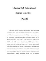
P: Meconium aspiration
#
____________________________________________________________
###
Mechanical obstruction Chemical inflammation Surfactant inactivation
##
Air trapping Atelectasis
## #
Air leaks Unevenly distributed ventilation Intrapulmonary shunting
## #
Respiratory acidosis$Pulmonary hypertension$Respiratory acidosis
I: CXR: hyperinflation, flattening of the diaphragm, cardiomegaly, gross patchy
shadowing.
ABG: #pO
2
occurs when there is right ! left shunting of blood through the
foramen ovale 28 to pulmonary hypertension.
M: Neonates born with meconium-stained liquor require a paediatrician to be
present at birth.
Immediate assessment of the infant:
. If infant is vigorous and crying no further management is required, as visual-
isation of the airway and ET suctioning will not prevent aspiration.
. If the infant is floppy and not breathing, the airway should be suctioned
under direct vision, either directly or via an ET tube/meconium aspirator.
. If at any stage during the resuscitation the HR falls bellow 60, oxygenation
including intermittent PPV overrides the desire to clear the airway.
Subsequent management in NICU: antibiotics, ventilatory assistance, and
exogenous surfactant therapy. Inhalation of NO and ECMO may be required.
C: Pulmonary hypertension (see separate chapter), pneumothorax.
P: Ultimate prognosis depends on the extent of asphyxial insult to the neonate
in utero, and degree of meconium aspiration.
115
CONDITIONS
Brough / Rapid Paediatrics and Child Health Final Proof 9.7.2004 2:45pm page 115
Meningitis
D: Infection of the subarachnoid space associated with an inflammatory response
of the meninges.
A: Bacterial:
Neonatal to 2 months:
GBS, gram-negative bacilli, e.g. Escherichia coli, Listeria monocytogenes.
1 month to 6 years:
Neisseria meningitidis (meningococcus), Streptococcus pneumoniae, Hib.
>> 6 years:
N. meningitidis, S. pneumoniae, mumps (pre-MMR).
Mycobacterium tuberculosis: can cause TB meningitis at all ages. Common-
est in children aged 6 months to 6 years.
Viral: enteroviruses (80%), CMV, arbovirus. HSV is more likely to cause enceph-
alitis (see chapter).
A/R: Impaired immunity: young age, defects of complement system (meningo-
coccal susceptibility), splenic defect from sickle-cell disease or asplenia (S.
pneumoniae and Hib susceptibility).
Factors associated with low socio-economic status: crowding, poverty
and close contact with affected individuals (transmission by respiratory secre-
tions).
E: Varies in different countries: UK 5.4–5.9% Republic of Ireland 10.5%,
France 0.7%, Germany 1.3%.
. N. meningitidis (meningococcus) causes majority of bacterial infections in
> 2 months. Has # since the introduction of vaccine Men. C.
. S. pneumoniae (pneumococcal) meningitis is rising in incidence. Majority
occur in < 2 years.
. Hib meningitis has fallen significantly since the introduction of the Hib
vaccination.
. TB meningitis: rare; 161 cases documented in the UK in 2001.
. Enteroviruses have seasonal variation and are more common in autumn
and summer.
H: Prodromal features of infection: otitis media/respiratory/GI symptoms.
Neonates: nonspecific symptoms such as fever (or hypothermia in severe ill-
ness), irritability, lethargy, seizures, shrill cry, or poor feeding.
Infants and children: fever, headache, neck stiffness, alteration in conscious-
ness (from lethargy to coma), nausea, vomiting, photophobia, anorexia, rash, or
seizures.
E: Neck stiffness: from meningeal irritation that prevents neck flexion.
Kernig’s sign: in the supine position extension of leg is painful when knee
and hip are flexed.
Rash: purpuric or petechial rash (usually non-blanching but may be blanching
in initial stages of disease). Characteristic of meningococcal infection.
Signs of raised ICP: papilloedema, #level of consciousness, focal neurology
(e.g. 6th nerve palsy), Cushing reflex ("BP, #HR), decerebrate posturing.
P: Bacteraemia precedes infection and bacteria enter the CSF via the choroid
plexus and rapidly multiply as local complement and antibody levels are low.
Inflammation follows with disruption of the blood–brain barrier, oedema, and
neutrophil infiltration.
I: Bloods: "WBC, "CRP, U&E, glucose, clotting studies, group and save.
Microscopy, culture and sensitivity: blood, stool, throat swab, urine, rapid
antigen screen.
116
CONDITIONS
Brough / Rapid Paediatrics and Child Health Final Proof 9.7.2004 2:45pm page 116
Meningitis continued
CT scan: if suggestive of "ICP to avoid coning on LP.
LP for CSF: contraind icated if focal neurological signs or "ICP suspected.
CSF in bacterial meningitis: neutrophils, "protein (1–5 g/L), #glucose (< 50%
serum level).
CSF in viral meningitis: lymphocytes (initially neutrophils), normal/mildly
elevated protein, normal glucose, PCR for diagnosis.
CSF: microscopy (Gram stain, e.g. for acid-fast bacilli in TB meningi tis), culture
and sensitivity.
M: Start empirical parenteral antibiotic treatment before the results of
investigations as meningococcal septicaemia is often rapidly fatal.
Bacterial meningitis:
. 3rd generation cephalosporin: ceftriaxone or cefotaxime IV.
. Resistant S. pneumoniae is becoming more prevalent worldwide, but
almost all strains in the UK are sensitive to third generation cephalosporins.
Therefore routine use of vancomycin for community acquired meningitis is
not justified in the UK.
. In infants < 3 months ampicillin should be started empirically to cover for
L. monocytogenes.
. TB meningitis requires 6–9 months therapy with isoniazid, rifampicin, pyra-
zinamide and streptomycin.
Culture and sensitivity will indicate subsequent antibiotic therapy.
Corticosteroids: studies of patients with Hib meningitis have shown some
improvement in morbidity (deafness or neurological deficit) with corticosteroid
treatment alongside antibiotics. Data is lacking in pneumococcal and meningo-
coccal meningitis.
Supportive care: analgesics, antipyretics.
Notification: meningitis is a notifiable disease (CCDC).
Contact prophylaxis: 2/7 of rifampicin for meningococcal infection.
Immunisations: BCG, men C, Hib.
Prevention: there are effective vaccines against N. meningitides C and
A. Pharmaceutical companies are developing a combination vaccine containing
B and C conjugate vaccines; this vaccine will prevent more than 90% of cases of
meningococcal meningitis in Europe.
C: During illness: convulsions, cerebral oedema, circulatory shock, DIC.
Neurological sequelae: hearing loss (10%), therefore all children must have
audiometry testing 2/3 weeks after infection is treated, visual impairment,
cerebral palsy, vasculitis may cause cranial nerve palsies, subdural effusions
that usually resolve spontaneously, hydrocephalus, cereb ral abscess, learning
disability. All children must have neurological follow-up to enable early detec-
tion and management of these complications.
P: Bacterial: overall mortality 5–10%; neurological complications 10–20%.
. Meningococcal meningitis: mortality 7–20%, with septicaemia 18–53%, usu-
ally have favourable neurological outcome if they survive the acute illness.
. Pneumococcal meningitis: has a lower mortality than meningococcal men-
ingitis but " association with adverse neurological sequelae.
. TB meningitis: 15–30% of those affected do not survive, and up to 25% of
people who recover may have long-term neurological sequelae.
Viral: 95% have complete recovery with no neurological sequelae.
117
CONDITIONS
Brough / Rapid Paediatrics and Child Health Final Proof 9.7.2004 2:45pm page 117
Mesenteric adenitis
D: Inflammation of the mesenteric lymph nodes, with systemic illness and ab-
dominal symptoms.
A: Virul URTIs: the commonest cause of mesenteric adenitis.
Bacterial URTIs: 28 to group A b-haemolytic streptococcus.
GI: Yersinia enterocolitica (commonest bacterial cause), Helicobacter jejuni,
Campylobacter jejuni, and Salmonella or Shigella species.
A/R: Most common DD for acute appendicitis.
E: Affects children of all ages.
H: Mesenteric adenitis is often a diagnosis of exclusion; however, may be a posi-
tive diagnosis if a patient undergoes an unnecessary laparotomy for suspected
appendicitis.
Recent infectious injury: URTI or GI infection; symptoms of coryza, pharyn-
gitis, or gastroenteritis may still be present (mainly diarrhoea).
GI: vague abdominal pain may vary intermittently.
E: Signs of acute appendicitis:
. excessive vomiting
. significant anorexia
. usually low-grade fever
. periumbilical region/RIF tenderness
. pain becomes continuous
. fixed tenderness in RIF
. once peritoneum is involved guarding and rebound tenderness
. localised tenderness of anterior rectal wall
. unusual to have signs of URT I.
Signs of mesenteric adenitis:
. little vomiting
. mild anorexia
. may have high fever
. periumbilical region/RIF tenderness may also be present
. pain is intermittent
. tenderness shifts depending on the position of the child
. no true signs of peritonism
. no localised tenderness on rectal examination
. often have signs of URTI.
P: Macro: enlargement of mesenteric lymph nodes.
Micro: proliferation of B and T cells 28 to infectious process.
I: Bloods: "WCC, U&E (may be dehydrated), "CRP.
USS of RIF: to visualise appendix and exclude acute appendicitis.
M: Careful observation:
. Admit for regular reassessment to differentiate between mesenteric adeni-
tis and acute appendicitis.
. Persisting local tenderness in the RIF lasting more than 3–6h warrants sur-
gical exploration.
C: None.
P: Mesenteric adenitis is a benign self-limiting disease.
118
CONDITIONS
Brough / Rapid Paediatrics and Child Health Final Proof 9.7.2004 2:45pm page 118
Myotonic dystrophy
D: Autosomal dominant multisystem disorder characterised by progressive muscle
wasting, muscle weakness, and myotonia (abnormal sustained contraction of
muscle).
A: Genetic defect: caused by expansion of CTG nucleotide triplet repeats at the
3
0
UTR of the myotonic dystrophy gene on chromosome 19. This gene codes for
DMPK.
Genetic anticipation: the disease has earlier onset or "severity in the off-
spring than in parents as a result of further triplet repeat expansion in succes-
sive generations.
A/R: Family history, antenatal history; polyhydramnios.
E: 2/10 000 live births. Severe congenital type is much rarer and almost always
inherited via the mother.
H: Depends on number of CTG repeats:
(1) Unaffected individuals (5–27 repeats).
(2) Mild myotonic dystrophy (50–100 repeats): cataracts, slight muscle weak-
ness in adulthood.
(3) Classical myotonic dystrophy (100–1000 repeats): myotonia, muscle
wasting, frontal balding, hypogonadism, cardiomyopathy, cardiac arrhyth-
mias, DM, respiratory impairment, adverse reactions to anaesthesia.
(4) Congenital myotonic dystrophy (1000–4000 repeats).
. At birth: hypoton ia, respiratory and feeding difficulties, marked facial
weakness. Myotonia is not a feature of the condition at this stage.
. If infant survives: gradual improvement in muscle strength and tone,
delayed motor development, persistence of facial weakness, severe
learning disability (6 0–70% of cases), classic features of myotonic dys-
trophy by the age of 10.
E: Peripheral weakness: hands (unable to release grip), forearms, and feet as
opposed to proximal weakness (hip, shoulder) in other dystrophies.
Myopathic facies: facial muscle weakness and wasting; bilateral ptosis,
wasting of frontalis and temporalis, and weakness of sternomastoids; all of
which result in lack of facial expression.
P: Disturbance in muscle fibre maturation with incomplete differentiation.
Infants: small undifferentiated muscle fibres.
Children: type 1 fibre atrophy; central nuclei, sarcoplasmic masses, ring fibres.
I: Bloods: CPK may be raised. Muscle biopsy: see P.
EMG: characteristic ‘diver bomber’ spontaneous electrical discharge by age 3.
DNA mutation analysis: sizing of the repeat array by PCR and/or Southern
blotting.
M: Multidisciplinary approach:
Medical: myotonia may be treated with quinine or procainamide. Support
respiratory and GI problems. Monitor for deformities.
Surgical: cataract operations.
Orthopaedic: ankle–foot arthroses for footdrop.
Occupational therapy: specially designed utensils for hand weakness.
Genetic counselling: for antenatal diagnosis.
Psychological support: for parent and child.
C: Joint contractures, foot deformities, early onset dementia.
P: Depends on the number of CTG repeats, extent of learning disability, and
early appropriate multidisciplinary involvement. The older the child is when
muscle weakness is first noticed, the slower the progression and less serious
the consequences. Most do not surv ive past 50 years of age.
119
CONDITIONS
Brough / Rapid Paediatrics and Child Health Final Proof 9.7.2004 2:45pm page 119