Surgical Atlas of pediatric otolaryngology - part 3 pps
Bạn đang xem bản rút gọn của tài liệu. Xem và tải ngay bản đầy đủ của tài liệu tại đây (873.82 KB, 57 trang )
Facial Nerve Exploration and Repair 159
• If placement of a sural nerve graft is anticipated, the lower leg, ankle,
and foot are similarly prepared. A surgical glove is placed over the toes,
and the foot and leg are draped with a sterile stockinette and placed on
a sterile sheet. An extremity drape is used to cover the leg.
• A thyroid or a split sheet is used to cover the patient and to drape the
entire surgical field.
• The scrub nurse stands next to the surgeon and in front of the back
scrub table. Surgical loupes and the microscope should be available to
help the surgeon locate and dissect along the nerve.
Procedure
Figure 7–18 overviews extratemporal exploration and repair for lesions.
Figure 7–18 Extratemporal
exploration and repair for
parotid and nonparotid lesions.
160 Surgical Atlas of Pediatric Otolaryngology
No 1. Incision and exposure
• The proposed preauricular incision (Figure 7–19) is marked with a
surgical marker.
♦
In older children and adolescents, the incision will be less noticeable
if curved posteriorly to conform to the anatomic indentation just
superior to the tragus, inferior to the tragus, or both. The incision
continues just inferior to the earlobe and posteriorly onto the mas-
toid process and is then curved inferiorly and anteriorly about 2
cm inferior to the mandible rim, preferably within a skin crease
(see Figure 7–19
A). Alternatively, the postauricular portion of the
incision can be continued into the hairline or directly posteriorly
into the hair as in a facelift incision.
♦
In infants (in whom the mastoid tip has not yet developed) and in
patients in whom the course of the nerve is considered anomalous,
an upper neck incision is made 2 cm inferior to the rim of the
mandible and curving onto the postauricular area. After the skin
incision is made, the skin flap is elevated superiorly with sharp and
blunt dissection until the facial nerve is located. The incision can
then be extended superiorly, if necessary (see Figure 7–19
B).
4
Figure 7–19 A, Incision line for
parotidectomy. Incision may be
extended (
arrows) as necessary to
the neck, hairline, or into the
hair.
B, Incision line for
parotidectomy in infant with
undeveloped mastoid tip. After
the facial nerve is identified, the
incision can be extended along
the dotted line as necessary.
(Reproduced and adapted
[
A only], with permission from
Farrior JB, Santini H. Facial
nerve identification in children.
Otolaryngol Head Neck Surg
1985;93:174–6.)
A
B
Facial Nerve Exploration and Repair 161
• The skin flap is elevated using sharp and blunt dissection to a point
about 1 cm superior, anterior, inferior, and posterior to the parotid
gland. Elevation usually is continued until the zygomatic arch, the
buccal fat pad, the upper one-third of the neck, and the mastoid tip
are exposed (Figure 7–20).
• The anterior border of the sternocleidomastoid muscle is identified.
The great auricular nerve is located at the posterior border of the
upper midportion of the sternocleidomastoid muscle and is exposed
superiorly until it branches just inferior to the auricle. The nerve is
kept in position as long as possible.
In smaller infants, transecting the
great auricular nerve may be unnecessary because the parotid gland is
more anterior than it is in older children and adults.
4
Figure 7–20 Elevation of parotidectomy skin flaps to expose zygoma, buccal fat pad, masseter muscle, and
sternocleidomastoid muscle.
162 Surgical Atlas of Pediatric Otolaryngology
• The anterior border of the sternocleidomastoid muscle is dissected
medially until the posterior belly of the digastric muscle is located
(Figure 7–21). The inferior border of the parotid gland is retracted
superiorly, and the digastric muscle is followed to its origin, medial to
the mastoid tip (the digastric muscle originates at the level of and
inferior to the stylomastoid foramen, through which the facial nerve
exits). This dissection
in infants may suffice to locate the main trunk
of the facial nerve, because the parotid gland
in infants is more ante-
rior (Figure 7–22).
4
• The preauricular area is dissected medially along the external canal
perichondrium. The parotid gland is retracted anteriorly. Sharp and
blunt dissection is continued medially until the cartilaginous “point-
er” is identified at the bony cartilaginous junction of the external
auditory canal (Figure 7–23).
• This dissection plane is continued superiorly to the level of the zygo-
matic arch. The preauricular dissection is continued medially, both
superior and inferior to the expected location of the facial nerve. This
technique facilitates eventual visualization of the nerve by widening
the surgical field.
Figure 7–21 Retraction of
inferior parotid gland and stern-
ocleidomastoid muscle to locate
posterior belly of digastric muscle.
164 Surgical Atlas of Pediatric Otolaryngology
No 2. Identifying the facial nerve
• The facial nerve is located about 1 cm medial to the tympanomastoid
suture line and the cartilaginous pointer at the external auditory
canal–bony cartilaginous junction, just anterior to the stylomastoid
foramen (Figure 7–24).
♦
This area is separated with blunt dissection parallel to the expect-
ed course of the nerve.
♦
Bleeding is controlled with clamps, 3-0 absorbable suture ties,
bipolar cautery, a Shaw scalpel, or light pressure using small or
large moist dissecting sponges.
Figure 7–24 Exposure of main
trunk of facial nerve and its tem-
porofacial and cervicofacial divi-
sions.
Facial Nerve Exploration and Repair 165
• Proper surgical technique is essential when working near the facial
nerve:
♦
The surgeon should minimize any contact with the facial nerve
and its branches.
♦
Visualization obscured by blood can be improved by lightly touch-
ing the facial nerve with small and large moist dissecting sponges.
Minimal pressure should be applied, and the nerve should not be
rubbed or suctioned.
♦
In most cases, knowledge of the anatomy of the facial nerve should
obviate the need for a facial nerve stimulator, which can further
traumatize the nerve.
♦
In revision surgery, post-traumatic exploration, and certain
patients with extensive neoplastic disease and infection, however,
the nerve may be identified through judicious use of a facial nerve
stimulator at its lowest setting.
• If the main trunk cannot be identified or if it is incorporated in scar
tissue or tumor, peripheral branches must be located and dissected in
retrograde fashion to the main trunk in the following areas: the
mandibular branch, temporal branch, buccal branches, and zygomat-
ic branch.
• The
mandibular branch, or ramus mandibulae, is located in the neck
lateral to the facial vessels and just superior to the submandibular gland.
♦
In many infants this branch is located more superiorly, lateral to the
mandible.
♦
In older children, if the mandibular branch is not located easily, the
fascia of the submandibular gland can be incised and elevated
superiorly along the posterior facial vein until the cervical or
mandibular branch is identified.
• The
temporal branch is usually found overlying or just superior to the
zygomatic arch deep to the superficial fascia, about halfway between
the anterior border of the auricle and the lateral bony orbital rim.
• A
buccal branch can be found coursing near and parallel to the parotid
duct about 1 to 1.5 cm inferior to the zygomatic arch.
• The
zygomatic branch can often be identified between the anterosu-
perior border of the parotid gland and the lateral inferior orbital rim.
• Rarely, for large neoplasms or when severe scarring has resulted from
prior surgery or trauma, mastoidectomy is indicated. In that circum-
stance the facial nerve is explored to locate the nerve and follow it
through the stylomastoid foramen.
• Once the main trunk has been identified, blunt dissection is contin-
ued anteriorly along the trunk until its bifurcation is located; the
facial nerve is then identified conclusively, and the parotid tissue lat-
eral to the nerve can be incised with a Shaw scalpel or with a No 11,
12, or 15 blade.
166 Surgical Atlas of Pediatric Otolaryngology
• Dissection along the nerve can be facilitated initially with a fine
hemostat such as a McCabe facial nerve dissector. Once the proper
plane has been established, curved mosquito-type forceps and small
Kelly clamps suffice. Each branch is dissected to a point distal to the
parotid gland (Figure 7–25).
No 3. Superficial and deep parotidectomy
• If possible, the parotid gland between the branches is removed with
the pathologic specimen. As large a margin of normal parotid tissue
as is possible is included around any tumor or cyst.
• If, after the superficial portion of the parotidectomy has been com-
pleted, the disease process is
medial to the facial nerve, the nerve is
elevated gently with sharp and blunt dissection. The main trunk and
each of the cervicofacial branches are retracted with vascular loops to
expose the underlying parotid tissue (Figure 7–26).
• The masseter muscle is identified anteriorly and medially to expose
and remove the tissue medial to the facial nerve. Dissection is begun
in a plane lateral to the masseter fascia and is continued to the poste-
rior border of the masseter muscle at the ascending ramus of the
mandible.
• A second dissection plane is begun on the mastoid tip inferior to the
stylomastoid foramen and the main trunk of the facial nerve. This
plane is continued medially and is connected with a plane beginning
on the bony canal superior to the facial nerve trunk.
• The remaining parotid tissue, which may be in the parapharyngeal
space, can be removed by advancing along the dissection planes.
♦
If necessary to protect the facial nerve, this portion of the excision
can be done in segments from between the branches of the nerve
or superior or inferior to the main trunk and to the superior or
inferior branches of the nerve.
♦
When dissecting superior to the trunk and posterior to the frontal
branch, care should be taken to avoid injuring the auriculotempo-
ral branch of the third division of the trigeminal nerve. This
branch lies near the superficial temporal artery between the auricle
and the mandible.
• In patients with large tumors of the parapharyngeal space, anterior
traction on the mandible may provide adequate additional operating
space. Otherwise, mandibular osteotomy and mandibular swing may
be necessary to adequately visualize and access the site so as to suffi-
ciently facilitate removal of tumor or parotid tissue.
168 Surgical Atlas of Pediatric Otolaryngology
• Once the pathology specimen has been removed, the nerve is inspect-
ed to ensure that it is intact:
♦
If transected at the trunk or at the temporalis, zygomatic, or
mandibular branches, the nerve is reapproximated using the fewest
9-0 or 10-0 monofilament nylon sutures needed to achieve coap-
tation (Figure 7–27).
♦
Anastomosis of severed buccal branches is usually unnecessary
because of the rich interanastomosis of buccal branch nerve fibers.
♦
The cervical branch is routinely sacrificed during parotidectomy,
and it does not require repair because loss of function is minimal.
• If a segment of the nerve has been removed, an immediate interposi-
tion nerve graft can be sutured in place (Figure 7–28).
♦
If delay of grafting is elected, the severed proximal and distal ends
should be marked with nonabsorbable (nylon) sutures to facilitate
localization at subsequent repair.
♦
Instead of using an interposition graft, less important branches
may be severed and anastomosed to more valuable branches—for
example, the buccal to the temporal or zygomatic branch, or the
buccal or cervical to the mandibular branch (Figure 7–29).
18
Figure 7–27 Sutured laceration
of main trunk of facial nerve.
170 Surgical Atlas of Pediatric Otolaryngology
• The wound is irrigated with saline solution, and the facial nerve is re-
examined for continuity.
• A Penrose or vacuum drain is placed inferior to the trunk, posterior
to the branches, and through either the inferior neck incision or a
separate incision.
• The preauricular skin can be approximated with 3-0 to 4-0 catgut
and 4-0 to 5-0 nylon sutures. The neck incision can be approximat-
ed with staples.
• Antibiotic ointment is applied to the incision. Fluffed gauze sponges
and 10 cm
× 10 cm dressing gauze sponges are used for a pressure dress-
ing. A Barton or nylon tubular net dressing can be applied for pressure.
Postoperative Care
• Facial function is checked when the patient is awakened from anesthesia.
♦
If paresis or paralysis is present and the nerve was left intact, the
patient is observed.
♦
If paralysis is present immediately after surgery and was not present
preoperatively, and if the status of the nerve was not checked, the
nerve should be explored to establish continuity, and any lacerations
should be repaired.
• The patient is observed for hematoma, seroma, salivary fistula, corneal
irritation, or new facial paralysis. Gustatory sweating may be a late com-
plication.
• Artificial tears are used, and if the eye is affected by facial paresis or
paralysis, the eyelid is closed before sleep.
• The dressing is re-inforced overnight to absorb bloody drainage. The
drain is removed on the first or second postoperative day or when
drainage is minimal or nonexistent.
• The pressure dressing is applied for 4 to 7 days, and the sutures or sta-
ples are removed on the seventh postoperative day.
• If the nerve was intact at the end of surgery, any postoperative paresis
should resolve in 4 to 6 weeks. If paralysis is present, recovery should
begin within 3 months and should continue for 12 months.
Facial Nerve Exploration and Repair 171
EXTRATEMPORAL EXPLORATION AND REPAIR AFTER
TRAUMA
Penetrating wounds of the face may affect the main trunk of the facial
nerve as well as any or all of its branches. Because facial nerve injury may
be part of more massive trauma, assessment of the patient’s airway and car-
diovascular status is important. Neurologic evaluation should be done after
the patient’s condition has stabilized and should include assessment of the
facial nerve. Computed tomography scans are appropriate when the tem-
poral bone has been traumatized. An MRI may help to locate injury to
either the nerve trunk or the larger nerve branches.
The type of trauma and duration of paralysis are also important. Differ-
entiating partial from complete facial paralysis may be difficult when facial
edema, ecchymosis, or lacerations exist and particularly if the patient is
uncooperative.
9,10,19
Any nerve branch lacerated anterior to the masseter
muscle is usually too small to approximate; in addition, function often
returns because of cross-anastomoses or neurotization.
A completely severed facial nerve continues to conduct impulses for
about 72 hours.
8–10
Wound exploration within 72 hours can thus be facil-
itated by use of nerve stimulation to help find the severed nerve branches.
The nerve stimulator is of no help if the patient is evaluated after 72 hours;
nonetheless, exploration and repair should be done as soon as it is feasible.
Dissection is facilitated when delayed until scarring has matured.
8–10
Nerve
grafts should be done before 18 months, by which time fibrosis of the neur-
al tissues and subsequent atrophy of muscle fibers is sufficiently advanced
to preclude a successful result from grafting.
Indications
• Facial paralysis immediately after a penetrating wound anterior or inferior
(or both) to the auricle and posterior to the anterior border of the mas-
seter muscle (repair is unnecessary for branches severed anterior to the
masseter muscle).
•
Facial paralysis immediately after blunt trauma to the head and neck.
Anesthetic Considerations
• General nasotracheal anesthesia is used as described above—see
Extratemporal Exploration and Repair for Lesions.
Preparation
• Preparation is made for possible parotidectomy, mastoidectomy with
facial nerve exploration, and grafting of the great auricular nerve or sural
nerve as described above —see
Intratemporal Exploration and Decompres-
sion
and Extratemporal Exploration and Repair for Lesions.
• The surgeon should have access to a nerve integrity monitor, facial nerve
stimulator, and microscope or surgical loupes.
172 Surgical Atlas of Pediatric Otolaryngology
Procedure
Figure 7–30 outlines extratemporal exploration and repair for facial nerve
trauma.
• For more peripheral cutaneous lacerations accompanied by segmental
paralysis, exploration of the wound and approximation of the severed
nerve endings may be possible.
• A parotidectomy approach is necessary to locate the facial nerve:
♦
If the lacerated peripheral branches cannot be located or approximated
♦
In more proximal facial lacerations
♦
In total facial nerve paralysis
♦
In cases of blunt trauma with no external laceration
Figure 7–30 Extratemporal explo-
ration and repair after trauma.
Facial Nerve Exploration and Repair 173
• As with the procedures described earlier in this chapter, a facial nerve
stimulator may facilitate locating the nerve if the surgical approach is
done within 72 hours after onset of paralysis.
• If the facial nerve trunk and its branches are intact, or if lacerations are
found only in the cervical or distal buccal branches, the nerve can be left
as is and the wounds can be cleaned, debrided, and sutured.
• In acute cases, once a nerve laceration or avulsion is located—and if the
ends can be approximated without causing tension—the nerve endings
are mobilized and sutured with the fewest number of 9-0 or 10-0
monofilament nylon sutures necessary to achieve coaptation. If the ends
cannot be approximated without the use of tension, underlying parotid
tissue may be removed to allow approximation without adding tension
to the suture line (Figure 7–31
A and B).
18
• When the laceration is situated at the stylomastoid foramen or at a pos-
terior location on the main trunk so that suturing of the proximal stump
is impossible, mastoidectomy with facial nerve exposure is necessary to
locate and mobilize the proximal nerve for reapproximation or grafting.
Figure 7–31 A, Avulsed parotid tissue and facial nerve. B, Superficial and underlying parotid tissue is removed to enable approxi-
mation of nerve branch ends after avulsion. n = nerve; m = muscle. (Adapted and reproduced with permission from Tucker HM.
The management of facial paralysis due to extracranial injuries. Laryngoscope 1978;88:348–54.)
174 Surgical Atlas of Pediatric Otolaryngology
• In cases of delayed repair, the nerve endings are mobilized, freshened
with a razor blade or with a 6500 Beaver Mini-Blade, and sutured with
the fewest 9-0 or 10-0 monofilament nylon sutures needed to achieve
coaptation (Figure 7–32
A to C). As in acute cases, underlying parotid
tissue may be removed to allow approximation without adding tension
to the suture line.
• Interposition nerve grafting is necessary if the severed segments cannot
be approximated or can be closed only under tension (see
Extratemporal
Repair, Rerouting, and Grafting
, below).
• If immediate grafting is impossible or inadvisable (eg, when gross cont-
amination or anesthetic complication is present), the severed proximal
and distal ends should be tagged with nonabsorbable nonreactive
(nylon) sutures for easier localization at subsequent repair.
• In cases of delayed repair, a frozen section of nerve margin should be
analyzed to determine whether neural fibrosis has occurred. If neural
fibrosis is detected, further nerve resection is needed until viable nerve
tissue is located.
• A Penrose or vacuum drain is placed inferior to the trunk and posterior
to the branches of the nerve, and it is passed either through the inferior
part of the incision or through a separate incision.
• Any wound lacerations are freshened; these lacerations, as well as the
parotidectomy incision, are approximated using 3-0 to 4-0 chromic
catgut subcutaneous sutures and 4-0 to 5-0 nylon cutaneous sutures.
• Antibiotic ointment is placed on the incision(s). Fluffed gauze sponges
and 10 cm
× 10 cm dressing gauze sponges are applied with either a Bar-
ton or nylon tubular net compression dressing.
Postoperative Care
• Postoperative care and nerve recovery time after surgical exploration and
repair are the same as discussed in the previous section on
Extratemporal
Exploration and Repair for Lesions
.
176 Surgical Atlas of Pediatric Otolaryngology
EXTRATEMPORAL REPAIR, REROUTING,AND
GRAFTING
Extratemporal facial nerve rerouting or grafting is done when primary
approximation of the nerve endings is impossible or cannot be done with-
out causing tension at the suture line. Extratemporal facial nerve rerouting
is done by removal of the existing parotid gland to enable approximation
without tension at the suture line.
8
Depending on length of defect, size of
the nerve, and condition of donor sites, nerve graft donor sites may include
the ipsilateral or contralateral great auricular nerve and the sural nerve.
Various nerve transfers, predominantly the hypoglossal-facial (XII-VII)
but also the cross-facial (VII-VII) and spinal accessory–facial (XI-VII), have
been advocated. The simplest and least debilitating nerve transfer can be
done by severing cervical or buccal branches and by approximating them to
the distal stump of the mandibular, zygomatic, or temporal branches.
9–11,18
Indications
• Approximation and suturing are indicated in any laceration at the trunk
or main branch posterior to the anterior border of the masseter, if this
approximation and suturing can be done without causing tension at the
suture line.
•
Rerouting of the extratemporal facial nerve is indicated if lacerated nerve
segments cannot be approximated without causing tension at the suture
line and in the presence of residual parotid tissue, which, if removed,
will enable approximation without causing tension at the suture line.
• For
interposition nerve grafting to be done, viable neural tissue must be
present at the proximal and distal segment margins. Grafting is indicat-
ed in three circumstances:
1. Extratemporal facial nerve laceration in which primary approxima-
tion and suturing are not possible
2. Primary approximation and suturing are achieved and the laceration
is under tension
3. Avulsion of more than 50% of the nerve has occurred
•
Nerve transfer of a cervical or buccal branch is indicated when lacerations
of mandibular, zygomatic, or temporal branches cannot be approximat-
ed without causing tension at the suture line and when sufficient length
and appropriate diameter of cervical or buccal branch are available for
primary anastomosis without causing tension at the suture line.
• Nerve transfer from another cranial nerve (XII, XI, or contralateral VII)
may be indicated as a last resort in patients with a flaccid face, func-
tioning distal nerve and neuromuscular junction, and no viable proxi-
mal nerve. An interposition nerve graft may be necessary to bridge the
gap. Because of the additional cranial nerve deficit that results after
nerve transfer, this procedure must not be undertaken unless the patient
has first given informed consent that takes into account not only the
result of nerve transfer but also the deficit that the patient should expect
after the loss of donor nerve.
Facial Nerve Exploration and Repair 177
Anesthetic Considerations
General nasotracheal anesthesia is used as described above—see Extratem-
poral Exploration and Repair for Lesions
.
Preparation
• Preparation is made for possible parotidectomy, mastoidectomy with
facial nerve exploration, and grafting of the great auricular nerve or sural
nerve as described above—see
Intratemporal Exploration and Decompres-
sion
and Extratemporal Exploration and Repair for Lesions.
• The surgeon should have access to a nerve integrity monitor, facial nerve
stimulator, and microscope or surgical loupes.
Procedure
• The extratemporal facial nerve is sutured and rerouted as described in
the two previous sections on
Extratemporal Exploration and Repair for
Lesions and Repair After Trauma
.
• Exposure and preparation of the proximal and distal facial nerve sites
before nerve grafting are done as described in the four previous sections.
• The nerve graft should be obtained after the recipient site is prepared. If
grafting is delayed after the nerve graft is obtained, the nerve should be
placed in Ringer’s lactate solution.
• The great auricular nerve (ipsilateral or contralateral) or the sural nerve
may be used.
No 1. Great auricular nerve graft
• Before grafting the great auricular nerve, the surgeon should test the
skin of the neck for sensation in the area of the C2/C3 distribution
on both the ipsilateral and the contralateral sides of the neck.
• The cervical portion of a parotidectomy incision can be used to locate
the great auricular nerve. Otherwise a horizontal incision is made par-
allel to (or, preferably, in) a skin crease about 2 cm inferior to the
mandibular margin and centered on the posterior border of the ster-
nocleidomastoid muscle. One long incision or two smaller stair-step
incisions can be made.
• Once isolated, the nerve can be dissected proximally toward its origin
in the cervical plexus to gain as much as 10 cm of additional length.
The cervical incision can be approximated in two layers by using 3-0
to 4-0 chromic catgut sutures and 4-0 to 5-0 nylon sutures.
• If the ipsilateral great auricular nerve is smaller than the severed
branch of the facial nerve, the contralateral great auricular nerve or
one or more fascicles of the sural nerve also can be grafted (see below).
Use of the great auricular nerve as a graft leaves a cutaneous sensory
deficit over the mastoid process and auricle.
No 2. Sural nerve graft
• The sural nerve is formed by the junction of the communicating
ramus of the lateral sural cutaneous nerve and the medial sural cuta-
neous nerve in the middle of the leg.
178 Surgical Atlas of Pediatric Otolaryngology
♦
The sural nerve is usually located just medial to the small saphe-
nous vein; it then continues inferiorly and curves anteriorly, infe-
rior to the lateral malleolus, where it divides into several branches.
The sural nerve ends as the lateral dorsal cutaneous nerve.
♦
The sural nerve is easiest to locate just posterior to the lateral
malleolus and next to the saphenous vein (Figure 7–33).
• The sural nerve can be exposed through a vertical incision that bisects
the plane between the lateral malleolus and the Achilles tendon. The
incision is then curved inferior to the lateral malleolus and onto the
lateral aspect of the foot (Figure 7–34
A).
• An alternative, more cosmetically acceptable way to expose the sural
nerve, is to first make a horizontal incision at the level of the lateral
malleolus and posterior to it (Figure 7–34
B).
• Lateral traction on the vein and the sural nerve reveals the nerve’s
superior and inferior course and thus allows easier dissection. Hori-
zontal incisions are then made at appropriate intervals to obtain a
longer graft. Up to 35 cm of nerve, consisting of two to four fascicles,
can be used.
• The leg incision is approximated in two layers by using 3-0 to 4-0
chromic catgut sutures and 4-0 to 5-0 nylon sutures. A compressive
leg dressing is applied using 10 cm
× 10 cm dressing gauze sponges
and elastic tape.
Figure 7–33 The sural nerve,
located adjacent to the saphenous
vein in the lower part of leg.
180 Surgical Atlas of Pediatric Otolaryngology
• An appropriate number of 9-0 or 10-0 monofilament nylon sutures
is used to approximate the perineurium of the graft to the epineuri-
um of the nerve (Figure 7–35). Depending on the width of the prox-
imal and distal nerve stump or stumps, either the entire nerve or one
or more fascicles can be used.
No 4. Hypoglossal facial anastomosis
• For hypoglossal facial anastomosis, the cervical portion of a
parotidectomy incision is made after the distal nerve stump has been
located.
• The hypoglossal nerve is found 1.5 cm superior to the carotid bifur-
cation, where the nerve courses laterally to the internal and external
carotid arteries and medially to the posterior belly of the digastric
muscle. To maximize the length of nerve available for transfer, the
nerve is dissected anteriorly as far as is possible before transection.
• Interposition nerve grafts may be necessary for approximation with-
out tension.
• Both the distal facial nerve segment and the hypoglossal stump are
prepared for perineural or epineural anastomosis, and the stump is
sutured to the recipient facial nerve by using an appropriate number
of 9-0 or 10-0 monofilament nylon sutures (Figure 7–36).
• Sacrifice of the hypoglossal nerve results in paralysis of the ipsilateral
side of the tongue and ipsilateral facial contraction during degluti-
tion. Facial function can be relearned.
• The wound is irrigated with sterile saline, and the skin incision is
approximated with 3-0 or 4-0 chromic catgut sutures and with 4-0 or
5-0 nylon sutures. Staples can be used to close the cervical portion of
the incision. Antibiotic ointment is applied to the incision line.
• To create pressure over the wound, a compressive Barton or tubular
net dressing or elastic tape is applied over fluffed gauze sponges and
10 cm
× 10 cm dressing gauze sponges.
Postoperative Care
• The facial and cervical dressings are re-inforced overnight to absorb
bloody drainage.
• The dressing is removed on the first or second postoperative day, and a
fresh compressive dressing is applied and is left in place for 4 to 7 days.
• The compressive leg dressing applied after the sural nerve grafting is
removed 1 to 3 days postoperatively; sutures or staples in the leg and
neck are removed 7 to 10 days postoperatively.
• Recovery of facial function should begin within 4 to 6 months postop-
eratively and may be expected to continue for at least 12 months post-
operatively.
182 Surgical Atlas of Pediatric Otolaryngology
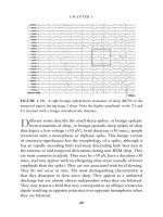
ELECTROPHYSIOLOGIC TESTING
Electrophysiologic (EP) testing can be used either to stimulate the affected
nerve-muscle complex or to record volitional muscle action potentials,
including fibrillation or defibrillation potentials. In patients with facial
paralysis, EP testing is sometimes used clinically to assess degree of nerve
damage, to help establish prognosis for recovery, and to determine need for
surgical intervention. However, because patients with partial paralysis have
complete recovery, EP testing can be useful only for patients who have com-
plete facial nerve paralysis. In addition, nerve action potentials remain near-
ly normal for as long as 72 hours, so nerve stimulation tests are useful only
after 72 hours and for no more than 2 or 3 weeks after injury. Volitional
muscle testing is not reliable until at least 2 or 3 weeks after injury.
20,21
EP Testing Procedures
Types of EP testing include nerve excitability testing, maximal stimulation
testing, electroneurography, electromyography, magnetic stimulation, and
testing of the stapedial muscle reflex.
Nerve excitability testing. Nerve excitability testing (NET) subjective-
ly compares movement of each side of the face after the facial nerve is stim-
ulated with the minimal amount of current necessary to elicit muscle con-
traction. A difference of at least 3.5 mA in stimulation intensity at the facial
nerve trunk or over its branches indicates progressive or impending degen-
eration.
22
As a clinical test, NET has largely been replaced by either maxi-
mal stimulation testing (MST) or electroneurography (ENOG).
Maximal stimulation testing. Maximal stimulation testing is a subjec-
tive test in which branches of the facial nerve are stimulated to determine
the level of current needed for maximal muscle contraction. The amount
of facial motion on each side of the face is compared subjectively, and the
response is recorded as a percentage of function of the unaffected side.
20
Electroneurography. Electroneurography consists of bipolar electrical
stimulation of the facial nerve trunk as well as bipolar recording (at periph-
eral branches) of two parameters: stimulation level needed for a maximal
response, and size and configuration of the compound muscle action
potential (CMAP).
Electroneurography can be performed using either of two methods. In
the standard method, electrodes for nerve stimulation and recording are
placed at fixed (standard) points on the face. In the optimized method, the
electrodes are moved to various points on the face to obtain the greatest
CMAP. Both techniques enable comparison between the CMAP on each
side of the face regardless of stimulation level.
The latent period between application of the stimulus and the start of
CMAP testing is sometimes used to indicate functional status of the facial
nerve. The recorded end point can be measured and compared statistically
with that of the other side. Use of ENOG varies from clinic to clinic, and
interpretation differs on the basis of both the pathology expected and the
treatment selected. Amplitude reduction of 90% or more within 1 to 3
weeks is interpreted as indicating need for surgical intervention.
20,21
Facial Nerve Exploration and Repair 183
Electromyography. When administered at least 2 to 3 weeks after nerve
injury, electromyography (EMG) can be useful for assessing facial re-inner-
vation. Volitional EMG can be used to monitor facial nerve recovery; if no
recovery is seen at a 2- to 3-week follow-up, fibrillation potentials become
established. Regeneration potentials become visible after about 12 weeks
and occur before facial motion becomes visible. In patients with acute
incomplete facial nerve paralysis, electromyographic evidence of fibrillation
potential is considered an indication of tumor, until this diagnosis is oth-
erwise disproved.
Magnetic stimulation. Magnetic stimulation is a technique in which
stimulation generated by a magnetic coil is applied at the stylomastoid fora-
men and is recorded retrograde at the vertex. Results of this test can indi-
cate facial nerve degeneration and damage.
21
Stapedial muscle reflex testing. Testing of the stapedial muscle reflex
can also be considered an electrophysiologic test because, as the first motor
branch of the facial nerve, the stapedial nerve is the first facial nerve branch
to recover. Recovery of an absent stapedius reflex within 21 days suggests
an excellent prognosis.
Appropriate Use
Basing treatment on the results of electrical tests alone is problematic for
two main reasons:
• Any indicated surgery will be delayed 3 days because a 3-day delay exists
between the time of injury and the time when nerve action potentials
become abnormal.
• Abnormal results of electrical tests remain abnormal and cannot be used
to monitor nerve regeneration.
Although electrical tests can be used to assess the prognosis and can
accurately indicate inappropriateness of surgical intervention, results of
these tests are not timely indicators for treatment. Members of our depart-
ment have used the clinical history, results of physical examination and,
when necessary, results of radiologic studies to assess the physical state of
the nerve and thus to serve as a guide toward surgical exploration of the
facial nerve.
ACKNOWLEDGMENTS
For reviewing an earlier draft of the manuscript, the author thanks Freder-
ick M. Byl, MD, E. Lila Jordan, RN, and Kedar K. Adour, MD, who also
helped with the “Electrophysiologic Testing” section for this edition. Jon
Coulter, MA, CMI, and Juan Domingo provided the illustrations. The
Medical Editing Department of Kaiser Foundation Research Institute pro-
vided editorial assistance.
184 Surgical Atlas of Pediatric Otolaryngology
REFERENCES
1. Proctor B. The anatomy of the facial nerve. Otolaryngol Clin North Am 1991;24:479–504.
2. Clemente CD, editor. Gray’s anatomy of the human body. 30th ed. Philadelphia: Lea &
Febiger; 1985.
3. Hollinshead WH. Anatomy for surgeons. 3rd ed. I. The head and neck. Philadelphia: Harper
& Row; 1982.
4. Farrior JB, Santini H. Facial nerve identification in children. Otolaryngol Head Neck Surg
1985;93:173–6.
5. Brackmann D, Shelton C, Arriaga M, editors. Otologic surgery. 2nd ed. Philadelphia: WB
Saunders; 2001.
6. Pellet W, Cannoni M, Pech A, et al. Otoneurosurgery. New York: Springer-Verlag; 1990.
7. May M. Facial paralysis in children. In: Bluestone CD, Stool SE, Arjona SK, editors. Pediatric
otolaryngology. Philadelphia: WB Saunders; 1983. p. 249–70.
8. Adkins WY, Osguthorpe JD. Management of trauma of the facial nerve. Otolaryngol Clin
North Am 1991;24:587–611.
9. Johns ME, Crumley RL. Facial nerve injury, repair, and rehabilitation [Otorhinolaryngology
self-instructional packages, 79200]. Washington (DC): American Academy of Otolaryngolo-
gy; 1979.
10. Papel ID. Rehabilitation of the paralyzed face. Otolaryngol Clin North Am 1991;24:727–38.
11. Fisch U, Lanser MJ. Facial nerve grafting. Otolaryngol Clin North Am 1991;24:691–708.
12. Hilsinger RL Jr. The facial nerve. In: Bluestone CD, Stool SE, editors. Atlas of pediatric oto-
laryngology. Philadelphia: WB Saunders; 1995. p. 129–66.
13. Batsakis JG. Tumors of the head and neck: clinical and pathological considerations. 2nd ed. Bal-
timore: Williams & Wilkins; 1979. p. 9–14, 64–6.
14. Gates GA. Diseases of the salivary glands. In: Bluestone CD, Stool SE, Arjona SK, editors. Pedi-
atric otolaryngology. Philadelphia: WB Saunders; 1983. p. 1023–36.
15. McRae RG, Lee KJ, Goertzen E. First branchial cleft anomalies and the facial nerve. Otolaryn-
gol Head Neck Surg 1983;91:197–202.
16. Schuller DE, McCabe BF. Salivary gland neoplasms in children. Otolaryngol Clin North Am
1977;10:399–412.
17. Harris JP, Davidson TM, May M, Fria T. Evaluation and treatment of congenital facial paraly-
sis. Arch Otolaryngol 1983;109:145–51.
18. Tucker HM. The management of facial paralysis due to extracranial injuries. Laryngoscope
1978;88:348–54.
19. Olsson JE, Shagets FW. Blunt trauma of the temporal bone [Otorhinolaryngology self-instruc-
tional package, 80386]. 2nd ed. Washington (DC): American Academy of Otolaryngolo-
gy–Head and Neck Surgery; 1986.
20. Adour KK. Facial nerve electrical testing. In: Jackeler RK, Brackmann DE, editors. Neurotol-
ogy. St. Louis (MO): Mosby; 1994. p. 1283–9.
21. Dobie RP. Tests of facial nerve function. In: Cummings CW, Frederickson JM, Harker LA, et
al, editors. Otolaryngology—Head & Neck Surgery. 3rd ed. St. Louis (MO): Mosby; 1998.
p. 2757–66.
22. Laumans EP, Jongkees LB. On the prognosis of peripheral paralysis of endotemporal origin.
Part II: Electrical tests. Ann Otol Rhinol Laryngol 1963;72:621–36.
CHAPTER 8
EAR CANAL STENOSIS
AND
ATRESIA
Simon C. Parisier, MD
Jose N. Fayad, MD
PRINCIPLES OF EAR CANAL SURGERY
Otologic surgery involving the ear canal is ideally performed when the skin
is not inflamed, and after any acute infectious processes are controlled.
Draining ears should be medically treated prior to surgery:
• Meticulous debridement of the ear canal is performed as an office pro-
cedure using an operating microscope and appropriate delicate instru-
ments. Wax, retained keratin debris, and secretions are cleansed to
expose the underlying skin and eardrum remnant.
• Granulation tissue is removed and sent for pathologic examination. The
resulting bleeding base is chemically cauterized.
• A culture of the ear canal may be obtained in selected refractory cases.
Appropriate antifungal or antibiotic topical drops and systemic oral
antibiotics are prescribed.
• The importance of preventing water entry into the ear when bathing or
swimming must be emphasized to the patient and family. Patients are
instructed to plug the affected ear with commercially available soft sili-
cone plugs or petroleum-impregnated lamb’s wool.
Inadequate attention to the external ear canal may cause an otherwise successful tympa-
nomastoid operation to fail. When performing ear surgery, a narrow canal or overhangs that
prevent adequate exposure may compromise the desired results. An understanding of the ear
canal anatomy, with analysis and correction of the structures producing narrowing or
obstruction, permits a systematic operative approach resulting in a patent meatus and canal.
A common iatrogenic complication of operations involving the external ear canal is par-
tial postoperative stenosis. A narrow meatal opening defeats the self-cleaning mechanism of
the external ear canal, leading to the “problem ear.” Following a canal wall–down mas-
toidectomy, the mastoid cavity, which becomes marsupialized into the external ear, must be
accessible. Failure to provide good access to the mastoid recess frequently results in prob-
lem ears that are difficult to manage.
186 Surgical Atlas of Pediatric Otolaryngology
• Parents and patients are admonished not to use cotton-tipped applica-
tors, which can impact debris in the canal and irritate the skin.
• Occasionally, when a patient is unable to comply with the recommend-
ed instructions or is refractory to the treatment, hospitalization is rec-
ommended for intravenous antibiotic and intensive local therapy.
Most “wet” ears can be converted to a dry state in 4 to 6 weeks using the
above recommendations. Surgery can be performed when the acute inflam-
matory ear process is quiescent. Occasionally, despite intensive therapy,
suppuration persists. In these cases, the indicated ear surgery might be
required, in spite of active drainage, in order to create a noninfected ear.
Generally, surgery performed in an actively inflamed ear may be accompa-
nied by increased bleeding that may obscure the underlying anatomy and
consequently compromise the surgical outcome. Also, in a small child,
blood loss may be significant, requiring blood transfusions.
Audiometric evaluations should always be performed prior to surgery on
an ear. The indications for obtaining radiographic imaging are determined
by the underlying existing clinical findings.
RECONSTRUCTION OF THE EXTERNAL EAR CANAL
Indications
Reconstruction of the external ear canal requires preserving the specialized,
cerumen-producing, migratory skin lining and managing its two anatomic
portions: lateral (cartilaginous) and medial (bony).
Lateral Cartilaginous Ear Canal
• Skin lining: contains hair follicles, sebaceous glands, and dermal layer.
This is the thicker layer of ear canal skin.
•
Procedure overview: perform meatoplasty by removing constricting carti-
lage from the anterior edge of the concha and by resecting cartilage that
forms the floor of the canal. The skin lining is preserved.
Medial Bony Ear Canal
• Skin lining: the lamina propria of the epidermis merges with the bony
periosteum. This is the thinner layer of ear canal skin.
•
Procedure overview: develop pedicled canal wall skin flaps posteriorly and
anteriorly. Expose bony overhangs, which are drilled away. Replace the
meticulously preserved canal wall skin, maintaining an epidermis-lined
canal. In cases for which the canal wall skin is deficient, use a split-thick-
ness skin graft to provide a stable epidermal canal lining.
Surgical Anatomy
• In the tympanic part of the temporal bone, two prominent sutures—the
anterior tympanosquamous suture and the posterior tympanomastoid
suture—may protrude and encroach on the canal lumen (Figure 8–1).
The resulting overhangs can prevent the eardrum margin and related
pathologic changes from being visualized.
• The suprameatal spine (see Figure 8–1), when prominent and anteriorly
oriented, can further narrow the canal at the bony-cartilaginous junction.
188 Surgical Atlas of Pediatric Otolaryngology
Procedure
The described approach is applicable to a wide variety of otologic proce-
dures, including tympanoplasty, mastoidectomy (wall–up or wall–down),
and repair of ear canal stenosis (acquired or congenital). The ear canal sur-
gical methods described include endaural and postauricular approaches
(Figures 8–2 and 8–3). Table 8–1 lists the advantages and limitations of
each approach.
Figure 8–2 An endaural
approach. (Reproduced with
permission from Johnson JT,
editor. American Academy of
Otolaryngology-Instruction
Courses. Vol 4. St. Louis (MO):
CV Mosby; 1991.)
Figure 8–3 A postauricular
approach. (Reproduced with
permission from Johnson JT,
editor. American Academy of
Otolaryngology-Instruction
Courses. Vol 4. St. Louis (MO):
CV Mosby; 1991.)
Ear Canal Stenosis and Atresia 189
No 1. Endaural approach
• The ear canal is injected with 1:100,000 epinephrine solution.
♦
Using a 25-gauge needle, the injection is placed into the dermal
portion of the ear canal where the last hair cells are located. Injec-
tion into the skin of the osseous canal results in blebs and tears of
the thin epidermis and should be avoided.
♦
The needle bevel is directed towards the bone. Fluid is injected
using digital pressure, forcing the anesthetic to hydrodissect
towards the eardrum. The skin is observed to blanch and thicken
as the anesthetic is slowly injected.
♦
The initial injection is made superiorly at 12 o’clock into the
loosely attached superior canal skin where the spread of the solu-
tion is limited by the skin’s fibrous attachments to the tympa-
nosquamous and tympanomastoid sutures. The second injection is
made into the skin inferiorly at 6 o’clock, the spread of the solu-
tion being confined to the tightly adherent skin overlying the tym-
panic bone.
Table 8–1 Endaural vs. postauricular approach for ear canal
reconstruction
Approach Advantages Disadvantages
Endaural ✓ Precise incision placement × Inadequate exposure to eradicate
✓ Facilitates development of a disease in a large pneumatized
rectangularly shaped, laterally mastoid cavity
based conchal-meatal skin flap
× Cartilage resection,
✓ Pedicled flap promotes healing, especially from the canal floor,
reduces granulations, and is technically difficult while
eliminates stenosis formation preserving canal wall–meatal
skin; this may hamper construction
of a large meatal opening
× Anterior sulcus area is more
difficult to access, especially when
there is a canal wall overhang
Postauricular
✓ Best exposure of extensively × Imprecise placement of canal skin
pneumatized mastoid bone incisions may result in skin loss,
✓ Enhanced visualization of the delayed healing, and partial stenosis
anterior canal-tympanic
× Additional surgical exposure
membrane sulcus and time are needed
✓ Direct exposure of conchal and
inferior canal cartilages facilitates
elevation of meatal and canal
wall skin
✓ Permits harvesting of areolar tissue
and temporalis fascia grafts, which
allow bone resurfacing after a canal
wall–down mastoidectomy