Thrombosis and thromboembolism - part 7 potx
Bạn đang xem bản rút gọn của tài liệu. Xem và tải ngay bản đầy đủ của tài liệu tại đây (251.45 KB, 39 trang )
216 Goldhaber
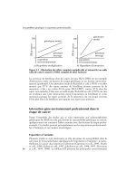
Figure 2 Homocysteine metabolism and possible mechanism of atherothrombotic dis-
ease. (Reprinted with permission from Ref. 30.)
tients with newly diagnosed VTE, I usually screen for factor V Leiden and the
prothrombin gene mutation, which are more common than other inherited throm-
bophilias (29), as well as acquired hyperhomocysteinemia (Fig. 2) and anticardio-
lipin antibodies. Elevated levels of homocysteine are usually easily treated with
folate (30,31), and the presence of anticardiolipin antibodies suggests the possible
need for prolonged and intensive anticoagulation (32,33).
B. Cancer
Data from the Danish National Registry of Patients were used to investigate the
risk of a diagnosis of cancer following the detection of VTE not associated with
Venous Thromboembolism 217
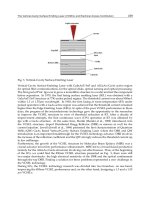
Figure 3 Cumulative probability of newly diagnosed cancer after a first VTE episode,
according to whether the VTE was idiopathic or nonidiopathic. (Reprinted with permission
from Ref. 35.)
surgery, known cancer, or pregnancy (34). There was a 30% increased risk of a
diagnosis of cancer among those with newly detected VTE. The risk was substan-
tially elevated only during the first 6 months of follow-up and declined rapidly
thereafter. Of those diagnosed with cancer within 1 year after the initial VTE
hospitalization, 40% had distant metastases at the time of the cancer diagnosis.
The association between cancer and VTE was most pronounced for cancers of
the pancreas, ovary, liver (primary hepatic cancer), and brain.
In the Swedish Cancer Registry, the risk of diagnosed cancer after a first
episode of VTE was elevated during at least the following 2 years (35). The
standardized incidence ratio for newly diagnosed cancer was 3.4 during the first
year after VTE and remained between 1.3 and 2.2 for the following 5 years. Of
the 854 patients with VTE, 534 had idiopathic VTE occurring in the absence of
surgery, trauma, temporary immobilization, or oral contraceptive use. These were
the patients in whom the association between VTE and the subsequent diagnosis
of cancer was apparent (Fig. 3).
IV. WOMEN’S HEALTH
A. Generations of Oral Contraceptives
First-generation oral contraceptives contained more than 50 µg of estrogen and
were associated with an alarming increase in the frequency of VTE, especially
218 Goldhaber
massive PE. Second-generation oral contraceptives contain less than 50 µg and
were introduced in the United States in 1967. Eventually, in 1989, first-generation
pills were withdrawn from the market.
Third-generation oral contraceptives utilize the new progestogens, desoges-
trel or gestodene. They cause acne and hirsutism less often and have a more
favorable effect on carbohydrate metabolism and lipid profiles than second-gener-
ation pills. Ironically, they are associated with a doubling or tripling of the VTE
rate compared with second-generation oral contraceptives (36,37). The explana-
tion for this surprising finding is that third-generation oral contraceptives lead to
acquired resistance to activated protein C, thus creating an effect similar to the
factor V Leiden mutation (38).
Despite the high relative risk of VTE from oral contraceptives, the absolute
risk is low. A New Zealand study of oral contraceptives and fatal PE estimated
the absolute risk of death from PE in current users as 1 per 10.5 million woman-
years. In this study, the risk of fatal PE was double among those taking third-
generation pills (39).
B. Oral Contraceptives and Thrombophilia
Oral contraceptives and thrombophilia appear to interact synergistically to in-
crease the risk of VTE. In a case-control study at Leiden University, women with
the factor V Leiden mutation who used oral contraceptives were at a 35-fold
greater risk of VTE than controls (40). In a subsequent analysis from the Leiden
Thrombophilia Study, which included cases with factor V Leiden, protein C or
S deficiency, the prothrombin gene 20210 A mutation, and antithrombin-III defi-
ciency, the overall risk of developing DVT during the first 6 months of oral
contraceptive use was increased 19-fold in thrombophilic women compared with
controls (41).
Whether women with a family history of VTE but no personal past history
of VTE should be screened prior to oral contraceptive use is controversial. For
women with known thrombophilia but no prior VTE, the safest policy is to use
alternative forms of contraception. However, no definitive ban on using oral con-
traceptives can be justified in this setting because the absolute risk of VTE re-
mains very low.
C. Pregnancy
PE is the leading cause of maternal death in the United Kingdom. Beginning in
the 1980s, the number of fatal PEs began to increase, especially following vaginal
delivery. In the mid-1990s, about two-thirds of the fatal PEs classified as maternal
deaths occurred postpartum, with cesarean section accounting for approximately
half of these catastrophic events (42).
Venous Thromboembolism 219
Two-thirds of DVT occur during pregnancy, and the remainder occur post-
partum. The risk of DVT is present throughout pregnancy and increases during
the third trimester. Of all antepartum DVT, about one-fifth occur during the first
trimester, one-third during the second trimester, and almost one-half during the
third trimester (43). After delivery, two of the most important risk factors for VTE
are increased maternal age and cesarean section. Emergency cesarean section in-
creases the VTE risk by about 50% compared with elective cesarean section.
Thrombophilia increases the risk of VTE during pregnancy and the puerpe-
rium. In a control study, the prevalence of factor V Leiden was 44% among women
with a history of VTE during pregnancy or the puerperium, and the prevalence of
the prothrombin gene mutation was 17%. Compared with controls, the Leiden mu-
tation increased the risk of VTE ninefold, and the prothrombin gene mutation in-
creased the risk by a factor of 15. The combination of the Leiden and prothrombin
gene mutations virtually multiplied the risk, estimated to be 107 times greater than
control. Fortunately, the absolute risk of VTE among carriers of each mutation
was low: 0.2% for Leiden and 0.5% for the prothrombin gene. However, among
those few women with both thrombophilic mutations, the absolute risk soared to
4.6% (44). Regardless of factor V Leiden, pregnancy itself causes hypercoagulabil-
ity because it induces a relative state of activated protein C resistance.
Thrombophilia has also been implicated in otherwise unexplained recurrent
pregnancy loss. The factor V Leiden mutation appears to double the risk of fetal
loss, possibly because of an increased frequency of placental vein thrombosis
(45,46). In addition to fetal demise, genetic thrombophilia appears to be associ-
ated with obstetrical complications, such as preeclampsia, abruptio placentae,
fetal growth retardation, and stillbirth (47).
It has been common practice to prophylax with heparin those pregnant
women who suffered a prior VTE. In a prospective study of 125 pregnant women
with a single prior VTE, antepartum heparin was withheld but postpartum antico-
agulation was administered for 4 to 6 weeks (48). Only 3 of the 125 women
(2.4%) developed antepartum VTE. Thus, VTE during pregnancy may be less
common in this population than had been previously thought.
D. Hormone Replacement Therapy
The traditional teaching used to be that hormone replacement therapy (HRT)
does not predispose to VTE. In 1996, this assumption was challenged when three
separate large data sets implicated HRT as doubling, tripling, or even quadrupling
the risk of VTE (49–51). As with oral contraceptives, the risk of VTE was highest
during the first year of HRT.
The Heart and Estrogen/progestin Replacement Study was a randomized
trial of 2763 postmenopausal women who had a history of coronary heart disease
but no previous VTE. They were allocated to conjugated equine estrogens, 0.625
220 Goldhaber
mg, plus medroxyprogesterone acetate, 2.5 mg, versus placebo. In results that
surprised the medical community, HRT did not reduce the rate of new coronary
events (52). Furthermore, the rate of VTE tripled among those women receiving
HRT (53). Certain subgroups were at especially high risk of increased VTE,
including women with lower extremity fractures (18-fold increase), cancer (4-
fold increase), postoperative state (5-fold increase), or nonsurgical hospitalization
(6-fold increase). Women with factor V Leiden seem to be at especially high risk
of VTE if they take HRT (54).
E. Selective Estrogen Receptor Modulators
An alternative to HRT is raloxifene, a selective estrogen receptor modulator that
has estrogenic effects on bone, lipid metabolites, and blood clotting but an estro-
gen antagonist effect on breast tissue. In a randomized controlled trial of 7705
osteoporotic postmenopausal women, raloxifene decreased the risk of breast can-
cer by 75% and decreased the rate of vertebral fractures, as had been hoped, but
it tripled the rate of VTE (55).
Tamoxifen, another selective estrogen receptor modulator, also acts as an
estrogen agonist on bone and an estrogen antagonist on breast tissue. In the British
Cancer Prevention Trial of women at high risk of breast cancer, 55 months of
treatment with tamoxifen 20 mg daily halved the rate of breast cancer. However,
the DVT rate increased by 60%, and the PE rate tripled (56).
V. CONCLUSIONS
In summary, VTE has enormous clinical impact. PE has a high mortality rate
despite advances in therapy, and VTE has a high recurrence rate after anticoagula-
tion is discontinued. DVT is often characterized by leg discomfort and postthrom-
botic venous insufficiency that adversely impacts the quality of life. Patients with
VTE often feel like they have a sword dangling over them because this disease
may be latent for many years and then recur. The etiologies of VTE are multifac-
torial, and we have arrived at an exciting juncture where we can identify with
increasing sophistication predisposing genetic, environmental, and hormonal fac-
tors that contribute to the risk of this illness.
REFERENCES
1. Oger E. Incidence of venous thromboembolism: a community-based study in West-
ern France. EPI-GETBP Study Group. Groupe d’Etude de la Thrombose de Bretagne
Occidentale. Thromb Haemost 2000; 83:657–660.
Venous Thromboembolism 221
2. Silverstein MD, Heit JA, Mohr DN, Petterson TM, O’Fallon WM, Melton LJ, III.
Trends in the incidence of deep vein thrombosis and pulmonary embolism: a 25-
year population-based study. Arch Intern Med 1998; 158:585–593.
3. Heit JA, Silverstein MD, Mohr DN, Petterson TM, O’Fallon WM, Melton LJ, III.
Risk factors for deep vein thrombosis and pulmonary embolism: a population-based
case-control study. Arch Intern Med 2000; 160:809–815.
4. Goldhaber SZ, Visani L, De Rosa M, for ICOPER. Acute pulmonary embolism:
Clinical outcomes in the International Cooperative Pulmonary Embolism Registry
(ICOPER). Lancet 1999; 353:1386–1389.
5. Heit JA, Silverstein MD, Mohr DN, Petterson TM, O’Fallon WM, Melton LJ, III.
Predictors of survival after deep vein thrombosis and pulmonary embolism: a popu-
lation-based, cohort study. Arch Intern Med 1999; 159:445–453.
6. Wicki J, Perrier A, Perneger TV, Bounameaux H, Junod AF. Predicting adverse
outcome in patients with acute pulmonary embolism: a risk score. Thromb Haemost
2000; 84:548–552.
7. Heit JA, Mohr DN, Silverstein MD, Petterson TM, O’Fallon WM, Melton LJ, III.
Predictors of recurrence after deep vein thrombosis and pulmonary embolism: a pop-
ulation-based cohort study. Arch Intern Med 2000; 160:761–768.
8. Hansson PO, Sorbo J, Eriksson H. Recurrent venous thromboembolism after deep
vein thrombosis: incidence and risk factors. Arch Intern Med 2000; 160:769–774.
9. Douketis JD, Foster GA, Crowther MA, Prins MH, Ginsberg JS. Clinical risk factors
and timing of recurrent venous thromboembolism during the initial 3 months of
anticoagulant therapy. Arch Intern Med 2000; 160:3431–3436.
10. Mohr DN, Silverstein MD, Heit JA, Petterson TM, O’Fallon WM, Melton LJ. The
venous stasis syndrome after deep venous thrombosis or pulmonary embolism: a
population-based study. Mayo Clin Proc 2000; 75:1249–1256.
11. Bergqvist D, Jendteg S, Johansen L, Persson U, Odegaard K. Cost of long-term
complications of deep venous thrombosis of the lower extremities: an analysis of a
defined patient population in Sweden. Ann Intern Med 1997; 126:454–457.
12. Walrath K, Berkovitz P, Morrison R, Goldhaber SZ. Frequently asked questions of
the Venous Thromboembolism Support Group. Brigham and Women’s Hospital.
1999. Available at: />13. Blaszyk H, Bjornsson J. Factor V Ieiden and morbid obesity in fatal postoperative
pulmonary embolism. Arch Surg 2000; 135:1410–1413.
14. Eekhoff EM, Rosendaal FR, Vandenbroucke JP. Minor events and the risk of deep
venous thrombosis. Thromb Haemost 2000; 83:408–411.
15. Klatsky AL, Armstrong MA, Poggi J. Risk of pulmonary embolism and/or deep
venous thrombosis in Asian-Americans. Am J Cardiol 2000; 85:1334–1337.
16. Goldhaber SZ, Grodstein F, Stampfer MJ, Manson JE, Colditz GA, Speizer FE et
al. A prospective study of risk factors for pulmonary embolism in women. JAMA
1997; 277:642–645.
17. Zornberg GL, Jick H. Antipsychotic drug use and risk of first-time idiopathic venous
thromboembolism: a case-control study. Lancet 2000; 356:1219–1223.
18. Guidelines on diagnosis and management of acute pulmonary embolism. Task Force
on Pulmonary Embolism, European Society of Cardiology. Eur Heart J 2000; 21:
1301–1336.
222 Goldhaber
19. Price DT, Ridker PM. Factor V Leiden mutation and the risks for thromboembolic
disease: A clinical perspective. Ann Intern Med 1997; 127:895–903.
20. Nguyen A. Prothrombin G20210A polymorphism and thrombophilia. Mayo Clin
Proc 2000; 75:595–604.
21. Bounameaux H. Factor V Leiden paradox: risk of deep-vein thrombosis but not of
pulmonary embolism. Lancet 2000; 356:182–183.
22. Ridker PM, Miletich JP, Stampfer MJ, Goldhaber SZ, Lindpaintner K, Hennekens
CH. Factor V Leiden and risks of recurrent idiopathic venous thromboembolism.
Circulation 1995; 92:2800–2802.
23. Simioni P, Prandoni P, Lensing AWA, Scudeller A, Sardella C, Prins MH, Villalta
S, Dazzi F, Girolami A. The risk of recurrent venous thromboembolism in patients
with an Arg
506
Gln mutation in the gene for factor V (factor V Leiden). N Engl
J Med 1997; 336:399–403.
24. Margaglione M, D’Andrea G, Colaizzo D, Cappucci G, del Popolo A, Brancaccio
V et al. Coexistence of factor V Leiden and Factor II A20210 mutations and recurrent
venous thromboembolism. Thromb Haemost 1999; 82:1583–1587.
25. De S, V, Martinelli I, Mannucci PM, Paciaroni K, Chiusolo P, Casorelli I et al. The
risk of recurrent deep venous thrombosis among heterozygous carriers of both factor
V Leiden and the G20210A prothrombin mutation. N Engl J Med 1999; 341:801–
806.
26. Miles JS, Miletich JP, Goldhaber SZ, Hennekens CH, Ridker PM. G20210A Muta-
tion in the prothrombin gene and the risk of recurrent venous thromboembolism.
JACC 2001; 37:1–4.
27. Meijers JC, Tekelenburg WL, Bouma BN, Bertina RM, Rosendaal FR. High levels
of coagulation factor XI as a risk factor for venous thrombosis. N Engl J Med 2000;
342:696–701.
28. Kyrle PA, Minar E, Hirschl M, Bialonczyk C, Stain M, Schneider B et al. High
plasma levels of factor VIII and the risk of recurrent venous thromboembolism. N
Engl J Med 2000; 343:457–462.
29. Rosendaal FR. Venous thrombosis: a multicausal disease. Lancet 1999; 353:1167–
1173.
30. Hankey GJ, Eikelboom JW. Homocysteine and vascular disease. Lancet 1999; 354:
407–413.
31. Langman LJ, Ray JG, Evrovski J, Yeo E, Cole DE. Hyperhomocyst(e)inemia and
the increased risk of venous thromboembolism: more evidence from a case-control
study. Arch Intern Med 2000; 160:961–964.
32. Greaves M. Antiphospholipid antibodies and thrombosis. Lancet 1999; 353:1348–
1353.
33. Schulman S, Svenungsson E, Granqvist S. Anticardiolipin antibodies predict early
recurrence of thromboembolism and death among patients with venous thromboem-
bolism following anticoagulant therapy. Duration of Anticoagulation Study Group.
Am J Med 1998; 104:332–338.
34. Sorensen HT, Mellemkjaer L, Steffensen FH, Olsen JH, Nielsen GL. The risk of a
diagnosis of cancer after primary deep venous thrombosis or pulmonary embolism.
N Engl J Med 1998; 338:1169–1173.
35. Schulman S, Lindmarker P. Incidence of cancer after prophylaxis with warfarin
Venous Thromboembolism 223
against recurrent venous thromboembolism. Duration of Anticoagulation Trial. N
Engl J Med 2000; 342:1953–1958.
36. Chasan-Taber L, Stampfer MJ. Epidemiology of oral contraceptives and cardiovas-
cular disease. Ann Intern Med 1998; 128: 467–477.
37. Jick H, Kaye JA, Vasilakis-Scaramozza C, Jick SS. Risk of venous thromboembo-
lism among users of third generation oral contraceptives compared with users of oral
contraceptives with levonorgestrel before and after 1995: cohort and case-control
analysis. BMJ 2000; 321:1190–1195.
38. Rosing J, Middeldorp S, Curvers J, Thomassen MCLGD, Nicolaes GAF, Meijers
JCM, Bouma BN, Bu
¨
ller HR, Prins MH, Tans G. Low-dose oral contraceptives and
acquired resistance to activated protein C: A randomized cross-over study. Lancet
1999; 354:2036–2040.
39. Parkin L, Skegg DCG, Wilson M, Herbison GP, Paul C. Oral contraceptives and
fatal pulmonary embolism. Lancet 2000; 355:2133–2134.
40. Vandenbroucke JP, Koster T, Brie
¨
t E, Reitsma PH, Bertina RM, Rosendaal FR.
Increased risk of venous thrombosis in oral-contraceptive users who are carriers of
factor V Leiden mutation. Lancet 1994; 344:1453–1457.
41. Bloemenkamp KWM, Rosendaal FR, Helmerhorst FM, Vandenbroucke JP. Higher
risk of venous thrombosis during early use of oral contraceptives in women with
inherited clotting defects. Arch Intern Med 2000; 160:49–52.
42. Greer IA. Thrombosis in pregnancy: maternal and fetal issues. Lancet 1999; 353:
1258–1265.
43. Ray JG, Chan WS. Deep vein thrombosis during pregnancy and the puerperium: A
meta-analysis of the period of risk and the leg of presentation. Obstet Gynecol Surv
1999; 54:265–271.
44. Gerhardt A, Scharf RE, Beckmann MW, Struve S, Bender HG, Pillny M, Sandmann
W, Zotz RB. Prothrombin and factor V mutations in women with a history of throm-
bosis during pregnancy and the puerperium. N Engl J Med 2000; 342:374–380.
45. Ridker PM, Miletich JP, Buring JE, Ariyo AA, Price DT, Manson JE, Hill JA. Factor
V Leiden mutation as a risk factor for recurrent pregnancy loss. Ann Intern Med
1998; 128:1000–1003.
46. Meinardi JR, Middeldorp S, de Kam PJ, Koopman MMW, van Pampus ECM, Ha-
mulya
´
k K, Prins MH, Bu
¨
ller HR, van der Meer J. Increased risk for fetal loss in
carriers of the factor V Leiden mutation. Ann Intern Med 1999; 130:736–739.
47. Kupferminc MJ, Eldor A, Steinman N, Many A, Bar-Am A, Jaffa A, Fait G, Lessing
JB. Increased frequency of genetic thrombophilia in women with complications of
pregnancy. N Engl J Med 1999; 340:9–13.
48. Brill-Edwards P, Ginsberg JS, Gent M, Hirsh J, Burrows R, Kearon C et al. Safety
of withholding heparin in pregnant women with a history of venous thromboembo-
lism. N Engl J Med 2000; 343:1439–1444.
49. Daly E, Vessey MP, Hawkins MM, Carson JL, Gough P, Marsh S. Risk of venous
thromboembolism in users of hormone replacement therapy. Lancet 1996; 348:977–
980.
50. Jick H, Derby LE, Myers MW, Vasilakis C, Newton KM. Risk of hospital admission
for idiopathic venous thromboembolism among users of postmenopausal oestrogens.
Lancet 1996; 348:981–983.
224 Goldhaber
51. Grodstein F, Stampfer MJ, Goldhaber SZ, Manson JE, Colditz GA, Speizer FE,
Willett WC, Hennekens CH. Prospective study of exogenous hormones and risk of
pulmonary embolism in women. Lancet 1996; 348:983–987.
52. Hulley C, Grady D, Bush T, et al. Randomized trial of estrogen plus progestin for
secondary prevention of coronary heart disease in postmenopausal women. Heart
and Estrogen/progestin Replacement Study (HERS) Research Group. JAMA 1998;
280:605–613.
53. Grady D, Wenger NK, Herrington D, Khan S, Furberg C, Hunninghake D, Vittingh-
off E, Hulley S, for the Heart and Estrogen/progestin Replacement Study Research
Group. Postmenopausal hormone therapy increases risk for venous thromboembolic
disease. The Heart and Estrogen/progestin Replacement Study. Ann Intern Med
2000; 132:689–696.
54. Lowe G, Woodward M, Vessey M, et al. Thrombotic variables and risk of idiopathic
venous thromboembolism in women aged 45–64 years. Relationships to hormone
replacement therapy. Thromb Haemost 2000; 83:530–535.
55. Cummings SR, Eckert S, Krueger KA, Grady D, Powles TJ, Cauley JA, Norton L,
Nickelsen T, Bjarnason NH, Morrow M, Lippman ME, Black D, Glusman JE, Costa
A, Jordan VC. The effect of raloxifene on risk of breast cancer in postmenopausal
women: results from the MORE randomized trial. Multiple Outcomes of Raloxifene
Evaluation. JAMA 1999; 281:2189–2197.
56. Fisher B, Costantino JP, Wickerham L, et al. Tamoxifen for the prevention of breast
cancer: report of the National Surgical Adjuvant Breast and Bowel Project P-1 Study.
J Natl Cancer Inst 1998; 90:1371–1388.
13
Integrated Diagnostic Approach to
Venous Thromboembolism
Henri Bounameaux
University Hospital of Geneva and Geneva School of Medicine,
Geneva, Switzerland
Diagnosis of deep vein thrombosis (DVT) depends mainly upon three clinical
tools: venous compression ultrasonography (US), assessment of prior clinical
probability (PCP), and measurement of fibrin D-Dimer (DD). In suspected pul-
monary embolism (PE), the same tools can be applied, in addition to ventilation/
perfusion lung scan (V/Q scan). In a few patients, venography (suspected DVT)
or pulmonary angiography (suspected PE) may also be required.
I. BRIEF DESCRIPTION OF THE DIAGNOSTIC TOOLS
Today venous compression ultrasonography (US) is the key diagnostic tool in
patients with a first episode of clinically suspected DVT. The sensitivity and
specificity exceed 90% for proximal DVT (1). The corresponding values are
definitely less (50% or less) for isolated distal DVT. Although these results may
be superior in experienced hands, they clearly highlight the need for integrating
the US result in a comprehensive approach.
A crucial issue in this regard is clinical assessment, which can be made
either by means of a standardized Wells score (2) or in an empirical way (3).
These two means have been compared in the case of suspected DVT (4). Both
the Wells score and the empirical evaluation can triage patients into a low, inter-
mediate, or high clinical probability category. However, the empirical method
performed slightly better in categorizing patients in the high-probability class,
while the Wells score categorized more patients in the low-probability class.
225
226 Bounameaux
Because of the very high sensitivity of some DD tests to the presence of
DVT and PE, they can be used alone or in combination with other findings (US
or clinical probability) to exclude venous thrombosis with a high negative pre-
dictive value (5). The rapid tests that have been validated in large patient popula-
tions include a rapid ELISA (VIDAS DD from bioMerieux), an automated turbi-
dimetric method (LIA test from Diagnostica STAGO), and the whole blood latex
test SimpliRED (from AGEN). Table 1 gives a summary of the performances of
these commercial tests. Other rapid tests (Turbiquant, Tinaquant, MDA D-Dimer)
are currently undergoing intensive clinical investigation.
Ventilation/perfusion lung scanning has been investigated in the large PIO-
PED study (6), which defined clear interpretation categories. Briefly, while a
normal/near-normal perfusion scan virtually rules out PE, a high-probability
ventilation/perfusion pattern allows one to diagnose PE. Low-probability and
intermediate-probability categories were also defined, but are considered in most
centers as nondiagnostic patterns.
In recent years, interest has developed in using spiral computed tomography
(CT) for PE diagnosis. Two systematic reviews have independently and simulta-
neously concluded that this new technique has not been adequately evaluated
(7,8) and that additional research is required to establish its place in clinical
practice. In particular, sensitivity of spiral CT to the presence of PE appears to
be lower than anticipated from the initial studies, especially if the embolus is
located in subsegmental vessels.
Gold standards for DVT and PE diagnosis are ascending venography and
pulmonary angiography, respectively. Both exams are invasive, costly, and not
devoid of risk. Moreover, their interpretation may be equivocal, as interob-
server agreement is far from optimal. The aim of any diagnostic strategy is to
reduce the number of invasive exams without increasing the number of false
diagnoses.
Table 1 Comparison of Three Rapid DD Tests for Diagnosing DVT and/or PE
Patients with DVT Sensitivity Specificity
DD test and/or PE (n) (%, 95% CI) (%, 95% CI)
VIDAS DD 1311, 305 98.7 (96.7–99.6) 39.6 (36.5–42.6)
LIA test 971, 310 99.4 (97.7–99.9) 39.6 (35.9–43.4)
SimpliRED 2393, 489 90.2 (87.2–92.7) 68.5 (66.5–70.6)
DD, D-dimer, DVT, deep vein thrombosis; PE, pulmonary embolism; 95% CI, 95% confidence in-
terval.
Venous Thromboembolism 227
II. BACKGROUND OF DIAGNOSTIC STRATEGIES
IN SUSPECTED DVT AND PE
Contemporary diagnostic strategies have been formulated according to two main
observations. First, while more than 50% of suspected patients had confirmed
DVT or PE in the early 1980s, this figure has decreased to 35% in the early
1990s and 20% in the late 1990s. This decrease in diagnostic yield most likely
reflects heightened clinical awareness of practitioners to the presence and danger
of venous thromboembolism. On the other hand, this trend means that the major-
ity of patients referred to a diagnostic center for evaluation will have the disorder
excluded rather than confirmed. Second, because DVT and PE are now recog-
nized as two clinical manifestations of a single disease, treatment algorithms and
diagnostic protocols have been streamlined.
As a consequence of these two observations, the first diagnostic step should
ideally be highly sensitive in order to detect the disease in a substantial proportion
of suspected patients. On the other hand, diagnosing DVT in a patient suspected
of PE suffices to rule in venous thromboembolism and to indicate anticoagulant
treatment.
The evaluation of a diagnostic tool or strategy culminates in management
trials or outcome studies in which the overall clinical strategy is applied. The
outcome at 3 or 6 months of follow-up is subsequently surveyed to determine
thromboembolic events that might have occurred in patients in whom anti-
coagulant treatment had been withheld on the basis of a negative diagnostic
workup (9).
III. DIAGNOSTIC STRATEGIES IN SUSPECTED DVT
Table 2 compares four recently proposed management strategies (2,3,10,11). One
Italian study relies on serial US (10) (i.e., repetition of compression US after 1
week in all patients with no DVT on the initial exam). A second Italian study
relies on serial US restricted to patients with a normal initial US and an abnormal
DD result (rapid latex test) (11). In the third strategy, serial US was restricted
to patients with a non-low-prior clinical probability (PCP) (2). In the fourth algo-
rithm, a single US was performed in patients with a DD result above the critical
cutoff (rapid ELISA method) (3), with PCP allowing the identification of patients
requiring venography (those with a high PCP, a positive DD, and a negative US).
These four strategies were assessed in management trials with long-term
(at least 3-month) follow-up. In the three trials in which DD (11), PCP (2), or
both (3) were added to the initial US exam, the proportion of patients requiring
a repeat US exam at 1 week was reduced from 76% in the study relying on US
228 Bounameaux
Table 2 Comparison of Four Validated Diagnostic Strategies in Patients Clinically
Suspected of DVT
Cogo et al. (10) Bernardi et al. (11) Wells et al. (2) Perrier et al. (3)
Tool/strategy RUS RUS ϩ DD RUS ϩ PCP US ϩ DD ϩ PCP
Patients (n) 1702 946 593 474
DVT prevalence 24% 28% 16% 24%
PCP assessment — — score empirical
DD test — Instant-IA — Vidas DD
Patients with US 1702 (100%) 946 (100%) 593 (100%) 346 (73%)
(n,%)
Patients with RUS 1302 (76%) 88 (9%) 166 (28%) 0
(n,%)
Yield of RUS 0.9% 5.7% 1.8% —
Venography (n, %) 0 0 33 (6%) 2 (0.4%)
3-month VTE risk 0.7% (0.3–1.2) 0.4% (0–0.9) 0.6% (0.1–1.8) 2.6% (0.2–4.9)
[% (95% CI)]
VTE, venous thromboembolism; DVT, deep vein thrombosis; FU, follow-up; US, compression ultra-
sonography; RUS, repeat US. (Instant-IA and Vidas DD are D-dimer tests marketed by Stago, As-
nie
`
res, France, and bioMe
´
rieux, Lyon, France, respectively.)
alone (10) to 9% when adding DD latex test (11), 28% when adding PCP (2),
or 0% when adding DD ELISA test and PCP assessment (3) (Table 2).
Compression venous US appears to be the key diagnostic tool in outpatients
clinically suspected of DVT. However, because the prevalence of DVT in the
suspected population is quite low (approximately 20%), using a highly sensitive
DD (ELISA) test as an initial screening would reduce the number of initial US
exams by one-fourth to one-third. Alternatively, PCP or DD may be used to
diminish the number of repeat US. However, the principle of repeat US after 1
week to pick up undiagnosed distal DVT that would have extended proximally,
albeit very attractive at a first glance, has a low yield and is not cost effective
(12). Last, the 3-month thromboembolic risk in these four studies (13) (Table 2)
was low and comparable to the 1.9% (95% CI; 0.4–5.4%) observed in clinically
suspected patients followed up after a negative venogram (14). Of note, these
results were obtained by means of a compression US technique limited to the
examination of the common and superficial femoral and popliteal veins. This at
least questions the affirmation by some investigators that a complete, more time-
consuming venous US examination (from the calf to the inferior vena cava)
should be performed in all patients clinically suspected of DVT (15).
Venous Thromboembolism 229
IV. DIAGNOSTIC STRATEGIES IN SUSPECTED PE
Recently, two sequential, mainly noninvasive strategies were applied in large
cohorts of patients with suspected PE (3,16). They are based on PCP, assessed
empirically (3) or by means of a clinical model (16); on a rapid DD ELISA test
(3); on venous compression US (3,16); on a V/Q lung scan (3,16); and, in some
cases, on pulmonary angiography. These strategies are depicted in Figures 1 and
Figure 1 Diagnostic algorithm for suspected pulmonary embolism. PE, pulmonary em-
bolism; US, lower limb venous ultrasonography; DVT, deep vein thrombosis; PCP, prior
clinical probability; near-N, near-normal; Rx, treatment; no Rx, no treatment. (From
Ref. 3.)
230 Bounameaux
Figure 2 Diagnostic algorithm for suspected pulmonary embolism. PE, pulmonary em-
bolism; US, lower limb venous ultrasonography; DVT, deep vein thrombosis; PCP, prior
clinical probability; near-N, near-normal; Rx, treatment; no Rx, no treatment. (From
Ref. 16.)
2. The 3-month thromboembolic risks that were associated with these algorithms
were 0.9% (95% CI; 0.2–2.7%) (3) or 0.5% (95% CI; 0.1–1.3%) (16), respec-
tively. Pulmonary angiography had to be performed in 11% (3) or 4% (16) of
patients. These strategies allow the management of the majority of patients with
widely available, noninvasive diagnostic tools. In a third, smaller Dutch study,
a sequential strategy of ventilation/perfusion lung scan, rapid whole-blood DD
test SimpliRED, US, and pulmonary angiography was used. The 3-month throm-
boembolic risk was 2.5% (95% CI; 0.5–7.1%) in the 121 patients in whom antico-
agulant treatment had been withheld on the basis of a normal perfusion or a
nondiagnostic lung scan pattern associated with a negative SimpliRED DD test.
(17).
Finally, in a secondary analysis of a database of 1034 consecutive patients
referred to a diagnostic center with clinically suspected PE, the diagnosis could
be ruled out by the combination of a low clinical probability and a nondiagnostic
lung scan (i.e., abnormal but non-high-probability pattern), provided no proximal
DVT had been detected on US exam. This combination was present in 175 pa-
tients of the cohort (22%), and was safe to rule out PE since the 3-month thrombo-
embolic risk was only 1.7% (95% CI; 0.4–4.9%) (18).
Venous Thromboembolism 231
Because these strategies have a similar efficacy, the less resource-intensive
strategy depicted in Figure 2 (3) is likely to be the most cost-effective one. How-
ever, formal cost-effectiveness studies comparing them are not yet available.
V. SCOPE AND LIMITATIONS OF THE PROPOSED
INTEGRATED APPROACH
The strategies described above can be combined in a single, integrated algorithm
(Fig. 3) that is, however, suitable for patients with clinical symptoms and/or
signs of venous thromboembolism who are referred to an outpatient clinic or an
emergency ward for ruling in/out the disease. It is not suited for patients who
are hospitalized in medical or surgical wards for longer periods of time, nor is
Figure 3 Integrated diagnostic algorithm for suspected venous thromboembolism. PE,
pulmonary embolism; US, lower limb venous ultrasonography; DVT, deep vein thrombo-
sis; PCP, prior clinical probability. (From Ref. 3.)
232 Bounameaux
it suitable for patients who present with hemodynamic instability. In the latter
case, the problem is therapeutic rather than diagnostic; moreover, emergency
treatment (e.g., thrombolysis or embolectomy) must be considered in these pa-
tients without delay. Thus, echocardiography and/or spiral CT may be first-line
diagnostic tools, because in this situation signs of right heart dysfunction or pres-
ence of proximal emboli are very likely to be present.
On the other hand, patients who are hospitalized in medical or surgical
wards for more than 24 h are unlikely to present with DD concentrations below
the critical cutoff due to comorbidities. Thus, in a diagnostic study of suspected
PE in hospitalized patients, the yield of a negative DD result was very low (about
5%) (19), which at least questions its utility in this category of patients.
Last, age considerably influences the performance of all diagnostic tools
(20). In an analysis of two large outcome studies that enrolled 1029 consecutive
outpatients presenting in the emergency ward with clinically suspected pulmo-
nary embolism (prevalence of pulmonary embolism 27%), the prevalence of pul-
monary embolism increased progressively from 12% below age 40 to 44% above
age 80. The positive predictive value of a high clinical probability of pulmonary
embolism was higher in the elderly (71 to 78% above 60 vs. 40 to 64% below
60). Sensitivity of DD testing was 100% in all age subgroups, but specificity
decreased markedly with age, from 67% below age 40 to 10% above age 80.
The diagnostic yield of lower limb venous compression US was higher in the
elderly [46% (95% CI; 26–67%) below 40 vs. 56% (95% CI; 44–68%) above
80]. The proportion of diagnostic (normal/near-normal or high probability) lung
scans also decreased from 68% to 42% with increasing age. This variable should
be kept in mind in the diagnostic approach of this disease.
VI. CONCLUSION AND PERSPECTIVES
Diagnosing DVT and PE has improved over the past 15 years primarily due to
the development of venous compression ultrasound of the lower limbs and D-
dimer measurement. If carefully applied, these strategies can reduce the need for
venography and pulmonary angiography to less than 10% of patients evaluated
and thus are likely to be cost effective. These newer diagnostic strategies should
thus be implemented in daily practice, taking into account local facilities and
expertise (21). In the near future, spiral CT might be included in the diagnostic
approach of PE, but its exact place remains to be determined.
REFERENCES
1. Kearon C, Julian JA, Math M, Newman TE, Ginsberg JS. Noninvasive diagnosis
of deep venous thrombosis. Ann Intern Med 1998; 128:663–677.
Venous Thromboembolism 233
2. Wells PS, Anderson DR, Bormanis J, Guy F, Mitchell M, Gray L, Clement C,
Robinson KS, Lewandowski B. Value of assessment of pretest probability of deep-
vein thrombosis in clinical management. Lancet 1997; 350:1795–1798.
3. Perrier A, Desmarais S, Miron MJ, de Moerloose P, Lepage R, Slosman D, Didier
D, Unger PF, Patenaude JV, Bounameaux H. Noninvasive diagnosis of venous
thromboembolism in outpatients. Lancet 1999; 353:190–195.
4. Miron MJ, Perrier A, Bounameaux H. Clinical assessment of suspected deep vein
thrombosis: comparison between a score and empirical assessment. J Intern Med
2000; 247:249–254.
5. Bounameaux H, de Moerloose P, Perrier A, Miron MJ. D-dimer testing in suspected
venous thromboembolism: an update. Q J Med 1997; 90:437–442.
6. The PIOPED Investigators. Value of the ventilation/perfusion scan in acute pulmo-
nary embolism. Results of the Prospective Investigation of Pulmonary Embolism
Diagnosis (PIOPED). JAMA 1990; 263:2753–2759.
7. Rathbun SW, Raskob GE, Whitsett TL. Sensitivity and specificity of helical com-
puted tomography in the diagnosis of pulmonary embolism: a systematic review.
Ann Intern Med 2000; 132:227–232.
8. Mullins MD, Becker DM, Hagspiel KD, Philbrick JT. The role of spiral volumetric
computed tomography in the diagnosis of pulmonary embolism. Arch Intern Med
2000; 160:293–298.
9. Bu
¨
ller HR, Lensing AWA, Hirsh J, tenCate JW. Deep vein thrombosis: new non-
invasive diagnostic tests. Thromb Haemost 1991; 66:133–137.
10. Cogo A, Lensing AWA, Koopman MMW, Piovella F, Siragusa S, Wells PS, Villalta
S, Bu
¨
ller HR, Turpie AGG, Prandoni P. Compression ultrasonography for diagnostic
management of patients with clinically suspected deep vein thrombosis: prospective
cohort study. BMJ 1998; 316:17–20.
11. Bernardi E, Prandoni P, Lensing AWA, Agnelli G, Guazzaloca G, Scannapieco G,
Piovella F, Verlato F, Tomasi C, Moia M, Scarano L, Girolami A. D-dimer testing
as an adjunct to ultrasonography in patients with clinically suspected deep vein
thrombosis: prospective cohort study. BMJ 1998; 317:1037–1040.
12. Perone N, Bounameaux H, Perrier A. Comparison of four strategies for diagnos-
ing deep vein thrombosis: a cost-effectiveness analysis. Am J Med 2001; 110:33–
40.
13. Bounameaux H, Perrier A. Rapid diagnosis of deep vein thrombosis: a comparison
between four strategies. Thromb Haemost 1999; 82:1360–1361.
14. Hull R, Hirsh J, Sackett DL, Taylor DW, Carter C, Turpie AGG, Powers P, Gent
M. Clinical validity of a negative venogram in patients with clinically suspected
venous thrombosis. Circulation 1981; 64:622–625.
15. Gena Frederick M, Hertzberg BS, Kliewer MA, Paulson EK, Bowie JD, Lalouche
KJ, DeLong DM, Carroll BA. Can the US examination for lower extremity deep
venous thrombosis be abbreviated? A prospective study of 755 examinations. Radi-
ology 1996; 199:45–47.
16. Wells PS, Ginsberg JS, Anderson DR, Kearon C, Gent M, Turpie AGG, Bormanis
J, Weitz J, Chamberlain M, Bowie D, Barnes D, Hirsh J. Use of a clinical model
for safe management of patients with suspected pulmonary embolism. Ann Intern
Med 1998; 129:997–1005.
17. de Groot MR, van Marwijk Kooy M, Pouwels JG, Engelage AH, Kuipers BF, Bu
¨
ller
234 Bounameaux
HR. The use of a rapid D-dimer blood test in the diagnostic work-up for pulmonary
embolism: a management study. Thromb Haemost 1999; 82:1588–1592.
18. Miron MJ, Perrier A, Bounameaux H, de Moerloose P, Slosman D, Didier D, Junod
A. Contribution of noninvasive evaluation to the diagnosis of pulmonary embolism
in hospitalized patients. Eur Resp J 1999; 13:1365–1370.
19. Perrier A, Miron MJ, Desmarais S, de Moerloose P, Slosman D, Didier D, Unger
PF, Junod A, Patenaude JV, Bounameaux H. Using clinical evaluation and lung scan
to rule out suspected pulmonary embolism: Is it a valid option in patients with normal
results of lower-limb venous compression ultrasonography? Arch Intern Med 2000;
160:512–516.
20. Righini M, Goehring C, Bounameaux H, Perrier A. Influence of age on performances
of common diagnostic tests in suspected pulmonary embolism. Am J Med 2000;
109:357–361.
21. Fennerty T. Pulmonary embolism. Hospitals should develop their own strategies for
diagnosis and management. BMJ 1998; 317:791–792.
14
The Management of Massive
Pulmonary Embolism
Jeremy P. Feldman
University of California at San Francisco, San Francisco, California
Samuel Z. Goldhaber
Brigham and Women’s Hospital and Harvard Medical School,
Boston, Massachusetts
Venous thromboembolism (VTE) remains a leading cause of cardiovascular mor-
bidity and mortality. The disease encompasses a spectrum of disorders including
distal calf vein thrombosis, proximal deep vein thrombosis, asymptomatic pulmo-
nary embolism, hemodynamically stable symptomatic pulmonary embolism
without right ventricular dysfunction, impending hemodynamically unstable pul-
monary embolism with right ventricular dysfunction, and hemodynamically un-
stable massive pulmonary embolism. This chapter will focus on massive and
submassive pulmonary embolism.
I. DEFINITION
Massive pulmonary embolism is accompanied by hemodynamic instability. Sub-
massive disease is associated with moderate or severe right ventricular dysfunc-
tion on echocardiography.
II. PATHOGENESIS AND PATHOPHYSIOLOGY
Virchow described three factors involved in the genesis of VTE: stasis, endothe-
lial injury, and hypercoagulability. These remain the major culprits. Progress has
235
236 Feldman and Goldhaber
come in our understanding of the myriad genetic thrombophilias. Clots formed in
the lower extremity veins (and occasionally the upper extremities) may propagate
proximally and embolize to the pulmonary vasculature. In the lungs, platelets,
vascular endothelium, and fibrin interact to increase pulmonary vascular resis-
tance due to loss of cross-sectional area of the pulmonary vascular tree, atelecta-
sis, and bronchoconstriction (1,2). Imbalances result in ventilation-perfusion mis-
matching, largely due to changes in regional perfusion that cause hypoxia, and
potentially right ventricular pressure overload, right-to-left shunting, and hypo-
tension. Right ventricular ischemia and bulging of the right ventricular septum
into the left ventricle have also been shown in animal models to contribute to
the hemodynamic effects of massive pulmonary embolism (3,4).
III. PROSPECTIVE REGISTRY
The International Cooperative Pulmonary Embolism Registry, a prospective
study of 2454 consecutive patients from 52 centers in seven countries, found that
88.9% of patients are symptomatic and hemodynamically stable; 4.2% of patients
presented in shock. Furthermore, of the patients undergoing echocardiography,
40% had right ventricular hypokinesis. The all-cause mortality rate of 11.4% at
2 weeks was highly correlated with more severe characteristics on presentation.
Hemodynamically unstable patients had a 58.3% mortality rate compared with
15.1% for patients who were stable at presentation (5).
IV. CLINICAL PRESENTATION
The clinical presentation of massive and submassive pulmonary embolism is
highly variable. In MAPPET (6), a multicenter registry that enrolled 1001 patients
from 204 centers with massive or submassive pulmonary embolism, dyspnea was
a near universal finding. Acute onset of symptoms and tachycardia were present
in almost 70% of patients. One-third of patients presented with syncope. A variety
of other findings may accompany massive pulmonary embolism (Table 1).
An important presentation in the emergency department is the patient with
cardiopulmonary symptoms and an elevated troponin level. Although the major-
ity of patients with markers of cardiac injury will have acute coronary syndromes,
pulmonary embolism may also cause this abnormality (7). The mechanism is
thought to be related to right ventricular ischemia due to acute dilatation (8).
Additionally, patients with elevated troponin levels are more likely to have elec-
trocardiographic findings suggestive of pulmonary embolism, including right-
bundle branch block and precordial T-wave abnormalities (9).
Management of PE 237
Table 1 Physical Findings in
Massive Pulmonary Embolism
Finding
Tachycardia
Fever
Distended neck veins
Tricuspid regurgitation
Right-sided heave
Right-sided S3
S1Q3T3
Right bundle branch block
Anterior precordial T-wave inversion
V. DIFFERENTIAL DIAGNOSIS
Table 2 lists common diseases that must be distinguished from massive pulmo-
nary embolism. Three diagnostic tests often are sufficient to distinguish pulmo-
nary embolism from the other possibilities. All patients should have an immediate
chest radiograph and an electrocardiogram, as well as blood pressures measured
in both arms and a pulsus paradoxus checked. Results obtained from these simple
and readily available tests should point the clinician toward pulmonary embolism
as a possibility.
The best emergent bedside imaging test is echocardiography. Highly sug-
gestive findings include: intracardiac thrombus, right ventricular septal bulging
into the left ventricle, McConnell’s sign (right ventricular hypokinesis with rela-
tive preservation of the apical region), pulmonary hypertension (estimated by the
velocity of the tricuspid regurgitation jet), and dilation of the right ventricle and
inferior vena cava (indicating elevated venous pressure) (10).
Table 2 Differential Diagnosis of Massive Pulmonary Embolism
Diagnosis Comments
Myocardial infarction Quality of pain, location, ECG
Cardiac tamponade CXR, pulsus paradoxus, distant heart
sounds, ECG
Tension pneumothorax Lung exam, CXR, ECG
Aortic dissection Quality of pain, pulse deficit, hyperten-
sion, CXR
238 Feldman and Goldhaber
d-Dimer, when measured using the ELISA method, is often helpful in the
evaluation of suspected pulmonary embolism. A quantitative d-dimer of less than
500 mg/mL makes pulmonary embolism unlikely. The negative predictive value
of the quantitative ELISA method exceeds 90%, and thus a negative result should
strongly suggest an alternative diagnosis (11–13). The converse, however, is not
true. An elevated d-dimer does not specifically point to pulmonary embolism.
Last, right heart catheterization may suggest the diagnosis. Elevated pulmo-
nary pressures with a normal pulmonary capillary wedge pressure, with a differ-
ence between the mean pulmonary artery pressure and the pulmonary capillary
wedge pressure of greater than 12 mmHg suggests the presence of pulmonary
vascular disease. Often, in patients who are critically ill, hypoxemia and arterial
hypotension are multifactorial, and assessment of the cardiac pressures may be
useful in combination with evaluation for lower extremity thrombus and more
conventional imaging modalities. Also, in patients receiving mechanical ventila-
tion, increasing dead space may be attributable to pulmonary embolism.
VI. DIAGNOSTIC IMAGING STUDIES
Bedside echocardiography has several clear advantages in hemodynamically un-
stable patients. Myocardial infarction and pericardial tamponade can readily be
identified and differentiated from pulmonary embolism. After the diagnosis of
pulmonary embolism is established, assessing right ventricular function also pro-
vides prognostic information in the setting of submassive disease. Several studies
have consistently shown that the presence of moderate or severe right ventricular
hypokinesis is a marker for increased mortality (14,15).
Additional studies include spiral computed tomography (CT), magnetic res-
onance pulmonary angiography (MRPA), ventilation-perfusion (V/Q) scanning,
and contrast pulmonary angiography (CPA). Each has advantages and disadvan-
tages (Table 3). As more emergency departments acquire CT scanning capability,
this modality is emerging as particularly useful, especially at times when echocar-
diography is not available. CT scanning is fast, provides high-quality images of
the proximal pulmonary vasculature, and is noninvasive. It also provides informa-
tion about the pericardial space and can be used (with a separate imaging acquisi-
tion protocol) to evaluate possible aortic dissection. However, in patients who are
breathing very rapidly, image quality is compromised. Furthermore, the patient is
exposed to intravenous contrast. In the hemodynamically unstable patient, chest
CT scanning is the best diagnostic study of choice to exclude large proximal
pulmonary emboli. Reported sensitivities range from 53 to 100%, and specificity
ranges from 81 to 100% (16–19). However, a negative CT scan does not exclude
peripheral emboli, and anticoagulation should not be withheld solely on the basis
Management of PE 239
Table 3 Diagnostic Imaging Tests
Test Comments
Echocardiogram Fast, portable, evaluates pericardium and left
ventricle, risk stratifies submassive PE.
Chest CT scan Readily available, can exclude massive disease,
may reveal alternative diagnoses.
MRPA Time-consuming, not widely available, not safe
for an unstable patient.
V/Q lung scan Widely available, no radiocontrast needed, often
nondiagnostic.
CPA Gold standard, invasive, limited availability.
MRPA, magnetic resonance pulmonary angiogram; CPA, contrast pulmonary
angiogram.
of a negative spiral study (19,20). Although several studies have attempted to
evaluate the safety of withholding anticoagulation after a negative spiral CT scan
(21,22), none of these studies actually included consecutive patients who only
were evaluated with spiral CT scanning. Rather, V/Q scanning and ultrasound
were widely used in addition to clinical prior probability. Whereas a negative CT
scan does not exclude peripheral pulmonary embolism, it does exclude proximal
disease. Thus in a hypotensive patient with a negative CT scan, an alternative
diagnosis must be sought.
Magnetic resonance pulmonary angiography (MRPA), although attractive
in principle, has not gained wide acceptance. Interobserver variability remains
high (23–25). Much as with CT angiography, proximal emboli are more easily
visualized than distal ones. The specificity appears high, but the resolution is not
as clear cut as with the most recent generation of chest CT scanners. In contrast to
CT, MRPA requires a longer period of breath-holding. Also, monitoring unstable
patients during MRPA is difficult. Settings where MRPA is particularly useful
include patients with life-threatening reactions to intravenous contrast, or patients
who refuse contrast pulmonary angiography. Currently, MRPA is not a suitable
diagnostic modality for the evaluation of possible massive PE in an unstable
patient.
In the more stable patient, V/Q scanning often provides enough information
to make a diagnosis of pulmonary embolism. The two main disadvantages are a
high frequency of nondiagnostic studies, especially in patients with coexisting
lung disease, and the difficulty in monitoring unstable patients during the test.
Spiral CT scanning of the chest may be at least as sensitive and specific. Further-
more, CT scanning has the advantage of identifying possible alternative diagnoses
240 Feldman and Goldhaber
such as pneumonia, aortic dissection, or atelectasis. The primary advantage of
V/Q lung scans is that no intravenous radiocontrast is required. Additionally, in
the pregnant patient, V/Q scans are still preferred.
In contrast to the often nondiagnostic results from lung scanning, contrast
pulmonary angiography (CPA) has long been considered the gold standard in the
evaluation of pulmonary embolism. In addition to providing useful diagnostic
information, catheter-directed therapy can be delivered once a diagnosis is estab-
lished. The disadvantages of angiography are related to the morbidity and mortal-
ity of the procedure. It is an invasive procedure that also employs radiocontrast
material. In experienced centers, however, the major morbidity rate is less than
1%. Zuckerman et al. reported no mortalities in a consecutive series of 547 pa-
tients (26).
VII. TREATMENT
The management of massive pulmonary embolism can be divided into two broad
categories, supportive measures and clot removal or dissolution. Ensuring ade-
quate oxygenation is paramount and may entail noninvasive positive pressure
ventilation or even endotracheal intubation. (However, positive end expiratory
pressure should be minimized as it imposes additional afterload to the right ventri-
cle.) In patients with arterial hypotension, diagnostic evaluation must proceed in
parallel with resuscitation.
Although data are sparse, several studies in animals suggest that fluid load-
ing in massive PE may result in further hemodynamic deterioration. Two mecha-
nisms have been set forth to explain this finding. The right and left ventricles
share an interventricular septum. As the right ventricle dilates, the septum bulges
into the left ventricle and limits diastolic volume and, thus, left ventricular stroke
volume and cardiac output. A second mechanism involves right ventricular isch-
emia that may be exacerbated by increased wall stress associated with further
increases in right ventricular end-diastolic volume caused by volume loading.
Despite the data generated from animal studies, there are few prospective studies
with humans. One study by Mercat et al. involving 13 patients did show that
cardiac index improved in response to fluid loading (27). Given the paucity of
data, a limited trial of fluids may be reasonable, but excessive volume resuscita-
tion should be avoided.
Is there an ideal pressor agent in arterial hypotension due to pulmonary
embolism? There have been no large prospective randomized controlled trials to
address this question. The literature is replete with case reports and cases series
describing the successful or unsuccessful use of a specific agent. Several small-
scale studies, mostly in animals, have evaluated norepinephrine, dobutamine, do-