Báo cáo y học: "Cerebral misery perfusion diagnosed using hypercapnic blood-oxygenation-level-dependent contrast functional magnetic resonance imaging: a case report" potx
Bạn đang xem bản rút gọn của tài liệu. Xem và tải ngay bản đầy đủ của tài liệu tại đây (560.88 KB, 5 trang )
CASE REPO R T Open Access
Cerebral misery perfusion diagnosed using
hypercapnic blood-oxygenation-level-dependent
contrast functional magnetic resonance imaging:
a case report
Adam L Gordon
1,2*
, Stephen Goode
3
, Olympio D’Souza
1
, Dorothee P Auer
4
, Sunil K Munshi
1
Abstract
Introduction: Cerebral misery perfusion represents a failure of cerebral autoregulation. It is an important
differential diagnosis in post-stroke patients presenting with collapses in the presence of haemodynamically
significant cerebrovascular stenosis. This is particularly the case when cortical or internal watershed infarcts are
present. When this condition occurs, further investigation should be done immedia tely.
Case presentation: A 50-year-old Caucasian man presented with a stroke secondary to complete occlusion of his
left internal carotid artery. He went on to suffer recurrent seizures. Neuroimaging demons trated numerous new
watershed-territory cerebral infarcts. No source of arterial thromboembolism was demonstrable. Hypercapnic blood-
oxygenation-level-dependent-contrast functional magnetic resonance imaging was used to measure his
cerebrovascular reserve capacity. The findings were suggestive of cerebral misery perfusion.
Conclusions: Blood-oxygenation-level-dependent-contrast functional magnetic resonance imaging allows the
inference of cerebral misery perfusion. This procedure is cheaper and more readily available than positron emission
tomography imaging, which is the current gold standard diagnostic test. The most evaluated treatment for
cerebral misery perfusion is extracranial-intracranial bypass. Although previous trials of this have been unfavourable,
the results of new stud ies involving extracranial-intracranial bypass in high-risk patients identified during cerebral
perfusion imaging are awaited.
Cerebral misery perfusion is an important and under-recognized condition in which emerging imaging and treat-
ment modalities present the possibility of practical and evidence-based management in the near future. Physicians
should thus be aware of this disorder and of recent developments in diagnostic tests that allow its detection.
Introduction
Cerebral misery perfusion (CMP) was first described by
Baron in 1981 [1] and represents a failure of cerebral
autoregulation. CMP has been associated with decreased
cerebral perfusion pressures in extracranial and intracra-
nial atheromatous diseases, complete carotid artery
occlusion, and Moyamoya disease [2]. Baron’ sinitial
description was of trans ient limb weakness and collapse
associated with changes i n patients’ posture [1]. Subse-
quent case reports have replicated this description.
Positron emission tomography (PET) scanning has
allowed us to follow the p rogression of cerebral haemo-
dynamic impairment in misery perfusion through the
measurement of regional cerebral blood flow (rCBF) and
tissue oxygen extraction fraction (OEF) [3]. The
hypothesized pathophysiology is outlined in Table 1.
The diagnosis of CMP should be considered in patients
with orthostatic stroke symptoms and significant cere-
brovascular stenosis. Patients with CMP have an
increased risk of progression to stroke as compared to
patients who have carotid occlusion without cerebral
haemodynamic impairment. The prospective St. Louis
Carotid Occlusion Study (STLCOS) followed 74 patients
with carotid occlusion, 32 with misery perfusion, and 42
* Correspondence:
1
Department of Stroke Medicine, Nottingham University Hospitals (City
Campus), Hucknall Road, Nottingham, UK
Gordon et al. Journal of Medical Case Reports 2010, 4:54
/>JOURNAL OF MEDICAL
CASE REPORTS
© 2010 Gordon et al; licensee BioMed Central Ltd. This is an Open Access article distributed under the terms of the Creative Commons
Attribution License ( which permits unrestricted use, distribution, and reproduction in
any medium, provided the or igina l work is properly cited.
with out any of the previous conditions. Over a mean fol-
low-up period of 31.5 months, 11 patients in the study
group went on to develop ipsilateral cerebral infarct com-
pared with t wo in t he control group (p = 0.04) [4]. This
association between cerebral infarction, particularly in
the watershed territories, and cerebral haemodynamic
impairment has since been described in a number of cer-
ebral perfusion imaging studies [5].
Case presentation
A 50-year-old Caucasian man was admitted with an acute
right hemiparesis, affecting his arm, leg and face, and a
right homonymous hemianopia. His vascular risk factors
included a 25 pack-year smoking history and a previous
myocardial infarction. An initial computed tomography
(CT) head scan and m agnetic resonance imaging (MRI)
scan showed a left parieto-occipital infarction in the pos-
terior cortical watershed territory. A carotid ultrasound
Doppler scan showed a complete occlusion of his left
internal carotid artery (ICA) which was confirmed on
magnetic resonance angiography (Figure 1). Left carotid
dissection was suspected but could not be proven as a
precipitant for his arterial occlusion because no intra-
mural haematoma was visible via a standard neck MRI.
He was discharged after making a good recovery at 12
days after he was admitted. He was started on aspirin,
dipyridamole and simvastatin.
Two months later he was readmitted following a pri-
mary focal seizure with secondary generalisation and
mild post-ictal right-sided weakness. The results of his
routine blood tests, renal function, glucose, erythrocyte
sedimentation rate (ESR), Chest X-ray, echocardiogram
(ECG) and 24-hour ECG were normal. A repeat MRI
showed a new infarct in his right anterior cortical
watershed territory. He made a reasonabl e recovery and
was independently mobile within one week of
admission.
Over the following year he suffered numerous col-
lapses. He described these as comprising right-sided
weakness and jerking movements prior to a loss of con-
sciousness. On each occasion he demonstrated good
recovery of physical function although subtle cognitive
impairment was noted by serial mini-mental state exam-
ination o n subsequent outpatient visits. A diagnosis of
post-stroke e pilepsy was made and he was commenced
on oral sodium valproate. Despite this he continued to
present with recurrent seizures.
A magnetic resonance scan was performed, which
confirmed the presence of numerous deep watershed
infarcts in addition to his two cortical infarcts. There
was persistent occlusion of his left I CA on this i maging.
In an attempt to understand the aetiology of our
patient’ s recurrent strokes, we applied a hypercapnia
BOLD fMRI technique to assess his cerebrovascular
reserve capacity (CVR). The technique is detailed else-
where [6]. In b rief, however, serial magnetic resonance
scans sensitised to tissue oxygenation (blood oxygena-
tion level dependent [BOLD] contrast) are obtained dur-
ing periods of normocapnia and hypercapnia (during
inhalation of 10% CO
2
). Using these images, CVR maps
are generated, where an increase in BOLD signal is used
as a marker of cerebral vasodilatory capacity.
Our patient de monstrated a significant loss of CO
2
-
indu ced BOLD reactivi ty in the hemisphere distal to his
carotid occlusion, suggesting maximal compensatory
vasodilation at rest (Figure 2). This represents stage 2
cerebral haemodynamic impairment. He was offered an
extracranial-intracranial vascular bypass surgery (EC-IC
bypass) but declined it.
Discussion
Our patient underwent a hypercapnia fMRI technique as
part of ongoing research at our institution. The use of
this technique has been validated in volunteers and,
increasingly, in patients [6]. The result o f this scan
showed a marked impairment in our patient’sCO
2
reac-
tivity in the hemisphere distal to his carotid artery
occlusion. It is not possible to comment on oxygen
extraction fraction from these CVR maps, but the
observed change allows us to infer stage 2 cerebral hae-
modynamic impairment (failure of cerebrovascular auto-
regulation). In the context of carotid occlusion and
normal BOLD increase in his contralatera l hemisphere,
we can further infer maximal compensatory vasodilation
at rest. Whether this vasod ilatation suffices to maintain
cerebral perfusion via increased oxygen extraction,
hence preventing progression to stage 3 cerebral haemo-
dynamic impairment, cannot be inferred from these
images.
Table 1 Pathophysiology of Cerebral Misery Perfusion
(Stage 1 to 3 Cerebral Haemodynamic Impairment)
Stage 1
(Cerebrovascular
autoregulation)
Any fall in regional cerebral perfusion pressure
(rCPP) is matched by a fall in regional
cerebrovascular resistance (rCBR) in order to
maintain regional cerebral blood flow (rCBF).
This is accommodated by vasodilatation and an
attendant increase in regional cerebral blood
volume (rCBV). Oxygen extraction factor (OEF)
remains constant.
Stage 2
(Misery Perfusion)
The capacity for compensatory vasodilatation is
exceeded (rCVR becomes a constant) and rCBF
therefore drops in tandem with rCPP. To meet
their metabolic demands, neurones must
“extract more oxygen” from the passing blood -
OEF increases.
Stage 3
(End-organ
compromise)
If rCBF continues to fall to the extent that the
brain can no longer compensate by increases in
OEF, end-organ dysfunction occurs (TIA). If this
situation persists, permanent end-organ damage
(stroke) occurs.
Gordon et al. Journal of Medical Case Reports 2010, 4:54
/>Page 2 of 5
PET scanning is unique because it provides absolute
measurements for both OEF and CBF, but is cumber-
some and not widely available. Other modalities, such as
the BOLD-contrast fMRI used here, establish a measure-
ment of rCBF either by compa ring the a ffected hemi-
sphere to the contralateral (normally perfused)
hemisphere or by utilising normative reference ranges.
Single Photon Emission CT (SPECT) and Xenon-
enhanced CT have also been validated for clinical diag-
nosis of misery perfusion. Dynamic contrast enhanced
CT, MRI perfusion imaging and arterial spin labelling
MR techniques are newer, promising and less invasive
techniques to assess rCBF [7].
There is some uncertainty surrounding the aetiology
of our patient’s recurrent collapses. Limb-je rking transi-
ent ischaemic attacks (TIAs) have been described in the
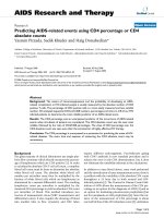
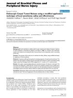
Figure 1 Watershed infarcts in the anterior cortical, posterior cortical and internal watershed territories of the left cerebral
hemisphere. Magnetic resonance angiogram shows complete occlusion of the left internal carotid artery.
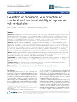
Figure 2 Cerebrovascular reserve map of our patient showing left internal caroti d artery occlusion and recurrent symptoms. Note the
loss of reactivity in the left hemisphere following CO2 inhalation. This is most prominent in the watershed territories.
Gordon et al. Journal of Medical Case Reports 2010, 4:54
/>Page 3 of 5
literature and are characterised by brief, involuntary,
coarse movements of the limbs. An association with car-
otid occlusion has been demonstrated and Han et al.[8]
have described the failure of cerebrovascular autoregula-
tion in thes e patients. However, TIAs are not associated
with the loss of consciousness or with secondary gener-
alisation and so would not fully explain this
presentation.
Post-stroke seizures, by contrast, are common and are
frequently characterised by generalisation and loss of
consciousness. Stroke is reported to be an aetiological
factor in 45% of seizures in patients over the age of 60
[9] and 5% to 20% of people who have strokes will go
on to develop seizures [10]. Early post-stroke seizures,
occurring up to two weeks after stroke, are postulated
to result from the accumulation of intracellular calcium
and sodium and extracellular glutamate during acute
ischemic injury. Late seizures occurring after this time
are thought to be a consequence of long-term scarring
and gliosis [10]. The accumulation of new ischaemic
events on repeat MRI, along with the demonstrated cer-
ebral haemodynamic impairment, raises the possibility
that our patient’ s seizures were a consequence of
ongoing cerebral ischaemia rather than of chronic gliotic
change, hence CMP is a possible explanation. It is, how-
ever, impossible to make this association with any
degree of certainty.
Attempts to treat patients with CMP have focused on
the a ugmentation of rCBF, predominantly by extracra-
nial-intracranial vascular anastomosis (EC-IC bypa ss).
Isolated case studies have reported the resolution of
symptomatic CMP following cerebral angioplasty for
intracranial arterial stenosis [11] but this procedure
remains largely untested. EC-IC bypass can be used to
describe a number of possible a pproaches but research
has focused predominantly on superficial temporal
artery to middle cerebral artery (STA-MCA) anastomo-
sis. The EC/IC bypass study was a large multicenter ran-
domized trial of STA-MCA a nastomosis with primary
end-points of 30-day stroke mortality and stroke inci-
dence. A total of 1377 patients were recruited and 663
were randomize d to receive EC-IC bypass. Patients ran-
domized to the treatment group performed less well on
all outcome measures despite having high graft patency
rates [12]. A fundamental weakness of the study was the
recruitment of patients with carotid or MCA occlusion
without reference to cerebral perfusion studies, which
has made it impossible to conduct subgroup analyses
evaluating the efficacy of the p rocedure in patients with
radiologically confirmed CMP.
The data from STLCOS regarding the prognosis in
misery perfusion and emergi ng imaging techniques have
provided justification for a reevaluation of EC-IC bypass.
Further support has come from imaging studies
demonstrating the resolution of CMP in small cohorts
undergoing EC-IC bypa ss [13]. Two large randomized
multicenter trials are underway and the publication of
resultsisawaited[14,15].Early intimations from the
Japanese Extracranial-intracranial-bypass Trial (JET),
however, have been positive, thus suggesting that data
from that trial may support the intervention [15].
Conclusion
CMP is an important differential diagnosis in post-
stroke patients presenting with recurrent collapses. The
hypercapnic-BOLD contrast fMRI procedure described
in this case report represents a promising new imaging
modality, without the resource implications of PET
scanning from which cerebral misery perfusion may be
inferred. Pending data from international trials may lead
to an increased profile for m isery perfusion in the near
future if EC-IC bypass is demonstrated to be effective.
Consent
Written informed consent was obtained from our
patient for publication of this case report and any
accompanying images. A copy of the written consent is
available for review by the Editor-in-Chief of this
journal.
Ethical Approval
The research project discussed in this article was
approved by the Derby NHS Research Ethics
Committee.
Author details
1
Department of Stroke Medicine, Nottingham University Hospitals (City
Campus), Hucknall Road, Nottingham, UK.
2
Division of Rehabilitation and
Ageing, University of Nottingham, Room B98, Medical School, Queens
Medical Centre, Nottingham, UK.
3
Division of Neuroradiology, University of
Nottingham, Medical School, Queens Medical Centre, Nottingham, UK.
4
Division of Academic Radiology, University of Nottingham, Medical School,
Queens Medical Centre, Nottingham, UK.
Authors’ contributions
AG, OD and SM reviewed our patient’s case notes and the current literature
on cerebral misery perfusion and drafted the manuscript. SG and DA
conducted the BOLD-MRI imaging, provided radiographic images, reported
on these, and provided the outlines of radiological procedures incorporated
in the manuscript. All authors read and approved the final manuscript.
Competing interests
The authors declare that they have no competing interests.
Received: 12 January 2009
Accepted: 18 February 2010 Published: 18 February 2010
References
1. Baron JC, Bousser MG, Rey A, Guillard A, Comar D, Castaigne P: Reversal of
focal “misery-perfusion syndrome” by extra-intracranial arterial bypass in
hemodynamic cerebral ischemia: a case study with 15O positron
emission tomography. Stroke 1981, 12(4):454-459.
2. Kirkpatrick PJ, Ng I: Cerebral revascularisation: where are we now?. J
Neurol Neurosurg Psychiatry 2005, 76(4):463-465.
Gordon et al. Journal of Medical Case Reports 2010, 4:54
/>Page 4 of 5
3. Powers WJ: Cerebral hemodynamics in ischemic cerebrovascular disease.
Annals of Neurology 1991, 29(3):231-240.
4. Grubb RL Jr, Derdeyn CP, Fritsch SM, Carpenter DA, Yundt KD, Videen TO,
Spitznagel EL, Powers WJ: Importance of hemodynamic factors in the
prognosis of symptomatic carotid occlusion. JAMA 1998,
280(12):1055-1060.
5. Momjian-Mayor I, Baron JC: The pathophysiology of watershed infarction
in internal carotid artery disease: review of cerebral perfusion studies.
Stroke 2005, 36:567-577.
6. Goode SD, Krishan S, Alexakis C, Mahajan R, Auer DP: Precision of
cerebrovascular reactivity assessment with use of different
quantification methods for hypercapnia functional MR imaging. Am J
Neuroradiol 2009, 30(5):972-977.
7. Wintermark M, Sesay M, Barbier E, Borbely K, Dillon W, Eastwood TC,
Grandin CB, Pedraza S, Soustiel J, Nariai T, Zaharchuk G, Caillé J, Dousset V,
Yonas H: Comparative overview of brain perfusion imaging techniques.
Stroke 2005, 36:2032-2033.
8. Han SW, Kim SH, Kim JK, Park CH, Yun MJ, Heo JH: Hemodynamic changes
in limb shaking TIA associated with anterior cerebral artery stenosis.
Neurology 2004, 63(8):1519-1521.
9. Forsgren L, Bucht G, Eriksson S, Bergmark L: Incidence and clinical
characterization of unprovoked seizures in adults: a prospective
population-based study. Epilepsia 1996, 37(3):224-229.
10. Silverman IE, Restrepo L, Matthews GC: Poststroke Seizures.”. Arch Neurol
2002, 59(2):195-201.
11. Derdeyn CP, Cross DT, Moran CJ, Dacey RGJ: Reversal of focal misery
perfusion after intracranial angioplasty: case report. Neurosurgery 2001,
48(2):436-440.
12. The EC/IC Bypass Study Group: Failure of extracranial-intracranial arterial
bypass to reduce the risk of ischemic stroke. Results of an international
randomized trial. N Engl J Med 1985, 313(19):1191-1200.
13. Gibbs JM, Wise RJ, Thomas DJ, Mansfield AO, Russell RW: Cerebral
haemodynamic changes after extracranial-intracranial bypass surgery. J
Neurol Neurosurg Psychiatry 1987, 50(2):140-150.
14. Wanebo JE, Amin-Hanjani S, Boyd C, Peery T: Assessing success after
cerebral revascularization for ischemia. Skull Base: An Interdisciplinary
Approach 2005, 15(3):215-227.
15. Mori E: Extracranial-intracranial arterial bypass surgery revisited.
International Journal of Stroke 2008, 3(s1):2-474.
doi:10.1186/1752-1947-4-54
Cite this article as: Gordon et al.: Cerebral misery perfusion diagnosed
using hypercapnic blood-oxygenation-level-dependent contrast
functional magnetic resonance imaging: a case report. Journal of Medical
Case Reports 2010 4:54.
Submit your next manuscript to BioMed Central
and take full advantage of:
• Convenient online submission
• Thorough peer review
• No space constraints or color figure charges
• Immediate publication on acceptance
• Inclusion in PubMed, CAS, Scopus and Google Scholar
• Research which is freely available for redistribution
Submit your manuscript at
www.biomedcentral.com/submit
Gordon et al. Journal of Medical Case Reports 2010, 4:54
/>Page 5 of 5