Báo cáo y học: " Two stage fracture of a polyethylene post in a 9-year-old posterior-stabilized knee prosthesis: a case report" pot
Bạn đang xem bản rút gọn của tài liệu. Xem và tải ngay bản đầy đủ của tài liệu tại đây (1.37 MB, 8 trang )
CAS E REP O R T Open Access
Two stage fracture of a polyethylene post
in a 9-year-old posterior-stabilized knee
prosthesis: a case report
Fabio D’Angelo
1*
, Daniele Marcolli
1
, Paolo Bulgheroni
1
, Luigi Murena
1
, Terenzio Congiu
2
, Paolo Cherubino
1
Abstract
Introduction: Several cases of tibial post breakage are reported in the literature. To the best of our knowledge,
only three cases of NexGen knee prosthesis (Zimmer, Warsaw, Indiana, USA) tibial post failure have been reported.
Case presentation: In November 1999, a 63-year-old Caucasian woman from Italy with a history of symptomatic
left knee osteoarthritis underwent a total knee arthroplasty. In March 2008, while rising from a chair, she felt a
sudden pain and instability in her left knee. She reported a fracture of the polyethylene post of the tibial insert. No
malposition or malalignment of either the femoral or tibial components were identified. The polyethylene tibial
insert was studied under light microscopy and scanning electron microscopy. The fracture was also noted to have
occurred without any notable polyethylene wear.
Conclusion: Scanning electron microscopy revealed two different damage patterns that could be explained with a
two-stage rupture of our patient’s polyethylene post. This could have been caused by a non-optimal ligamentous
balancing during first implant surgery. Her knee probably developed a varus instability that weakened the post,
and then a posterior anterior stress finally broke the polyethylene.
Introduction
The interaction between the polyethylene post of the
tibial tray and t he femoral cam is necessary f or the
proper functioning of posterior stabilized (PS) knee
prosthesis [1]. PS total knee arthroplasty (TKA) was
developed to grant stability, to achieve a higher range of
motion due to rollback, and to prevent posterior sub-
luxation of the implant [2]. The polyethylene spine con-
tacts the cam at approxim ately 7 0° of flexion, thus
preventing posterior subluxa tion. Mediolateral stability,
however, is dependent only on a well balanced and
aligned knee [3].
Polyethylene wear is a complication that could contri-
bute to aseptic loosening and osteolysis after TKA [4].
Acknowledged factors that can influence polyethylene
wear include prosthesis design, manufacturing, and poor
surgical technique [5,6].
Several cases of tibial post breakage are reported in
the literature [7-13]. To the best of our knowledge,
three cases of NexGen PS knee prosthesis (Zimmer,
Warsaw, Indiana, USA) tibial post failure have been
reported [14-16]. This case report focuses on light
microscopy and scanning electron microscopy (SEM)
evaluation of the broken polyethylene insert. This report
also aims to explain a possible mechanism for the failure
of tibial post.
Case presentation
In November 1999, a 63-year-old Caucasian woman
from Italy (weight = 100 kg, height = 1.60 m, body mass
index = 39) with a history of symptomatic left knee
osteoarthritis underwent a TKA in another hospital. The
implant used was a NexGen PS knee prosthesis (Zim-
mer, Warsaw, Indiana, USA) with a tibial component
size of 4, a femoral component size of D, and a poly-
ethylene insert 10 mm in thickness. No problem was
reported during the follow-up examination, and the
patient was able to perform normal life activities for the
next nine years.
In March 2008, while rising from a chair, she felt a
sudden pain and instability in her left knee. After this
* Correspondence:
1
Department of Orthopaedics and Traumatology, University of Insubria,
Ospedale di Circolo - Fondazione Macchi, V le Borri 57, 21100 Varese, Italy
D’Angelo et al. Journal of Medical Case Reports 2010, 4:65
/>JOURNAL OF MEDICAL
CASE REPORTS
© 2010 D’Angelo et al; licensee BioMed Central Ltd. This is an Open Access article distributed under the terms of the Creative
Commons Attribution License ( which permits u nrestricted use, distributio n, and
reproduction in any medium, provided the ori ginal work is properly cited.
acute event she was unable to bear weight on her left
knee, and was thus forced to use crutches. On physical
examination she presented a mild effusion of the knee, a
flexion of 90°, and knee hyperextension. The joint pre-
sented signs of both anteroposterior and varus to valgus
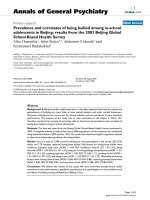
instability. X-ray examinations showing the anteropos-
terior view of the knee did not indicate any remarkable
alteration in polyethylene wear, while the lateral view
showed a hyperextension of the tibia with a posterior
subluxation of the femur (Figure 1). The hypothesis of
post breakage was thus made.
Our patient underwent diagnostic knee arthroscopy
and was scheduled to have her polyethylene insert chan-
ged. The procedure granted a clear view of the polyethy-
lene and the broken post in articulation. However,
actual findings showed that the polyethylene had no
relevant wear areas or alterations. Based on these find-
ings, we performed an anterior approach with medial
parapatellar arthrotomy. During surgery, samples of the
periprosthetic tissue were taken in order to obtain a his-
tological evaluation. These specimens were treated with
haematoxylin-eosin and von Kossa staining, and were
then studied under light microscopy using polarized
light in order to detect the typical birefringence of the
polyethylene debris.
The total knee components appeared to be well fixed
intraoperatively. The polyethylene insert was substitu ted
with a 12-mm CD LPS Flex articular surface (Zimmer,
Warsaw, Indiana, USA). The stability in full extension,
mid-flexion, and full flexion of the knee was tested
intraoperatively, and appeared to be good. The patient
had no postoperative complications and recovered well.
Thepolyethyleneinsertandthebrokenpostwere
both prepared for scanning electron microscopy (SEM)
evaluation (Figure 2).
At six months follow-up the patient had recovered
complete function of her left knee. She was free from
pain and could walk normally without any support
(Figure 3).
Discussion
The design feature common to all PS knee prosthesis is
the cam-and-post mechanism that is incorporated into
the femoral and tibial components. The ca m on the
femoral compon ent is designed to engage the post of
the tibial polyethylene during knee flexion. This interac-
tion provides a functional substitute for the posterior
cruciate ligament (PCL), thus resulting in femoral roll-
back as flexion increases. In addition, the cam and the
post work to limit posterior displacement of the tibia
relative to the femur in extension [1].
In some cases in which the resulting laxity in flexion
is greater than the so-called “ jump distance” ,orthe
height of the post, acute dislocation may occur. Another
Figure 1 Preoperative X-rays. The hyperextension of the tibia in relation to the posterior subluxation of the femur can be seen.
D’Angelo et al. Journal of Medical Case Reports 2010, 4:65
/>Page 2 of 8
potential cause of flexion instability in a knee with PS
prosthesis is the failure of the polyethylene post. This
can be caused by either polyethylene acute fracture or
fatigue fracture, which is a consequence of repetitive
anterior impingement between the metal femoral cam
and the polyethylene post [7].
No malposition or malalignment of both the femoral
and tibial components were identified in our patient. Con-
firming the findin gs of Colizza et al., [17], polarized light
microscopy did not reveal any notable polyethylene wear.
Scanning electron microscopy, as reported in the lit-
erature [14,18], is an effective modality for analyzing the
surface of fatigue frac tures. An evaluation of the
retrieved tibial polyethylene insert via SEM revealed two
different damage patterns, considering the medial
part and the lateral aspect (Figure 4). The medial part
(Figure2areas1,2and3,Figure4,Figure5,Figure6)
presented a fracture line laminated in front and smooth
behind and with the tear lines with a mediolateral and
anterior posterior orientation.
The medial part of the fracture edge appears to be
smooth (Figure 2 areas 2 and 3) and with a different
orientation of the fracture lines. These characteristics
suggest a chronic instability of the varus valgus knee
prosthesis that slowly weakened the polyethylene post.
Meanwhi le, the lateral part (Figure 2 areas 4, 5 and 6,
Figure 7, Figur e 8, Figure 9) of the fracture presented a
sharp line that ends anteriorly with a laminated tear
(Figure 2 area 6, Figure 9) parallel to the anterior edge
of the polyethylene insert. This implies that this area
could be the terminal acute failure area of the fractured
post. The final rupture occurred after the chronic weak-
ening of the polyethylene due to the mediolateral stress
on the tibial post. These features could be explained
with a two-stage rupture of the polyethylene post. First,
a varus and anterioposterior force caused partial rupture
and instability of the post, which caused progressive
smoothening of the medial and posterior fracture edges.
Consequently, an anterioposterior lift-off force led to
the complete rupture of the post. This could have been
Figure 2 Numerated areas of the polyethylene insert.
Figure 3 X-rays at six months follow-up examination.
D’Angelo et al. Journal of Medical Case Reports 2010, 4:65
/>Page 3 of 8
Figure 4 Area 1 of Figure 2.
Figure 5 Area 2 of Figure 2.
D’Angelo et al. Journal of Medical Case Reports 2010, 4:65
/>Page 4 of 8
Figure 6 Area 3 of Figure 2.
Figure 7 Area 4 of Figure 2.
D’Angelo et al. Journal of Medical Case Reports 2010, 4:65
/>Page 5 of 8
Figure 8 Area 5 of Figure 2.
Figure 9 Area 6 of Figure 2.
D’Angelo et al. Journal of Medical Case Reports 2010, 4:65
/>Page 6 of 8
caused by a non-optimal ligamentous balancing during
the first implant surgery. Our patient’ s knee probably
developed progressive va ru s instability that slowly wea-
kened the post, and then an anterioposterior stress
finally broke the polyethylene.
Light microscopy evaluation showed a typical chronic
inflammatory reaction. Rare polyethylene flakes were
identifiable under polarized light. These particles
appeared to be well-contro lled by giant cells. No metal-
losis was observed (Figure 10). Von Kossa staining
returned negative results. Such findings can be consid-
ered normal sinovia in TKA. The absence of polyethy-
lene particles confirmed the macroscopic evidence of
the absence of wear, which could have caused the
breakage.
Conclusion
Especially in posterior stabilized designs, it is important
to achieve a well-balanced and aligned knee in order to
reduce stress on the polyethylene spine that could
otherwise lead to fatigue fracture [3].
We believe that the major failure mechanism of the
polyethylene post in our patient was the mild varus
valgus instability related to a non-optimal ligamentous
balancing during her first imp lant surgery. This aspect,
together with our patient’s weight, produced a progres-
sive weakening of the polyethylene post, which finally
broke due to hyperextension mechanism.
Based on the experience of Callaghan et al.[19],
proper femoral component positioning and avoiding
excessive posterior tibial slope during implant surgery is
crucial to reduce the anterior impingement of the post.
Our patient’s tibial slope was only 2°, which indicates a
good compromise between ROM and tibial post
impingement.
For most patients, once the diagnosis has been estab-
lished the revision of the polyethylen e insert is manda-
tory when components are well-fixed and in good
alignmen t. In our patient, an insert that was only 2 mm
thicker was enough to restore the stability of her knee.
However, if the components are loose or malpositioned,
complete revision surgery is recommended.
When sudden pain and instability appear in a func-
tioning knee PS TKA, a tibial post breakage must be
considered.
Consent
Written informed consent was obtained from the patient
for publication of this case report and accompanying
images. A copy of the written consent is available for
review by the Editor-in-Chief of this journal.
Author details
1
Department of Orthopaedics and Traumatology, University of Insubria,
Ospedale di Circolo - Fondazione Macchi, V le Borri 57, 21100 Varese, Italy.
2
Department of Normal Human Morphology “L Cattaneo”, University of
Insubria, Via O Rossi 9, 21100 Varese, Italy.
Authors’ contributions
FD performed the surgery, was involved in the bibliographic research, and
was a major contributor in writing the manuscript. DM was involved in the
bibliographic research and was also a major contributor in writing the
manuscript. PB performed diagnostic knee arthroscopy. LM was involved in
the bibliographic research. TC performed scanning electron microscopy
evaluation and light microscopy of the samples from the patient. PC also
performed surgery and contributed in writing the manuscript. All authors
read and approved the final manuscript.
Competing interests
The authors declare that they have no competing interests.
Received: 4 November 2009 Accepted: 23 February 2010
Published: 23 February 2010
References
1. Clarke HD, Math KR, Scuderi GR: Polyethylene post failure in posterior
stabilized total knee arthroplasty. J Arthroplasty 2004, 19:652-657.
2. Insall JN, Lachiewicz PF, Burnstein AH: The posterior stabilized condylar
prosthesis: two of four-year clinical experience. J Bone Joint Surg 1982,
64A:1317-1323.
3. Hendel D, Garti A, Weisbort M: Fracture of the central polyethylene tibial
spine in posterior stabilized total knee arthroplasty. J Arthroplasty 2003,
18:672-674.
4. Lonner JH, Siliski JM, Scott RD: Prodromes of failure in total knee
arthroplasty. J Arthroplasty 1999, 14:488-492.
5. Moreland JR: Mechanisms of failure in total knee arthroplasty. Clin Orthop
1988, 226:49-64.
6. Kilgus DJ, Moreland JR, Finerman GA, Funahashi TT, Tipton JS: Catastrophic
wear of tibial polyethylene inserts. Clin Orthop 1991, 273:223-231.
7. Lombardi AV Jr, Mallory TH, Vaughn BK, Krugel R, Honkala TK, Sorscher M,
Kolczun M: Dislocation following primary posterior-stabilized total knee
arthroplasty. J Arthroplasty 1993, 8:633-639.
8. Mestha P, Shenava Y, D’Arcy C: Fracture of the polyethylene tibial post in
posterior stabilized (Insall Burnstein II) total knee arthroplasty. J
Arthroplast 2000, 15:814-815.
9. Ng TP, Chiu KY: Recurrent dislocation of total knee arthroplasty. J
Arthroplasty 2003, 18:1067-1070.
10. Mariconda M, Lotti G, Milano C: Fracture of posterior-stabilized tibial
insert in a Genesis knee prosthesis. J Arthroplasty 2000, 15:529-530.
Figure 10 Dense connective tissues with typical inflammator y
cells and rare polyethylene flakes (arrow) are identifiable.
D’Angelo et al. Journal of Medical Case Reports 2010, 4:65
/>Page 7 of 8
11. Mauerhan D: Fracture of the polyethylene tibial post in a posterior
cruciate-substituting total knee arthroplasty mimicking patellar clunk
syndrome. J Arthroplasty 2003, 7:942-945.
12. Bal BS, Greenberg D: Failure of a metal-reinforced tibial post in total knee
arthroplasty. J Arthroplasty 2007, 22:464-467.
13. Ridgeway S, Moskal JT: Early instability with mobile bearing total knee
arthroplasty. J Arthroplasty 2004, 19:686-693.
14. Chiu YS, Chen WM, Huang CK, Chiang CC, Chen TH: Fracture of the
polyethylene tibial post in a NexGen posterior-stabilized knee
prosthesis. J Arthroplasty 2004, 19 :1045-1049.
15. Shih KC, Chou LC: Fracture of the polyethylene tibial spine in NexGen
posterior stabilized flex knee prosthesis: a case report. J Orthop Surg
Taiwan 2007, 24:30-34.
16. Lee CS, Chen WM, Kou HC, Lo WH, Chen CL: Early non-traumatic fracture
of the polyethylene tibial post in a NexGen LPS flex posterior stabilized
knee prosthesis. J Arthroplasty 2009, 24:1292-1299.
17. Colizza WA, Insall JN, Scuderi GR: The posterior stabilized total knee
prosthesis.: assessment of polyethylene damage and osteolysis after a
10-year minimum follow-up. J Bone Joint Surg Am 1977, 11:1713-1720.
18. Lee EW, Kim HT: Early fatigue failures of cemented, forged, cobalt-
chromium femoral stems at the neck-shoulder junction. J Arthroplasty
2001, 16:236-238.
19. Callaghan JJ, O’Rourke MR, Goetz DD, Schmalzried TP, Campbell PA,
Johnston RC: Tibial post impingement in posterior stabilized total knee
arthroplasty. Clin Orthop 2002, 404:83-88.
doi:10.1186/1752-1947-4-65
Cite this article as: D’Angelo et al.: Two stage fracture of a polyethylene
post in a 9-year-old posterior-stabilized knee prosthesis: a case report.
Journal of Medical Case Reports 2010 4:65.
Submit your next manuscript to BioMed Central
and take full advantage of:
• Convenient online submission
• Thorough peer review
• No space constraints or color figure charges
• Immediate publication on acceptance
• Inclusion in PubMed, CAS, Scopus and Google Scholar
• Research which is freely available for redistribution
Submit your manuscript at
www.biomedcentral.com/submit
D’Angelo et al. Journal of Medical Case Reports 2010, 4:65
/>Page 8 of 8