Báo cáo y học: "A rare case of a swollen knee due to disseminated synovial chondromatosis: a case report" pptx
Bạn đang xem bản rút gọn của tài liệu. Xem và tải ngay bản đầy đủ của tài liệu tại đây (710.88 KB, 5 trang )
CAS E REP O R T Open Access
A rare case of a swollen knee due to
disseminated synovial chondromatosis: a case
report
Hugh Mackenzie
*
, Vivek Gulati, Samantha Tross
Abstract
Introduction: A synovial chondromatosis is a rare benign neoplasm on the synovium. Although described as a
benign disease, it can be very destructive and can cause severe osteoarthritis and pain. To the best of our
knowledge, we report the first known case of an extensive presentation of this intra-articular and extra-articular
disease of the knee joint.
Case presentation: A 49-year-old Caucasian man presented with right knee pain and stiffness caused by diffuse
intra-articular and extra-articular synovial chondromatosis. He underwent careful preoperative imaging and
planning followed by a two-stage arthroscopic and open procedure in order to completely eradicate the disease.
He has regained full range of movement, but continues to experience residual pain due to severe osteoarthritis.
Conclusions: Although synovial chondromatosis is described as a benign disease, it can be very destructive and
debilitating. A challenging management dilemma arises when confronted with both synovial chondromatosis and
osteoarthritis.
Introduction
A synovial chondromatosis is a rare benign neoplasm
that is caused by metaplasia of the synovium into chon-
drocytes [1]. The aetiology of the disease is uncertain.
Milligram classified the disease into three phases: early
(activ e intrasynov ial disease but no loose bodies), transi-
tional disease (active disease and loose b odies), and late
(multiple loose bodies but no intrasynovial disease) [2].
Thediseaseiscommonlymono-articularandmostly
affects the knee [3]. It occurs twice as frequently in men
than women and usually presents with incre asing joint
pain and swelling during the third to fifth decade of a
patient’s life [4]. A patient with synovial chondromatosis
experiences a decreased range of motion, palpable swel-
ling, effusion, and crepitus [4].
The disease is usually intracapsular, but can also be
extracapsular on rare occasions [5]. In this case report,
we describe a patient with both intra- and extra-articu-
lar diseases. To the best of our knowledge, this is the
first case with such an extensive presentation of intra-
and extra-articular disease of the knee joint.
Case presentation
A 49-year-old Cauc asian man presented with a six-
month history of progressively worsening right knee
pain with associated swelling. The pain was present
when the patient was at rest, and worsened when the
leg was bearing weight, thus restricting his walking to
short distances. His knee had become increasingly swol-
len. He denied any symptomatic night pain, locking, or
a giving way of his knee. The patient was otherwise fit
and well. His medical history was unremarkable and he
was only taking ibuprofen for the pain.
Upon examination, the patient was seen to have marked
quadriceps wasting of his right lower limb and a visibly
swollen popliteal fossa. On palpation the swelling was
hard, non-mobile, well defined, and measured 4 × 8 cm.
The swelling was non-tender and there were no associated
skin changes. Conversely, the patient had tenderness over
the med ial joint line. He could fully extend his knee, but
flexion was restricted to only 115 degrees. There was no
ligamentous instability and a McMurray test proved
* Correspondence:
Department of Orthopaedics, Ealing Hospital, Uxbridge Road, Southall,
Middlesex UB1 3HW, UK
Mackenzie et al. Journal of Medical Case Reports 2010, 4:113
/>JOURNAL OF MEDICAL
CASE REPORTS
© 2010 Mackenzie et al ; licensee BioMed Central Ltd. This is an Open Access article distributed under the terms of the Creativ e
Commons Attribution License ( which permits unrestricted use, distribution, and
reproduction in any medium, provided the original work is properly cited.
equivocal. An examination of the patient’s hip revealed no
abnormality.
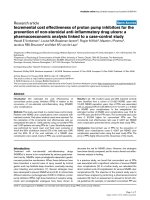
A plain radiograph of the patient’s knee revealed multi-
ple calcific densities within the soft tissues surrounding it
(Figure 1). Although some of these appeared to lie within
the capsule, the majority appeared to be outside of it.
These appearances were thought to be consistent with
idiopathic tumoral calcinosis. However, to further scruti-
nize these calcifications, a magnetic resonance imaging
(MRI) scan was recommended. It showed an extensive
thickening of the patient’s synovium, multiple intra-
articular calcific and ossific loose bodies, and large calci-
fied bursal extensions. The bursal component extended
into the patient’s posterior distal thigh and his proximal
calf. These findings were thought to be consistent with
very extensive synovial chondromatosis (Figure 1).
The patient’s blood tests were normal: corrected cal-
cium was 2.24 mmol/l, parathyroid hormone 2.5 pmol/l,
inorganic p hosphate 1.17 mmol/ l, serum urate
296 μmol/l, white cell count 7.2 × 10
9
/l, a nd C-reactive
protein 4 mg/l.
A two-stage proc edure was planned following the
findings of the MRI scan. The first stage was arthro-
scopy, which was able to note Grade IV osteoarthritis
alongside florid synovial chondromatosis in the medial
compartment (Figure 2). There were multiple loose
bod ies within this compartment and nod ules were fixed
to the synovium. A synovectomy with debridement and
excision of these bodies was thus performed.
The second stage involved an open exploration of the
patient’s popliteal fossa. Three large calcified masses were
found, all enclosed in bursal sacs (Figure 3). The first was
just medial to the posterior tibial nerve; the second was
deep into the medial head of the gastrocnemius muscle;
and the third was lateral to the semimembranosus at the
level of the oblique popliteal ligament. All three masses
were excised and the sacs were closed with purse string
sutures. A histological review at the Royal National Ortho-
paedic Hospital in Stanmore, UK confirmed our diagnosis
of synovial chondromatosis. The sections showed nests of
chondrocytes with focal ossification and focally attenuated
synovium overlying the nodules.
After the operation, the patient underwent weekly
physiotherapy sessions focusing on quadriceps strength-
ening, with a daily exercise regime to supplement this.
He recovered well and three months after the operation,
has regained his right knee’s full range of movement
with flexion incre ased to 130 degrees, which is equal to
that of his left knee. He has residual medial joint line
tenderness, undoubtedly due to osteoarthritis.
Discussion
Cartilage cells are absent inside the synovial mem brane.
It follows therefore that the development of synovial
Figure 1 Plain radiograph and magnetic resonance imagin g scans showing multiple soft tissu e calcifications within and outside the
joint capsule of the right knee.
Mackenzie et al. Journal of Medical Case Reports 2010, 4:113
/>Page 2 of 5
Figure 2 An arthroscopic photograph showing nodules of chondromatosis fixed to the synovium.
Figure 3 An intraoperative photograph showing the extent of the popliteal disease.
Mackenzie et al. Journal of Medical Case Reports 2010, 4:113
/>Page 3 of 5
chondromatosis depends on metaplastic transformation
of the synovial c ells into chondrocytes via an unknown
stimulus [1]. These chondrocytes become pedunculated
and encrusted inside the synovium and eventually
expelled into the joint as loose bodies [6].
Extra-articular synovial chondromatosis is rare, but
the combination of intra- and extra-articular diseases
described he re is an extremely rare condition. Given the
initial X-ray image of large extra-articular calcification,
we felt that the patient was more likely t o have idio-
pathic tumoral calcinosis. However, tumoral calcinosis
usually only affects people from Africa and the Carib-
bean in their second decade of life. Moreover, the calci-
fications are usually bilateral, affecting multiple sites,
and are ver y rarely intra-articular [7]. Our patient, how-
ever, was Caucasian and the MRI scan showed a single
lesion with an intra-articular component. Florid syn ovial
chondromatosis was thus a more likely diagnosis. This
was also confirmed by a histological examination.
Extra-articular diseases can be classified as tenosyno-
vial chondromatosis or bursal chondromatosis depend-
ing on the origin [8]. In this case, we propose that
either intra-articular synovial chondromatosis had pene-
trated the patient’s popliteal bursas, or bursal chondro-
matosis had infiltrated his knee joint. To the best of our
knowledge, this pattern o f disease in the knee has only
been reported twice in t he literature and never to this
extent [5,9]. This obviously raises concerns regarding a
possible transformation to synovial chondrosarcoma.
However, histological investigation revealed no signifi-
cant nuclear atypia, thus ruling out malignancy. The
literature reports only 33 cases of malignant transforma-
tion in the setting of histologically confirmed synovial
chondromatosis [6]. A key feature of all these cases is
therecurrenceofbenigndiseasepriortoadiagnosisof
malignant disease.
The extent of the disease and the presence of severe
osteoarthritis also presented a challenging management
problem. The combination of synovial chondromatosis
and degenerative a rthritis is a common finding in the
advanced stage of the disease [3]. Primary synovial
chondromatosis over time can lead to cartilage degen-
eration by mechanical wear via the loose bodies and
through nutrient deprivation to the a rticular cartilage
[3]. However, degenerative ar thritis can lead to second-
ary synovial chondromatosis [3]. As radiotherapy and
chemotherapy have no effe ct on synovial cho ndromato-
sis, surgical excision is the preferred treatment [4]. In
cases that involve localized intra-articular disease, com-
plete e xcision of the abnormal synovium seems to pro-
vide a cure. Generalized intra-articular disease with pain
and swelling requires total synovectomy and a removal
of the loose bodies. Extra-articular disease treatment
aims for complete excision [10].
Three surgical options were considered, namely high
tibial osteotomy (HTO), excision of the synovial and
bursal chondromatosis alone, or excision combined wit h
a total knee replacement. T he ideal treatment for severe
arthritis limited to the medial compartment in someone
within the same age range as our patient is a unicom-
partmental knee replacement. However, without
complete synovectomy, our patient’ssynovialchondro-
matosis could recur and thus compromise his joint
replacement. H TO with realignment of the joint forces
may lengthen the lifespan of the joint and delay the
need for joint re placement. Total knee arthroplasty
(TKA) has been proven to be an effective treatment for
synovial chondromatosis. However, even with complete
synovectomy alongside a TKA, recurrence of the disease
has been repo rted [3]. This is probably due to incom-
plete synovectomy at the time of operation, which leaves
remnants of pathological synovium [3]. Excision of the
chondromatosis formed the initial surgical treatment
plan, leaving us thus with the scope to perform an
arthroplasty in should the need arise the future. To
achieve full excision of the disease our patient required
arthroscopic debridement to treat the intra-articular dis-
ease, as well as a n open posterior approach to remove
the disease from the popliteal bursas.
The residual pain experienced by the patient causes a
further management dilemma. Although the pain is cur-
rently being contr olled by analg esia, the p ossibility of
HTO or TKA is being discussed with the patient.
Conclusions
A synovial chondromatosis is a rare condition but one
which can be highly aggressive and destructive. This
case, with its rare presentation of intra- and extra-
articular disease, highlights the importance of careful
clinical assessment, lateral thinking, appropriate use of
investigation, and careful pre-operative planning.
Consent
Written informed consent was obtained from the patient
for publicatio n of this case report and any accompany-
ing images. A copy of the writ ten consent is available
for review by the Editor-in-Chief of this journal.
Abbreviations
CRP: C-reactive protein; HTO: high tibial osteotomy; MRI: magnetic resonance
imaging; TKA: total knee arthroplasty.
Authors’ contributions
ST was the operating surgeon involved in the case. HM was the major
contributor in writing the manuscript. VG edited the manuscript and assisted
in reviewing the literature. All authors read and approved the final
manuscript.
Competing interests
The authors declare that they have no competing interests.
Mackenzie et al. Journal of Medical Case Reports 2010, 4:113
/>Page 4 of 5
Received: 4 November 2009 Accepted: 23 April 2010
Published: 23 April 2010
References
1. Jeffreys TE: Synovial chondromatosis. J Bone Joint Surg 1967, 3:530-534.
2. Miligram JW: Synovial osteochondromatosis. J Bone Joint Surg 1977,
59-A:792.
3. Ackerman D, Lett P, Galat DD Jr, Parvizi J, Stuart MJ: Results of total hip
and total knee arthroplasties in patients with synovial chondromatosis.
J Arthroplasty 2008, 23(3):395-400.
4. Temple HT, Gibbons CL: Tumors and tumor-related conditions about the
knee. Oxford Textbook of Orthopaedics and Trauma Oxford: Oxford
University PressBulstrode C, Buckwalter J, Carr A, Marsh L, Fairbank J,
Wilson-Macdonald J, Bowden G, 1 2002, 2:1153-1154.
5. Sim FH, Dahlin DC, Ivins JC: Extra-articular synovial chondromatosis.
J Bone Joint Surg 1977, 4:492-495.
6. Sah AP, Geller DS, Mankin HJ, Rosenberg AE, Delaney TF, Wright CD,
Hornicek FJ: Malignant transformation of synovial chondromatosis of the
shoulder to chondrosarcoma: a case report. J Bone Joint Surg Am 2007,
89(6):1321-1328.
7. Bullough P: Benign Soft-Tissue Tumours. Orthopaedic Pathology New York:
MosbyBullough P, 4 2004, 290-293.
8. Symeonides PJ: Bursal chondromatosis. Bone Joint Surg Br 1966,
48(2):371-373.
9. Dunn AW, Whisler JH: Synovial chondromatosis of the knee with
associated extracapsular chondromas. J Bone Joint Surg Am 1973,
55(8):1747-1748.
10. Maurice H, Crone M, Watt I: Synovial chondromatosis. J Bone Joint Surg
(Br) 1988, 70-B:807-811.
doi:10.1186/1752-1947-4-113
Cite this article as: Mackenzie et al.: A rare case of a swollen knee due
to disseminated synovial chondromatosis: a case report. Journal of
Medical Case Reports 2010 4:113.
Submit your next manuscript to BioMed Central
and take full advantage of:
• Convenient online submission
• Thorough peer review
• No space constraints or color figure charges
• Immediate publication on acceptance
• Inclusion in PubMed, CAS, Scopus and Google Scholar
• Research which is freely available for redistribution
Submit your manuscript at
www.biomedcentral.com/submit
Mackenzie et al. Journal of Medical Case Reports 2010, 4:113
/>Page 5 of 5