Báo cáo y học: "Successful medical management of emphysematous gastritis with concomitant portal venous air: a case report" docx
Bạn đang xem bản rút gọn của tài liệu. Xem và tải ngay bản đầy đủ của tài liệu tại đây (712.07 KB, 4 trang )
JOURNAL OF MEDICAL
CASE REPORTS
Paul et al. Journal of Medical Case Reports 2010, 4:140
/>Open Access
CASE REPORT
BioMed Central
© 2010 Paul et al; licensee BioMed Central Ltd. This is an Open Access article distributed under the terms of the Creative Commons At-
tribution License ( which permits unrestricted use, distribution, and reproduction in any
medium, provided the original work is properly cited.
Case report
Successful medical management of
emphysematous gastritis with concomitant portal
venous air: a case report
Manju Paul
1
, Savio John*
1
, Madhav C Menon
1
, Nazar H Golewale
2
, Stan L Weiss
2
and Uma K Murthy
1
Abstract
Introduction: The causes of diffuse abdominal pain following pelvic surgery are numerous. We present a rare case of
acute abdominal pain in a woman in the post-partum period.
Case presentation: A 25-year-old Caucasian woman with neurofibromatosis type 1 presented to our hospital with
diffuse abdominal pain immediately after a cesarean section. The patient was acutely ill and toxic with a fever of 38.8°C,
a pulse of 120 beats per minute and a distended abdomen with absent bowel sounds. A computed tomography scan
showed air in the wall of the stomach and portal venous system. The patient was successfully treated with intravenous
antibiotics, bowel rest and total parenteral nutrition.
Conclusion: It is rare for a case of emphysematous gastritis associated with portal venous air to be treated successfully
without surgery. To the best of our knowledge, to date there has been no reported association of emphysematous
gastritis with neurofibromatosis.
Introduction
The causes of diffuse abdominal pain following pelvic
surgery are numerous. A strong consideration of a serious
intra-abdominal pathology needs to be entertained when
imaging studies demonstrate air in the wall of the gastro-
intestinal organs in patients with fever, distended abdo-
men and absent bowel sounds in the immediate post-
operative period. The importance of early identification
of the underlying disease process is illustrated in this rare
case of acute abdominal pain in a young female in the
post-partum period.
Case presentation
A 25-year-old Caucasian female with a history of type 1
neurofibromatosis was brought to our hospital with dif-
fuse abdominal pain, nausea, vomiting and fever follow-
ing cesarean section for fetal distress. She was transferred
to our institution within 18 hours of the onset of symp-
toms for surgical intervention in view of the ominous
findings on computed tomography (CT) scan and endos-
copy done at the peripheral hospital. She had not passed
flatus or stool since surgery and denied hemetemesis,
melena, shortness of breath, or chest pain. There was no
history of tobacco or alcohol abuse, ingestion of corrosive
substances or non-steroidal anti inflammatory drugs
(NSAIDs).
Our patient appeared acutely ill and toxic. She had a
temperature of 38.8°C, pulse of 120/min, blood pressure
of 154/90 mmHg, respiratory rate of 24/min, and oxygen
saturation of 97% on 2 L of oxygen. The cardiac and respi-
ratory exams were otherwise unremarkable. Her abdo-
men was markedly distended. There was diffuse
tenderness on palpation of the abdomen with no perito-
neal signs. The cesarean section incision appeared clean
with no tenderness or discharge. Bowel sounds were
absent on auscultation. There were multiple neurofibro-
mas on our patient's neck and anterior chest consistent
with her diagnosis of neurofibromatosis. Her white blood
cell count was 25,000/mm
3
, with 91% neutrophils. The
initial electrolytes, amylase, lipase and liver function tests
were within normal limits.
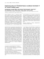
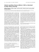
CT scan of our patient's abdomen showed marked gas-
tric dilation and air in the wall of the stomach along the
entire greater curvature and portal venous system (Fig-
* Correspondence:
1
Department of Medicine, State University of New York Upstate Medical
University, 750 E Adams Street, Syracuse, NY 13202, USA
Full list of author information is available at the end of the article
Paul et al. Journal of Medical Case Reports 2010, 4:140
/>Page 2 of 4
ures 1 and 2). There was marked dilatation of the small
and large bowel. Esophagogastroduodenoscopy (EGD) of
our patient showed areas of diffuse mucosal congestion
and extreme pallor as well as ulceration on the posterior
wall and greater curvature of the stomach. Gastric biopsy
revealed transmural necrosis. Streptococcus viridans was
isolated from gastric biopsy. Blood cultures did not grow
any pathogenic bacteria and nasogastric cultures were
not obtained.
Our patient was diagnosed with emphysematous gastri-
tis and promptly started on intravenous clindamycin and
piperacillin/tazobactam, nasogastric decompression and
intravenous hydration. Total parenteral nutrition was ini-
tiated from day two and our patient was closely moni-
tored in the intensive care unit for three days. She
improved with the above measures and tube feedings
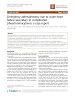
were initiated from day seven. Follow-up CT scan on day
eight showed resolution of the gastric and portal venous
air (Figure 3). Our patient was finally discharged home on
oral proton pump inhibitors on day 10. A follow-up EGD
two months later showed no sequelae and our patient
remained asymptomatic.
Discussion
Differential diagnoses for gas in the wall of the stomach
are emphysematous gastritis and gastric emphysema or
gastric pneumatosis. Theories suggested for gastric wall
air include mechanical, pulmonary, ischemic and bacte-
rial sources [1].
The mechanical theory suggests that gas is forced into
the bowel wall through a mucosal defect such as with air
insufflation during endoscopy. Our patient had gastric
pneumatosis evident on the CT scan even before endos-
copy, thus ruling out air insufflation at endoscopy as the
source of pneumatosis. The rupture of emphysematous
bullae in some patients can cause alveolar air to enter the
mediastinum, dissect along the great vessels to the retro-
peritoneum and through the mesenteric perivascular
spaces to reach the bowel wall. Important clues to this
clinical situation are the concomitant presence of pneu-
momediastinum and advanced chronic obstructive pul-
monary disease (COPD), which were absent in our
patient, thus making a pulmonary process unlikely. Slow-
healing mucosal ulcerations caused by ischemia, peptic
ulcer disease or inflammatory bowel disease, may also
lead to dissection of the luminal gas into the bowel wall
Figure 1 Scout film showing air along the greater curvature of
the stomach.
Figure 2 CT of the abdomen showing air in the stomach wall and
portal venous system. Black arrow: Portal venous air. White arrows:
Air in the stomach wall.
Figure 3 Follow-up abdominal CT on day eight showing resolu-
tion of stomach wall and portal venous air.
Paul et al. Journal of Medical Case Reports 2010, 4:140
/>Page 3 of 4
[2]. Our patient did not have any episode of peri-opera-
tive hypotension and the overall clinical picture did not
support an underlying ischemic process.
Emphysematous gastritis is a rare but grave variant of
phlegmonous gastritis. It is generally caused by local
infection through a mucosal defect by gas-forming
microorganisms or via hematogenous dissemination
from a distant focus. The stomach is a very uncommon
site of involvement, due to its abundant blood supply,
acidic pH and efficient mucosal barrier [3]. Most fre-
quently isolated organisms are streptococci, Escherichia
coli, Enterobacter species, Pseudomonas aeruginosa and
Clostridium perfringens [1]. It has been associated with
alcohol abuse, ingestion of corrosive substances, gastro-
enteritis, diabetes, NSAIDs [4], abdominal surgery, gas-
tric infarction, phytobezoar [5], adenocarcinoma of the
stomach [6], leukemia, pancreatitis, disseminated
strongyloidiasis in a patient receiving chemotherapy for
lymphoma [7], all of which can breach the integrity of the
mucosa. Our patient had none of these conditions except
the history of recent pelvic surgery. It is possible that a
sub-clinical uterine or pelvic sepsis resulting from sur-
gery could have resulted in a hematogenous or transperi-
toneal infection of the stomach.
Patients with emphysematous gastritis usually present
with severe abdominal pain, nausea, vomiting, hemetem-
esis, low grade fevers and tachycardia [8] as our patient
did. Patients with gastric emphysema or gastric pneuma-
tosis generally do not present with acute abdomen, and
the prognosis is excellent [1]. Currently, CT scan is the
most accurate diagnostic exam [9], although a plain
abdominal X-ray can be used as the initial imaging study
[10].
It is important to differentiate emphysematous gastritis
from gastric emphysema. Early institution of antibiotic
therapy covering anaerobes and gram negative bacilli,
intravenous hydration and appropriate nutrition is the
mainstay of treatment. Emphysematous gastritis usually
has a fulminant course with a mortality rate of 60% and
gastric strictures are as common as 25% [9]. Surgery
should be avoided during the acute phase in the absence
of bowel perforation due to friability of the mucosa and
the delay in healing of the sutured margins [1]. Air in the
portal vein or its radicals occurs when intraluminal or
bacterial gas enters the portomesenteric circulation [11-
13]. Necrotic bowel wall from infection, inflammation or
ischemia and/or markedly increased intraluminal pres-
sures seem to favor the entry of air into the venous radi-
cals. In a large series of 64 patients with this finding, the
reported mortality was 75%, nearly all patients requiring
surgery [11]. Recent reviews have suggested that the mere
finding of portal venous air by itself does not require sur-
gery; it is important to treat patients based on their clini-
cal condition [14].
Conclusions
A case is presented in which emphysematous gastritis
with portal venous air complicates cesarean section.
Although this condition often requires surgery, this case
resolved with appropriate medical management. To the
best of our knowledge, this is the second report of
emphysematous gastritis associated with portal venous
air that was successfully treated without surgical inter-
vention [15]. To date, there has been no reported associa-
tion of emphysematous gastritis with neurofibromatosis.
Consent
Written informed consent was obtained from the patient
for publication of this case report and any accompanying
images. A copy of the written consent is available for
review by the Editor-in-Chief of this journal.
Competing interests
The authors declare that they have no competing interests.
Authors' contributions
SJ and MP evaluated the patient, reviewed the literature and drafted the article.
MCM drafted the article, reviewed the literature and revised it critically. NHG
and SLW interpreted the imaging studies, reviewed the literature and drafted
the article pertinent to their filed of expertise. UKM supervised patient care,
revised the articled critically and provided valuable inputs with regard to the
management of the patient and final version of the draft. All authors read and
approved the final manuscript.
Author Details
1
Department of Medicine, State University of New York Upstate Medical
University, 750 E Adams Street, Syracuse, NY 13202, USA and
2
Department of
Radiology, State University of New York Upstate Medical University, 750 E
Adams Street, Syracuse, NY 13210, USA
References
1. van Mook WN, Geest S van der, Goessens ML, Schoon EJ, Ramsay G: Gas
within the wall of the stomach due to emphysematous gastritis: case
report and review. Eur J Gastroenterol Hepatol 2002, 14(10):1155-1160.
2. Berends M, Bodewes HW, Netten PM: Rare localization of air. Neth J Med
2007, 65(5):191-195.
3. Bashour CA, Popovich MJ, Irefin SA, Esfandiari S, Ratliff NB, Hoffman WD,
Averbook AW: Emphysematous gastritis. Surgery 1998, 123(6):716-718.
4. Yalamanchili M, Cady W: Emphysematous gastritis in a hemodialysis
patient. South Med J 2003, 96:84-88.
5. Lagios MD, Suydam MJ: Emphysematous gastritis with perforation
complicating phytobezoar. Am J Dis Child 1968, 116:202-204.
6. Smith TJ: Emphysematous gastritis associated with adenocarcinoma of
the stomach. Am J Dig Dis 1966, 11:341-345.
7. Williford ME, Foster WL Jr, Halvorsen RA, Thompson WM:
Emphysematous gastritis secondary to disseminated strongyloidiasis.
Gastrointest Radiol 1982, 7:123-126.
8. Allan K, Barriga J, Afshani M, Davila R, Tombazzi C: Emphysematous
gastritis. Am J Med Sci 2005, 329(4):205-207.
9. Hadas-Halpren I, Hiller R, Guberman D: Emphysematous gastritis
secondary to ingestion of large amounts of Coca-Cola. Am J
Gastroenterol 1993, 88:127-129.
10. Farfel B, Eichhorn R: Emphysematous gastritis. Am J Gastroenterol 1956,
25:125-130.
11. Liebman PR, Patten MT, Manny J, Benfield JR, Hechtman HB: Hepatic-
portal venous gas in adults: etiology, pathophysiology and clinical
significance.
Ann Surg 1978, 187(3):281-287.
Received: 21 October 2009 Accepted: 19 May 2010
Published: 19 May 2010
This article is available from: 2010 Paul et al; licensee BioMed Central Ltd. This is an Open Access article distributed under the terms of the Creative Commons Attribution License ( which permits unrestricted use, distribution, and reproduction in any medium, provided the original work is properly cited.Journal of Medical Case Repo rts 2010, 4:140
Paul et al. Journal of Medical Case Reports 2010, 4:140
/>Page 4 of 4
12. See C, Elliot D: Pneumatosis intestinalis and portal venous gas. N Engl J
Med 2004, 350:e3.
13. Karaosmanoğlu D, Oktar SÖ, Arac M, Erbas G: Portal and systemic venous
gas in a patient after lumbar puncture. Br J Radiol 2005, 78:767-769.
14. Paran H, Epstein T, Feinberg MS, Zissin R: Mesenteric and portal vein gas:
computerized tomography findings and clinical significance. Dig Surg
2003, 20:127-132.
15. Wissam Al-Jundi, Ali Shebl: Emphysematous gastritis: Case report and
literature review. Int J Surg 2008, 6:e63-e66.
doi: 10.1186/1752-1947-4-140
Cite this article as: Paul et al., Successful medical management of emphyse-
matous gastritis with concomitant portal venous air: a case report Journal of
Medical Case Reports 2010, 4:140