báo cáo khoa học: " LINE dancing in the human genome: transposable elements and disease" ppt
Bạn đang xem bản rút gọn của tài liệu. Xem và tải ngay bản đầy đủ của tài liệu tại đây (356.91 KB, 8 trang )
Belancio et al.: Genome Medicine 2009, 1:97
Abstract
Transposable elements (TEs) have been consistently under-
estimated in their contribution to genetic instability and human
disease. TEs can cause human disease by creating insertional
mutations in genes, and also contributing to genetic instability
through non-allelic homologous recombination and introduction
of sequences that evolve into various cis-acting signals that
alter gene expression. Other outcomes of TE activity, such as
their potential to cause DNA double-strand breaks or to
modulate the epigenetic state of chromosomes, are less fully
characterized. The currently active human transposable elements
are members of the non-LTR retroelement families, LINE-1, Alu
(SINE), and SVA. The impact of germline insertional muta-
genesis by TEs is well established, whereas the rate of post-
insertional TE-mediated germline mutations and all forms of
somatic mutations remain less well quantified. The number of
human diseases discovered to be associated with non-allelic
homologous recombination between TEs, and particularly
between Alu elements, is growing at an unprecedented rate.
Improvement in the technology for detection of such events, as
well as the mounting interest in the research and medical
communities in resolving the underlying causes of the human
diseases with unknown etiology, explain this increase. Here, we
focus on the most recent advances in understanding of the
impact of the active human TEs on the stability of the human
genome and its relevance to human disease.
Introduction to mammalian transposable
elements
Transposable elements (TEs) occupy almost half, 46%, of the
human genome, making the TE content of our genome one of
the highest among mammals, second only to the opossum
genome with a reported TE content of 52% [1,2]. The total
representation of TE-related sequences in the human genome
is probably even higher, as many of the sequences of the most
ancient TEs have deteriorated beyond recognition [3]. The
human genome contains two major classes of TEs, DNA and
RNA transposons, defined by the type of molecule used as an
intermediate in their mobilization.
DNA TEs encode a transposase that re-enters the nucleus
to specifically recognize transposon sequences in chromo-
somal DNA. The transposase excises these sequences from
their genomic location and inserts them into a new
genomic site (reviewed in [4]); this is also referred to as
‘cut and paste’ transposition. Human DNA TE activity
subsided over 37 million years ago [5]; as a result, DNA
TEs no longer contribute significantly to the ongoing
mutagenesis in humans.
Retrotransposons or retroelements make use of an RNA-
mediated transposition process. Retroelements are sub-
divided into two major groups: those containing long-
terminal repeats, LTR retroelements, and all others, lumped
into the category of non-LTR retroelements. Although
inactive in humans for millions of years, the best known
LTR retrotransposons, the endogenous retro viruses, make
up approximately 8% of the human genome [1]. This
contrasts with rodent genomes, in which LTR elements
continue to contribute a high proportion of the germline
TE-associated mutations (reviewed in [6]).
Non-LTR retrotransposons include autonomous and non-
autonomous members. The autonomous long interspersed
element-1 (LINE-1 or L1), and its non-autonomous partners,
such as ‘SINE-R, VNTR, and Alu’ (SVA) and the short
interspersed element (SINE) Alu, are the only mobile
elements with clear evidence of current retrotrans-
positional activity in the human genome [7] and will
therefore be the primary focus of this article.
The human L1 is about 6 kb long and encodes two open
reading frames, ORF1 and ORF2, which are both required
for L1 retrotransposition (Figure 1a) [8]. ORF2 encodes
endonuclease and reverse transcriptase activities that are
crucial for the insertion mechanism [8,9]. SINEs and SVA
elements do not encode any proteins [10], instead they
Review
LINE dancing in the human genome: transposable elements and
disease
Victoria P Belancio
†
, Prescott L Deininger* and Astrid M Roy-Engel*
Addresses: *Department of Epidemiology, School of Public Health and Tropical Medicine, Tulane Cancer Center, Tulane University, SL-49
1430 Tulane Ave, New Orleans, LA 70112, USA.
†
Department of Structural and Cellular Biology, School of Medicine, Tulane Cancer Center
and Tulane Center for Aging, Tulane University, SL-49 1430 Tulane Ave, New Orleans, LA 70112, USA.
Correspondence: Prescott L Deininger. Email:
AML, acute myelogenous leukemia; BRCA1, breast cancer-1 gene; CGH, comparative genomic hybridization; DSB, double-strand break;
LINE-1, L1, long interspersed element-1; LTR, long terminal repeat; NAHR, non-allelic homologous recombination; NHEJ, non-homologous
end joining; ORF, open reading frame; SINE, short interspersed element; SVA, SINE-R, VNTR, and Alu element; TE, transposable element.
97.2
Belancio et al.: Genome Medicine 2009, 1:97
depend on the presence of the functional L1s, and they are
therefore often referred to as L1 parasites [11]. In contrast
to L1, Alu elements require only ORF2 of L1 for their
mobilization [11,12]. Alu elements are transcribed by RNA
polymerase III and encode a variable length adenosine-
rich region at their 3’ end, a critical feature for retro-
transposition [10]. SVA is a composite element containing
a complex sequence composed of a (CCCTCT)
n
hexamer
repeat region, an Alu-derived region, a variable number
tandem repeat (VNTR) region and a retroviral-derived
sequence (Figure 1a) [13]. The requirements for SVA
mobilization are still poorly understood [13,14].
TE activity has often been assumed to be confined to the
germline, early embryogenesis, and potentially cancer cells
[15-18]. The most recent reports indicate that expression of
L1 RNA (VP Belancio, A Roy-Engel, R Pochampally and
P Deininger, personal communication) and L1 protein [16]
occurs in human somatic tissues and that somatic L1
retrotransposition takes place in transgenic mouse models
[19,20]. Interestingly, L1 transgenic mice show higher L1
mobilization in somatic tissues than in the germline
[19,21]. Other evidence of somatic L1 mobilization comes
from a somatic L1 insertion that inactivates the adeno-
matous polyposis coli (APC) gene, leading to colon cancer
[22]. There are currently very limited data on the somatic
expression of Alu and SVA elements, and we do not have a
true appreciation of the level of somatic insertion that is
occurring from endogenous elements.
Human diseases caused by TE-mediated
insertional mutagenesis
The most obvious form of mutagenesis common to all TEs
is the disruption of gene function or regulation resulting
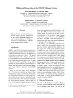
Figure 1
L1 expression leads to different types of DNA damage. Schematic structures of an SVA element (labeled SVA), showing the CCCTCT repeat,
the Alu-derived (A-like) region, the variable number tandem repeat (VNTR) region, and the long terminal repeat (LTR)-derived region; an Alu
element (labeled Alu (SINE)), showing left (purple) and right (pink) halves separated by the A-rich region (A) and the variable length A-tail
((A)
n
) followed by the 3’ region (white), which has a variable length and sequence; and an L1 element (labeled LINE-1), showing open
reading frame (ORF)1 (light blue) and ORF2 (dark blue) and the 5’ untranslated region, inter-ORF region and 3’ untranslated region (white).
(a) The typical insertion of these elements into the genome, which can lead to insertional mutagenesis. (b) Dispersed repetitive elements
such as Alu elements can undergo non-allelic homologous recombination, which can cause a deletion (shown) or duplication (not shown).
The dashed arrow indicates the potential site of DNA damage by an L1 endonuclease that may help initiate these recombination events.
(c) Potential outcomes of the repair of the L1-induced double-strand breaks (DSBs). The L1 recognition site is in black; surrounding
sequence is in blue; inserted nucleotides are in red. The associated changes are typical of what might be seen with repair of the DSB by
non-homologous end joining. It is also possible that the sites are simply re-ligated with no mutation occurring, or alternatively, these sites
may cause recombination, as shown in (b).
Alu1
Alu1/2
NNT T TTNNAANN
NNAAAANNTTNN
NNTTANN
NNAATNN
NNT T TNAANN
NNAAANTTNN
Small insertions
LINE-1
NNT T TTAANN
NNAAAAT TNN
L1 endonuclease site
ORF1
(A)
N
(A)
N
Alu (SINE)
SVA
(A) (A)
N
(A)
(A)
N
(A)
N
(CCCTCT)
n
A-like VNTR LTR-derived region
Deletions
Point mutations
Alu2
ORF2
(a) (b) (c)
97.3
Belancio et al.: Genome Medicine 2009, 1:97
from the insertion of new element copies (Figure 1b). The
fortuitous discovery of the first known active human L1
was the result of its retrotransposition into the factor VIII
gene, causing a de novo case of hemophilia [23]. L1, Alu,
and SVA are reported to cause a broad range of human
diseases (reviewed in [7,10,24]). Examples include a
diverse collection of diseases, such as neurofibromatosis,
choroideremia, cholinesterase deficiency, Apert syndrome,
Dent’s disease, β-thalassemia, and Walker-Warburg syn-
drome. Because of the relatively random insertion process,
there is great diversity in the type of genetic diseases
associated with TE insertions. However, there is a very
strong overrepresentation of X-chromosome-linked diseases
caused by TEs that could be a result of ascertainment bias
(that is, X-linked genetic defects are more easily detected
because of the single X-chromosome in males or could also
reflect the higher density of L1 elements on the X
chromosome). Compilations of the known human diseases
attributed to TE insertions (33 Alu, 11 L1, and 4 SVA) are
provided in recent reviews [7,10]. Most of these diseases are
due to germline insertions and have been detected as rare
recessive diseases. However, some cases of cancers have
been identified that are probably somatic mutations in
which a TE insertion has disrupted a critical gene, such as
BRCA1 and BRCA2 in breast cancer or APC in colon cancer.
Interference with gene expression
Almost all of the reported retroelement insertions that
cause human diseases have either interrupted the ORF or
inserted in close proximity to a splice site, leading to a
major disruption of gene function [7,10]. However, many
insertions that do not cause disease may still influence the
expression of the genes in which they insert, thus pre-
disposing cells or individuals to disease by slightly
changing gene expression. For example, insertion of TE
elements might introduce functional splice and poly-
adenylation sites [25-29], resulting in aberrant processing
of some of the transcripts produced by a gene. In addition,
they might introduce regulatory regions that would
influence the strength of its promoter, or even add
promoter sequences [30,31]. It has been suggested that L1
elements inserted in the intron of a gene could cause ‘gene
breaking’ [25,28] that could create proteins truncated from
either end, possibly leading to altered functions or
dominant-negative effects. In contrast to L1, Alu elements
need to accumulate a critical mutation(s) that creates an
appropriate functional cis-acting sequence (both splicing
and polyadenylation) to have this effect [32-34].
Human disease caused by post-insertional TE
mutagenesis
Recombination
TEs continue to contribute to genetic instability after
insertion through non-allelic homologous recombination
(NAHR). The presence of multiple closely related
sequences throughout the genome facilitates misalignment
of repeated sequences, allowing uneven genetic exchange
between alleles that contribute to deletions and dupli-
cations (Figure 1c; reviewed in [35,36]). Comparisons of
the human and chimpanzee genomes have shown that L1
and Alu recombination deletions caused over a megabase
of difference in more than 100 individual deletions [37-39].
Alu elements not only cause deletions, but also seem to
contribute to the formation of segmental duplications. A
genome-wide set of 2,366 duplication alignments demon-
strated the enrichment of Alu elements near the junction
between the two duplicated sequences in all cases,
suggesting Alu involvement in these rearrangements [40].
These segmental duplications lead to altered expression of
the genes located in these regions and result in further
instability by promoting non-allelic recombination between
duplicated segments, leading to recurrent genetic disease.
TE-mediated NAHR (in particular, recombination between
Alu elements) contributes directly to a large variety of
genetic diseases. The frequency of this type of genetic
rearrangement varies depending on the affected gene
(reviewed in [35,41]). Genes such as MLL-1 (which is
involved in acute myelogenous leukemia (AML)) [42], VHL
(von Hippel-Lindau syndrome) [43], and BRCA1 (familial
breast cancer) [44] seem to be hotspots for Alu-Alu
recombination, with a series of independent recombination
events occurring with different Alu elements in the region.
BRCA1 has 137 Alus in its introns, making up over 40% of
its gene sequence. Studies of BRCA1 mutations have shown
that, in 23 different individuals, 44 of these Alu elements
were involved in duplication/deletion events in this gene.
VHL is also subject to extensive Alu-Alu recombination,
with almost a third of its germline mutations resulting
from large deletions, and 90% of the mapped events
involving Alu-Alu NAHR [45]. Of 30 Alu-Alu recombi na-
tion events mapped, seven involve one particular
Y-subfamily Alu element recombining with other Alus in
the gene. The Y-subfamily is young and therefore shows
lower than average divergence relative to other genomic
Alus, which might explain its high recombination rate.
Similar observations implicating a particular ‘hotspot’ Alu
element were reported for multiple cases of rearrange-
ments in the LDL receptor gene causing familial hyper-
cholesterolemia [46], and also for the SLC7A7 gene, where
one Alu accounted for 38% of all rearranged chromosomes
in patients with lysinuric protein intolerance [47].
The MLL1 gene, which is associated with AML, is often
involved in chromosomal translocations causing expres-
sion of an oncogenic fusion gene. Of the cases of AML
without a visible translocation, seven out of nine cases
studied involved a duplication caused by Alu-Alu recom-
bination events in intron 1 and 6, which resulted in a
duplication of exons 2 to 6 of the gene [42]. Similar
duplications have consistently been found in the blood of
healthy individuals [48], suggesting that these types of
97.4
Belancio et al.: Genome Medicine 2009, 1:97
recombination events occur spontaneously and regularly
throughout the lifespan of an individual. The cellular
environment can potentially increase Alu-Alu NAHR.
Mutants in TP53 (which encodes the tumor-suppressor
p53) increase these Alu-Alu recombination events, possibly
contributing to malignancy. Although it is clear that active
human TEs contribute to spontaneous genetic diseases, the
exact extent of their involvement in this process remains
elusive. This uncertainty makes the contribution of TEs to
human diseases difficult to assess, for the most part due to
the absence of uniform and reliable diagnostic methods.
Detection and diagnosis of diseases caused
by TE insertion
The introduction of PCR technology for diagnostics
revolutionized the field of human genetic testing. Within a
clinical setting, PCR across the exons of a gene and
sequence analysis is commonplace. This approach is great
for identifying point mutations and small insertion/
deletion events, but will detect only small TE insertions
very near the exons. Most PCR-based tests are inadequate
for the detection of the large deletions, rearrangements,
and duplications often associated with TE-induced muta-
genesis. Awareness of this bias led to the use of alternative
methods that can detect copy number variations (CNVs),
such as long-range PCR, targeted array comparative
genome hybridization (array-CGH) analysis, and multiplex
ligation-dependent probe assays (MLPAs). These approaches
are better at detection of duplicated or deleted exons that
occur from Alu-Alu recombination events. New tests using
either MLPA or array-CGH are becoming more common-
place, particularly for diagnostics in cancer, and can detect
most genomic duplications and deletions but not larger TE
insertions. Traditional Southern blot analysis is still one of
the few robust methods for detecting large TE insertions
but it is rarely used in diagnostic tests today. The fact that
the majority of sporadic human diseases have a subset of
cases of unknown etiology leaves a possibility that
TE-induced DNA damage may be responsible for at least
some of them. In fact, genomic analysis by methods
specifically targeting potential involvement of TEs in the
sporadic human diseases revealed that a significant
proportion of them are, indeed, caused by TEs [43]. One of
the most promising technologies for characterizing all of
the TE-based variation with minimal ascertainment bias is
the potential usage of some of the upcoming next-
generation DNA sequencing approaches for random
sequencing of the entire genome of an individual. However,
this approach is still some years from clinical usefulness.
L1-associated DNA double-strand breaks
Recent publications from several laboratories have
reported the formation of DNA double-strand breaks
(DSBs) associated with L1 expression [49-51]. These DSBs
depend on the enzymatic activity of the L1 ORF2
endonuclease domain [51], and their formation triggers
various cellular responses [51,52], including apoptosis,
cellular senescence, cell-cycle checkpoints, and DNA repair
responses. DSBs are highly mutagenic and can lead to
small deletions or insertions if repaired by the non-
homologous end-joining (NHEJ) repair machinery (Figure 1d).
L1-induced DSBs may also cause recombination events
when repaired by homology-driven repair, potentially
leading to large genomic rearrangements (reviewed in
[41]). Homologous recombination (HR) repairs damaged
genomic sequence either by using the unaltered
counterpart as a template in a gene conversion event or
through non-allelic homologous interactions that lead to
deletions or duplications between the homologous
sequences, as described above for Alu element-mediated
NAHR. Given that all L1, Alu, and SVA copies in the human
genome are generated with target site duplications that
contain an L1 endonuclease recognition site, there are
roughly 3 million potential cleavage sites adjacent to these
elements that may help them contribute to NAHR-
mediated events. Because many L1-endonuclease-mediated
events may lack the typical hallmarks of L1 involvement
(such as the target site duplications that normally flank
mobilized sequences and a run of adenosines), we cannot
currently assess the relative contribution of this process to
genetic instability.
Modulators of TE activity
The TE amplification cycle involves complex interactions
with various cellular factors and compartments, any of
which can be positively or negatively regulated by intrinsic
or extracellular environmental factors. The L1 lifecycle and
some of its known modulators are depicted in Figure 2.
Modulations by the genomic environment
Levels of TE activity can vary both because of the
polymorphism of these elements between different
individuals, as well as variations in epigenetic regulation of
TE loci. Even though each human genome averages
500,000 L1 copies, of which about 3,000 are full-length
and roughly 200 are potentially functional [1,53], only a
handful of elements have high levels of activity in each
genome [53]. The rest have mutated sufficiently to lose
most or all retrotransposition potential. All of the highly
active elements found to date are polymorphic in the
population, with each individual probably having a
different assortment of active elements [53]. Because these
loci consist of the youngest L1 integration events, they have
had the least time to accumulate inactivating mutations
and are more likely to remain active. In addition to the
presence/absence polymorphism of these ‘hot’ elements,
the same L1 loci accumulate distinct point mutations in
various individuals that contribute to the diversity in their
potential activity [54,55]. Thus, there may be as much as
several hundred-fold variability in L1 activity in different
individuals [55]. Recent advances in understanding of the
sequence components controlling Alu activity [56-58]
97.5
Belancio et al.: Genome Medicine 2009, 1:97
indicate that its retrotransposition is also likely to vary in
individual genomes.
DNA methylation of the CpG island in the 5’ region of L1
[59] is one of the powerful mechanisms controlling L1
promoter activity that minimizes the exposure of genomic
DNA to L1-associated damage. The genome-wide hypo-
methy lation of repetitive sequences observed during
malignant transformation unleashes L1 expression that is
usually tightly regulated in untransformed cells [60].
Methylation of genomic DNA often triggers specific histone
modifications, resulting in chromatin remodeling. The role of
epigenetic control in L1 expression has recently attracted
significant interest, particularly because little is known about
the effects of the intronic or near-genic full-length L1
insertions on the epigenetic state of the affected human genes.
Figure 2
Modulators of the L1 lifecycle. The L1 amplification cycle can be divided into several steps. (a) Transcription. L1 amplification initiates with
transcription, and regulation of L1 at this step can be modified by epigenetic modifications, DNA methylation, and recruitment of transcription
factors. (b) Before leaving the nucleus, the number of retrocompetent full-length L1 transcripts can be reduced by RNA processing through
premature polyadenylation and splicing. (c) Translation. Full-length L1 enters the cytoplasm to be translated, producing ORF1 and ORF2
proteins for retrotransposition. The two proteins interact with the L1 transcript to form an L1 ribonucleoprotein particle (RNP). RNA
interference can affect this step. (d) Insertion of a new L1 copy. The L1 RNP reaches the nucleus, where the DNA is cleaved by the L1 ORF2
endonuclease activity. It is proposed that reverse transcription occurs through a process referred to as target primed reverse transcription
(TPRT) [71]. The L1 ORF2 reverse transcriptase activity generates the first strand of DNA. DNA repair proteins are likely to be involved in
inhibiting the L1 integration step. (e) Effects of external stimuli. Ionizing radiation or heavy metals can affect L1 at multiple steps, such as
transcriptional activation or altering DNA repair pathways.
AAAA
AAAA
AAAA
AAAA
AAAA AAAA
L1 RNP
Translation
TPRTIntegration
RT
(a)
(b)
(e)
(d)
DNA repair
(c)
and
Functional L1 locus
L1 ORF1 protein
L1 ORF2 protein
Cytoplasm
L1-induced DSBs
97.6
Belancio et al.: Genome Medicine 2009, 1:97
Modulations by the cellular environment
Among the multiple cellular pathways influencing L1
expression and activity are DNA methylation, tissue-
specific transcription factors (Figure 2a), RNA processing
(Figure 2b), and RNA interference (Figure 2c) [25-27, 29,
61-63]. In addition, some cellular proteins greatly influence
integration (Figure 2d) of L1 and Alu elements; these
include DNA repair proteins, such as the ataxia telangiec-
tasia mutated kinase (ATM) and the endonuclease dimer
composed of excision repair complementing protein 1
(ERCC1) and xeroderma pigmentosum complement group
(XPF) [51,64,65], and also viral defense proteins, such as
the apolipoprotein B mRNA editing enzyme, catalytic
polypeptide-like 3C (APOBEC3) family of proteins [66,67]
(Figure 2). L1 mobilization in NHEJ-negative hamster cells
causes the element to lose the endonuclease dependence
that it shows in a wild-type genetic background, and it then
requires only functional L1 reverse transcriptase to achieve
wild-type retrotransposition levels [68]. Because of the
diversity of the L1-associated mutagenesis, it will not be
surprising if additional DNA repair pathways are reported
to modulate L1 activity.
Given the multitude of cellular factors influencing L1
activity, it is easy to imagine that polymorphisms or
mutations in any of the genes whose function is important
for suppressing L1 activity may have an impact on its
contribution to genetic instability. One of the most
profound examples is the mouse knockout of DNA-methyl-
transferase-3-like protein (Dnmt3L), a modulator of de
novo DNA methylation in the germline, which results in
upregulation of the expression of endogenous L1 and LTR
elements that coincides with meiotic catastrophe during
spermatogenesis [69,70].
Modulations by the extracellular environment
TE activity is influenced not only by the intrinsic cellular
environment, but also by external stimuli (Figure 2e).
Ionizing radiation, heavy metals (present in cigarette
smoke and workplace exposures), anti-cancer therapies,
air pollutants, and DNA demethylation agents can locally
or systemically cause increases in endogenous TE activity
(reviewed in [50,70]), potentially leading to new health
problems (such as sporadic cancers) or exacerbating
preexisting conditions (such as the rise of a more
aggressive cancer phenotype). The mechanisms of the
environmental influences on human TE activity are only
just beginning to emerge as we are learning more about
their interaction with various cellular pathways. Some of
the environmental factors enhance TE expression by
changing the epigenetic state of the genome; others, such
as heavy metals, probably exert their effect by influencing
cellular enzymes that are important for keeping TE activity
at bay. Because of the early stage of this area of
investigation, no diseases have yet been directly associated
with increased activity of TEs due to exposure to
environmental toxicants. However, with the new advances
in whole-genome studies, some of these crucial questions
are likely to be answered in the near future.
Conclusions
TE activity can generate a wide-spectrum of genomic
mutations, ranging from point mutations to gross
rearrangements with gain of genomic information, as well
as interference with normal gene processing and expres-
sion after insertion. These mutations contribute to idio-
pathic human disease. Because of the intimate relationship
between L1 activity and multiple cellular processes, it is
likely that people with genetic backgrounds that produce
defects in any of the pathways influencing the L1 lifecycle
are more vulnerable to insult from TEs. Thus, to evaluate
the impact of these elements on the stability of the human
genome and human disease, it is crucial to take into
account their cumulative activity in a specific genetic
background as well as the potential modulating effects of
the extracellular environment.
The increasing ease of sequencing genomes is likely to help
clarify the extent of the contribution of mobile elements to
genetic instability in many human diseases. This infor ma-
tion is critical in determining the full spectrum of mutations
contributing to human disease. However, the full impact of
these ubiquitous, high-copy-number elements on the
biology of the cell may remain elusive for some time.
Competing interests
The authors declare that they have no competing interests.
Authors’ contributions
All authors participated equally in the conception and
writing of this article.
Acknowledgements
This article was made possible by grants P20RR020152 (PLD, VPB,
and AMR-E), R01GM45668 (PLD), and R01GM079709A (AMR-E)
from the National Institutes of Health (NIH) and an EPSCOR grant
from the National Science Foundation (PLD). VPB is supported by
NIH/NIA grant 5K01AG030074 and an Ellison Medical Foundation
New Scholar in Aging award (AG-NS-0447-08). The contents of the
article are solely the responsibility of the authors and do not
necessarily represent the official views of the National Center for
Research Resources or the NIH. Competitive Advantage Funds
(2006) from the Louisiana Cancer Research Consortium (LCRC)
were also awarded to AMR-E.
References
1. Lander ES, Linton LM, Birren B, Nusbaum C, Zody MC,
Baldwin J, Devon K, Dewar K, Doyle M, FitzHugh W, Funke R,
Gage D, Harris K, Heaford A, Howland J, Kann L, Lehoczky J,
LeVine R, McEwan P, McKernan K, Meldrim J, Mesirov JP,
Miranda C, Morris W, Naylor J, Raymond C, Rosetti M, Santos
R, Sheridan A, Sougnez C, et al.: Initial sequencing and anal-
ysis of the human genome. Nature 2001, 409:860-921.
2. Warren WC, Hillier LW, Marshall Graves JA, Birney E, Ponting
CP, Grutzner F, Belov K, Miller W, Clarke L, Chinwalla AT, Yang
SP, Heger A, Locke DP, Miethke P, Waters PD, Veyrunes F,
Fulton L, Fulton B, Graves T, Wallis J, Puente XS, Lopez-Otin
C, Ordonez GR, Eichler EE, Chen L, Cheng Z, Deakin JE,
97.7
Belancio et al.: Genome Medicine 2009, 1:97
Alsop A, Thompson K, Kirby P, et al.: Genome analysis of the
platypus reveals unique signatures of evolution. Nature
2008, 453:175-183.
3. Gu W, Castoe TA, Hedges DJ, Batzer MA, Pollock DD:
Identification of repeat structure in large genomes using
repeat probability clouds. Anal Biochem 2008, 380:77-83.
4. Sinzelle L, Izsvak Z, Ivics Z: Molecular domestication of
transposable elements: from detrimental parasites to
useful host genes. Cell Mol Life Sci 2009, 66:1073-1093.
5. Pace JK, Feschotte C: The evolutionary history of human
DNA transposons: evidence for intense activity in the
primate lineage. Genome Res 2007, 17:422-432.
6. Maksakova IA, Romanish MT, Gagnier L, Dunn CA, van de
Lagemaat LN, Mager DL: Retroviral elements and their
hosts: insertional mutagenesis in the mouse germ line.
PLoS Genet 2006, 2:e2-
7. Chen JM, Stenson PD, Cooper DN, Ferec C: A systematic
analysis of LINE-1 endonuclease-dependent retrotranspo-
sitional events causing human genetic disease. Hum Genet
2005, 117:411-427.
8. Moran JV, Holmes SE, Naas TP, DeBerardinis RJ, Boeke JD,
Kazazian HH Jr: High frequency retrotransposition in cul-
tured mammalian cells. Cell 1996, 87:917-927.
9. Feng Q, Moran JV, Kazazian HH Jr, Boeke JD: Human L1 ret-
rotransposon encodes a conserved endonuclease
required for retrotransposition. Cell 1996, 87:905-916.
10. Belancio VP, Hedges DJ, Deininger P: Mammalian non-LTR
retrotransposons: for better or worse, in sickness and in
health. Genome Res 2008, 18:343-358.
11. Dewannieux M, Esnault C, Heidmann T: LINE-mediated retro-
transposition of marked Alu sequences. Nat Genet 2003,
35: 41-48.
12. Wallace N, Wagstaff BJ, Deininger PL, Roy-Engel AM: LINE-1
ORF1 protein enhances Alu SINE retrotransposition. Gene
2008, 419:1-6.
13. Ostertag EM, Goodier JL, Zhang Y, Kazazian HH Jr: SVA ele-
ments are nonautonomous retrotransposons that cause
disease in humans. Am J Hum Genet 2003, 73:1444-1451.
14. Wang H, Xing J, Grover D, Hedges DJ, Han K, Walker JA,
Batzer MA: SVA elements: a hominid-specific retroposon
family. J Mol Biol 2005, 354:994-1007.
15. Branciforte D, Martin SL: Developmental and cell type spe-
cificity of LINE-1 expression in mouse testis: implications
for transposition. Mol Cell Biol 1994, 14:2584-2592.
16. Ergun S, Buschmann C, Heukeshoven J, Dammann K,
Schnieders F, Lauke H, Chalajour F, Kilic N, Stratling WH,
Schumann GG: Cell type-specific expression of LINE-1
open reading frames 1 and 2 in fetal and adult human
tissues. J Biol Chem 2004, 279:27753-27763.
17. Martin SL: Ribonucleoprotein particles with LINE-1 RNA in
mouse embryonal carcinoma cells. Mol Cell Biol 1991, 11:
4804-4807.
18. Martin SL, Branciforte D: Synchronous expression of LINE-1
RNA and protein in mouse embryonal carcinoma cells. Mol
Cell Biol 1993, 13:5383-5392.
19. An W, Han JS, Schrum CM, Maitra A, Koentgen F, Boeke JD:
Conditional activation of a single-copy L1 transgene in
mice by Cre. Genesis 2008, 46:373-383.
20. Kano H, Godoy I, Courtney C, Vetter MR, Gerton GL, Ostertag
EM, Kazazian HH Jr: L1 retrotransposition occurs mainly in
embryogenesis and creates somatic mosaicism. Genes
Dev 2009, 23:1303-1312.
21. Babushok DV, Ostertag EM, Courtney CE, Choi JM, Kazazian
HH Jr: L1 integration in a transgenic mouse model. Genome
Res 2006, 16:240-250.
22. Miki Y, Nishisho I, Horii A, Miyoshi Y, Utsunomiya J, Kinzler KW,
Vogelstein B, Nakamura Y: Disruption of the APC gene by a
retrotransposal insertion of L1 sequence in a colon cancer.
Cancer Res 1992, 52:643-645.
23. Dombroski BA, Mathias SL, Nanthakumar E, Scott AF,
Kazazian HH Jr: Isolation of an active human transposable
element. Science 1991, 254:1805-1808.
24. Babushok DV, Kazazian HH Jr: Progress in understanding
the biology of the human mutagen LINE-1. Hum Mutat 2007,
28: 527-539.
25. Belancio VP, Hedges DJ, Deininger P: LINE-1 RNA splicing
and influences on mammalian gene expression. Nucleic
Acids Res 2006, 34:1512-1521.
26. Belancio VP, Roy-Engel AM, Deininger P: The impact of mul-
tiple splice sites in human L1 elements. Gene 2008, 411:38-
45.
27. Perepelitsa-Belancio V, Deininger P: RNA truncation by pre-
mature polyadenylation attenuates human mobile element
activity. Nat Genet 2003, 35:363-366.
28. Wheelan SJ, Aizawa Y, Han JS, Boeke JD: Gene-breaking: a
new paradigm for human retrotransposon-mediated gene
evolution. Genome Res 2005, 15:1073-1078.
29. Han JS, Szak ST, Boeke JD: Transcriptional disruption by
the L1 retrotransposon and implications for mammalian
transcriptomes. Nature 2004, 429:268-274.
30. Matlik K, Redik K, Speek M: L1 antisense promoter drives
tissue-specific transcription of human genes. J Biomed
Biotechnol 2006, 2006:1-16.
31. Speek M: Antisense promoter of human L1 retrotranspo-
son drives transcription of adjacent cellular genes. Mol Cell
Biol 2001, 21:1973-1985.
32. Chen C, Ara T, Gautheret D: Using Alu elements as polyade-
nylation sites: a case of retroposon exaptation. Mol Biol
Evol 2009, 26:327-334.
33. Lee YK, Chew A, Phan H, Greenhalgh DG, Cho K: Genome-
wide expression profiles of endogenous retroviruses in
lymphoid tissues and their biological properties. Virology
2008, 373:263-273.
34. Sorek R, Ast G, Graur D: Alu-containing exons are alterna-
tively spliced. Genome Res 2002, 12:1060-1067.
35. Deininger PL, Batzer MA: Alu repeats and human disease.
Mol Genet Metab 1999, 67:183-193.
36. Deininger PL, Batzer MA: Mammalian retroelements.
Genome Res 2002, 12:1455-1465.
37. Han K, Sen SK, Wang J, Callinan PA, Lee J, Cordaux R, Liang
P, Batzer MA: Genomic rearrangements by LINE-1 inser-
tion-mediated deletion in the human and chimpanzee line-
ages. Nucleic Acids Res 2005, 33:4040-4052.
38. Han K, Lee J, Meyer TJ, Wang J, Sen SK, Srikanta D, Liang P,
Batzer MA: Alu recombination-mediated structural dele-
tions in the chimpanzee genome. PLoS Genet 2007, 3:1939-
1949.
39. Han K, Lee J, Meyer TJ, Remedios P, Goodwin L, Batzer MA:
L1 recombination-associated deletions generate human
genomic variation. Proc Natl Acad Sci USA 2008, 105:19366-
19371.
40. Bailey JA, Liu G, Eichler EE: An Alu transposition model for
the origin and expansion of human segmental duplica-
tions. Am J Hum Genet 2003, 73:823-834.
41. Hedges DJ, Deininger PL: Inviting instability: transposable
elements, double-strand breaks, and the maintenance of
genome integrity. Mutat Res 2006, 616:46-59.
42. Strout MP, Marcucci G, Bloomfield CD, Caligiuri MA: The
partial tandem duplication of ALL1 (MLL) is consistently
generated by Alu-mediated homologous recombination in
acute myeloid leukemia. Proc Natl Acad Sci USA 1998, 95:
2390-2395.
43. Franke G, Bausch B, Hoffmann MM, Cybulla M, Wilhelm C,
Kohlhase J, Scherer G, Neumann HP: Alu-Alu recombination
underlies the vast majority of large VHL germline dele-
tions: molecular characterization and genotype-phenotype
correlations in VHL patients. Hum Mutat 2009, 30:776-786.
44. Mazoyer S: Genomic rearrangements in the BRCA1 and
BRCA2 genes. Hum Mutat 2005, 25:415-422.
45. Casarin A, Martella M, Polli R, Leonardi E, Anesi L, Murgia A:
Molecular characterization of large deletions in the von
Hippel-Lindau (VHL) gene by quantitative real-time PCR:
the hypothesis of an Alu-mediated mechanism underlying
97.8
Belancio et al.: Genome Medicine 2009, 1:97
VHL gene rearrangements. Mol Diagn Ther 2006, 10:243-
249.
46. Kim SH, Bae JH, Chae JJ, Kim UK, Choe SJ, Namkoong Y,
Kim HS, Park YB, Lee CC: Long-distance PCR-based
screening for large rearrangements of the LDL receptor
gene in Korean patients with familial hypercholestero-
lemia. Clin Chem 1999, 45:1424-1430.
47. Font-Llitjos M, Rodriguez-Santiago B, Espino M, Sillue R,
Manas S, Gomez L, Perez-Jurado LA, Palacin M, Nunes V:
Novel SLC7A7 large rearrangements in lysinuric protein
intolerance patients involving the same AluY repeat. Eur J
Hum Genet 2009, 17:71-79.
48. Schnittger S, Wormann B, Hiddemann W, Griesinger F: Partial
tandem duplications of the MLL gene are detectable in
peripheral blood and bone marrow of nearly all healthy
donors. Blood 1998, 92:1728-1734.
49. Belgnaoui SM, Gosden RG, Semmes OJ, Haoudi A: Human
LINE-1 retrotransposon induces DNA damage and apopto-
sis in cancer cells. Cancer Cell Int 2006, 6:13.
50. Farkash EA, Kao GD, Horman SR, Prak ET: Gamma radiation
increases endonuclease-dependent L1 retrotransposition
in a cultured cell assay. Nucleic Acids Res 2006, 34:1196-
1204.
51. Gasior SL, Wakeman TP, Xu B, Deininger PL: The human
LINE-1 retrotransposon creates DNA double-strand breaks.
J Mol Biol 2006, 357:1383-1393.
52. Wallace NA, Belancio VP, Deininger PL: L1 mobile element
expression causes multiple types of toxicity. Gene 2008,
419: 75-81.
53. Brouha B, Schustak J, Badge RM, Lutz-Prigge S, Farley AH,
Moran JV, Kazazian HH Jr: Hot L1s account for the bulk of
retrotransposition in the human population. Proc Natl Acad
Sci USA 2003, 100:5280-5285.
54. Lutz SM, Vincent BJ, Kazazian HH Jr, Batzer MA, Moran JV:
Allelic heterogeneity in LINE-1 retrotransposition activity.
Am J Hum Genet 2003, 73:1431-1437.
55. Seleme MC, Vetter MR, Cordaux R, Bastone L, Batzer MA,
Kazazian HH Jr: Extensive individual variation in L1 retro-
transposition capability contributes to human genetic
diversity. Proc Natl Acad Sci USA 2006, 103:6611-6616.
56. Comeaux MS, Roy-Engel AM, Hedges DJ, Deininger PL:
Diverse cis factors controlling Alu retrotransposition: what
causes Alu elements to die? Genome Res 2009, 19:545-555.
57. Roy-Engel AM, Salem AH, Oyeniran OO, Deininger L, Hedges
DJ, Kilroy GE, Batzer MA, Deininger PL: Active Alu element
“A-tails”: size does matter. Genome Res 2002, 12:1333-
1344.
58. Bennett EA, Keller H, Mills RE, Schmidt S, Moran JV,
Weichenrieder O, Devine SE: Active Alu retrotransposons in
the human genome. Genome Res 2008, 18:1875-1883.
59. Hata K, Sakaki Y: Identification of critical CpG sites for
repression of L1 transcription by DNA methylation. Gene
1997, 189:227-234.
60. Weisenberger DJ, Campan M, Long TI, Kim M, Woods C, Fiala
E, Ehrlich M, Laird PW: Analysis of repetitive element DNA
methylation by MethyLight. Nucleic Acids Res 2005, 33:
6823-6836.
61. Yang N, Zhang L, Zhang Y, Kazazian HH: An important role
for RUNX3 in human L1 transcription and retrotransposi-
tion. Nucleic Acids Res 2003, 31:4929-4940.
62. Yang N, Kazazian HH Jr: L1 retrotransposition is sup-
pressed by endogenously encoded small interfering RNAs
in human cultured cells. Nat Struct Mol Biol 2006, 13:763-
771.
63. Yu F, Zingler N, Schumann G, Stratling WH: Methyl-CpG-
binding protein 2 represses LINE-1 expression and retro-
transposition but not Alu transcription. Nucleic Acids Res
2001, 29:4493-4501.
64. Gasior SL, Roy-Engel AM, Deininger PL: ERCC1/XPF limits
L1 retrotransposition. DNA Repair (Amst) 2008, 7:983-989.
65. Suzuki J, Yamaguchi K, Kajikawa M, Ichiyanagi K, Adachi N,
Koyama H, Takeda S, Okada N: Genetic evidence that the
non-homologous end-joining repair pathway is involved in
LINE retrotransposition. PLoS Genet 2009, 5:e1000461.
66. Hulme AE, Bogerd HP, Cullen BR, Moran JV: Selective inhibi-
tion of Alu retrotransposition by APOBEC3G. Gene 2007,
390: 199-205.
67. Stenglein MD, Harris RS: APOBEC3B and APOBEC3F inhibit
L1 retrotransposition by a DNA deamination-independent
mechanism. J Biol Chem 2006, 281:16837-16841.
68. Morrish TA, Gilbert N, Myers JS, Vincent BJ, Stamato TD,
Taccioli GE, Batzer MA, Moran JV: DNA repair mediated by
endonuclease-independent LINE-1 retrotransposition. Nat
Genet 2002, 31:159-165.
69. Bourc’his D, Bestor TH: Meiotic catastrophe and retrotrans-
poson reactivation in male germ cells lacking Dnmt3L.
Nature 2004, 431:96-99.
70. Belancio V, Roy-Engel AM: Xenobiotics-modulation of
human mobile elements and genetic instability. In
Encyclopedia of Environmental Health. Edited by Nriagu JO.
Amsterdam: Elsevier; 2009:1-10.
71. Cost GJ, Feng Q, Jacquier A, Boeke JD: Human L1 element
target-primed reverse transcription in vitro. EMBO J 2002,
21: 5899-5910.
Published: 27 October 2009
doi:10.1186/gm97
© 2009 BioMed Central Ltd