BIOLOGICALLY ACTIVE NATURAL PRODUCTS: AGROCHEMICALS - CHAPTER 10 potx
Bạn đang xem bản rút gọn của tài liệu. Xem và tải ngay bản đầy đủ của tài liệu tại đây (460.04 KB, 10 trang )
10
Sequestration of Phytotoxins by Plants:
Implications for Biosynthetic Production
S. O. Duke, A. M. Rimando, M. V. Duke, R. N. Paul, J. F. S. Ferreira,
and R. J. Smeda
CONTENTS
10.1 Introduction
10.2 Examples
10.2.1 Glandular Localization
10.2.2 Subepidermal Localization
10.3 Conclusions
References
10.1 Introduction
The function of most secondary plant products or phytochemicals is unknown. Clearly, the
chemical composition of plant species varies considerably and this variation is largely the
result of selection pressure from many environmental and biotic factors. Understanding
the biological function of a phytochemical in the producing plant provides a strong clue as
to how humans may benefit from the products of secondary plant metabolism.
Each plant species has co-evolved with a large number of other plant species, insects,
plant pathogens, soil microbes, nematodes, grazing animals, birds, and other organisms.
The response of the plant to evolutionary pressures caused by these interactions is some-
times expressed as the production of specific phytochemicals that influence the interaction
in favor of the plant. For example, synthesis of certain phytoalexins has evolved in
response to selection pressures caused by particular plant pathogens. The selection pres-
sures that forced the evolution of secondary biochemical pathways that produce potent
chemical defenses,
1,2
also resulted in the evolution of mechanisms and structures, such as
secretory glands, to localize and sequester the compounds associated with these defenses.
Sequestration can be advantageous in delivery of the phytochemical at the proper time,
place, and event, as well as in protecting the producing plant from cytotoxicity of the
sequestered compound.
The tissue and/or cellular localization of phytochemicals can be useful in predicting func-
tion and potential biological activity. Furthermore, this information can be useful in discov-
ery of human uses of these compounds and in planning production of the compounds by
© 1999 by CRC Press LLC
biosynthesis.
3
We provide several examples of such relationships of the localization or
sequestration of plant secondary products to their biological activities.
10.2 Examples
Only two types of sequestration will be discussed in this chapter, glandular and subepider-
mal localization — the cases with which we are most familiar. Secondary products can be
sequestered by other mechanisms and structures, including idioblasts (anomalous, lone
cells located in an otherwise homogeneous tissue), lactifers, nectaries, resin canals, special-
ized cell layers other than subepidermal layers, or secretion into cell walls.
10.2.1 Glandular Localization
The aerial surfaces of 20 to 30% of vascular plant species contain glandular trichomes.
4,5
A
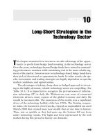
common type of glandular trichome, the peltate gland, is characterized by the swollen,
globular appearance of the cuticle which has split from the secretory cell walls as the sub-
cuticular space engorges with secretory product
e.g.,6,7
(Figure 10.1). The secretory product
often includes highly concentrated secondary metabolites with biological activities of
interest to the pesticide, pharmaceutical, and flavor and fragrance industries.
In some cases, plants have been grown to produce such compounds (e.g., the pyrethroids
of Chrysanthemum species
8
or essential oils of mints and other culinary herbs
9
). Attempts to
produce compounds found in glandular trichomes in undifferentiated plant cell or tissue
cultures have not been very successful.
3,10,11
This may be because many of the compounds
FIGURE 10.1
Light micrograph of an Artemisia annua trichome with the subcuticular space (→) filled with essential oils. (From
Ferreira, J.F.S. and Janick, J., Intl. J. Plant Sci., 156, 801, 1995. With permission.)
© 1999 by CRC Press LLC
found in glandular trichomes are autotoxic to the producing plant if they are not seques-
tered in the subcuticular space of the peltate glands.
The general cytotoxicity of many of these compounds necessitated concomitant evolu-
tion of secretory mechanisms and/or sequestration to avoid autotoxicity and to deliver the
compounds to the proper site for their ecological function. One can speculate that peltate
glandular trichomes are largely the result of this evolutionary interaction between chemis-
try and anatomical structure, in response to biotic selection pressures.
The functions of glandular trichomes and the compounds contained within them are often
unclear. There is no evidence to suggest that these structures and their contents help the
plant to grow faster or provide a direct advantage in plant–plant competition. Nor do they
give it a direct reproductive advantage. Unlike other types of trichomes, the relatively
sparse coverage of mature plant surfaces by glandular trichomes precludes a direct function
such as a physical hindrance to insects or as a means of reducing solar heating, evaporative
loss, or damaging ultraviolet irradiation.
4
However, some glandular trichomes produce
sticky materials that entrap insects, and others can secrete sufficient resinous material to
coat the plant surface, particularly in immature tissue, thus indirectly performing these
functions.
4,12
Considering the biological activities of many of the compounds sequestered in
or exuded from these structures, the selection pressures that favored evolution of secreting
glandular trichomes in many species were likely those of interspecies interactions. Such
interactions could occur with other plant species, microbes, or insects and other herbivores.
Secretion of phytotoxins by glandular trichomes could be of secondary benefit to the pro-
ducing plant. For example, one of the first compounds demonstrated to play a role in
plant–plant allelopathy was 1,8-cineole.
13
This highly phytotoxic monoterpene is probably
entirely localized in the glandular trichomes of Salvia species,
14,15
and together with other
terpenoids, inhibits growth and development of surrounding plants.
13,16
Such compounds
apparently reach competitors through volatilization and/or leaching from ground litter.
This delivery system for phytotoxins does not appear to be very efficient.
Glandular trichomes often contain potent phytotoxins that play no apparent role in
plant–plant allelopathy. For example, artemisinin, a sesquiterpenoid antimalarial drug,
17
is
highly phytotoxic to most plants, including the producing species, Artemisia annua L.
18-21
Nevertheless, to our knowledge, A. annua has not been reported to be allelopathic toward
other plant species. The efforts to devise weed management methods for commercial
monocultures of this drug plant species indicates that it is not a good competitor.
22
Artemisinin is found only in the subcuticular space of the glands of A. annua,
23,24
thus,
sequestering it to avoid autotoxicity. These glands clearly play no essential role for the
plant, because glandless biotypes are similar to glanded biotypes in most respects.
23
We
think that the most likely function of the glandular trichomes of A. annua and many other
species is as repositories of chemicals that defend against insects, herbivores, and microbial
pathogens. Many of the compounds stored in these glands are often suggested as alle-
lochemicals involved in plant–plant interactions without rigorous proof of such a role.
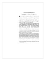
Developing and very young leaves are particularly vulnerable to attack by insects, her-
bivores, and pathogens. Glandular trichomes develop rapidly on leaf primordia
(Figure 10.2A). As trichomes mature and become more sparsely distributed over the
expanding leaf surface (Figure 10.2B), they may spread their contents on the leaf surface by
leakage through the cuticle
e.g.,4,25
or by rupturing of the cuticle.
e.g., 6,24
Some of the com-
pounds may play roles in plant–insect interactions other than protective functions, such as
attraction of pollinators or other beneficial insects.
Another property of many of the compounds produced by glandular trichomes, partic-
ularly the terpenoids, is their antimicrobial activity.
15,26
Essential oils containing these com-
pounds have been used for many years as preservatives and disinfectants. It follows that
© 1999 by CRC Press LLC
the antifungal and antibacterial properties of these compounds must have some role in
plant defenses against pathogens. However, constitutive defenses of plants against patho-
gens have not received as much study as inducible defenses.
Plant species that produce glandular trichomes generally produce relatively large
amounts of bioactive secondary products. The glandular trichomes are the primary pro-
duction sites of many of these compounds and perhaps the only site of synthesis of some.
There are a few cases, such as that of certain tobacco diterpenes,
27
in which the trichome
has conclusively been demonstrated to be the exclusive site of biosynthesis.
Phenolic compounds from the shikimate pathway are common constituents of secretory
glands. Many phenolics found in glands have biological activities. However, there has been
relatively little interest in commercial exploitation of these compounds. In some Solanum
species, phenolic compounds are released by breakage of specialized glands by insects
which results in rapid oxidation of the phenolics to quinones by polyphenol oxidase (PPO)
released from gland cell plastids.
28
The phenolic quinones polymerize rapidly enough to
“glue” insects to the leaf surface or to render their mouth parts unusable. Other secondary
compounds of glandular trichomes, such as sugar esters of fatty acids,
12
also play a role in
combating plant pests.
Extracting secondary compounds from producing plants has been the traditional
method of obtaining these compounds. This has apparently been done with little or no con-
sideration of the biochemistry or anatomy of the plant. An example of this is the pharma-
ceutical industry’s approach to artemisinin production from plants.
29
Artemisinin is an
effective antimalarial drug that is desperately needed to fight Plasmodium falciparum strains
that have become highly resistant to drugs currently in use.
17,30
The plant has been selected
and bred for the most productive biotypes and varieties.
31
Effects of agricultural chemicals,
soil types, and cultural methods of farming on artemisinin production have been deter-
mined.
32
The relative artemisinin yields of different plant parts and the same plant parts at
different developmental stages have been studied. This has evidently been done with no
consideration of the functioning of the secretory glands, in which all of the artemisinin
appears to be synthesized. Furthermore, development of extraction methods for the com-
pound has been conducted without regard to this knowledge.
e.g.,33,34
Pharmaceutical chem-
ists have apparently assumed that all of the plant cells must be extracted. However,
maceration of the entire tissues breaks cellular compartments, blending oxidative enzymes
FIGURE 10.2
Scanning electron micrographs of trichomes on (A) leaf primordia and (B) mature leaves of Artemisia annua.
(From Duke, S.O. and Paul, R.N., Intl. J. Plant Sci., 154, 107, 1993. With permission.)
© 1999 by CRC Press LLC
with secondary compound substrates, often leading to enzymatic “browning”. Part of the
browning process is caused by the generation of quinones from hydroxylated phenolic
compounds. The highly reactive quinones react with each other and other compounds to
generate brown, polymeric compounds.
We found that all of the drug and one of its precursors, artemisitene, could be extracted
by a brief dip in organic solvent to extract only the subcuticular space of the gland.
23
This
procedure minimizes the possibility of reactions of these relatively unstable compounds
with degrading enzymes or other plant constituents.
Our studies with fresh leaf tissue of Artemisia annua support this view. Leaf tissues (stems
excluded), were cut from 8-week-old potted plants and weighed. A half portion of the fresh
plant material was extracted by brief immersion in CHCl
3
and the other half was extracted
by homogenizing in CHCl
3
. Extraction by immersion was done by steeping 5 g of leaves in
100 ml CHCl
3
for 20 sec. The extract was filtered and the process repeated two more times.
The extracts were pooled and dried in a vacuum. Complete extraction of artemisinin was
ascertained by homogenizing the dipped leaf tissue in CHCl
3
(3x) using the same volume
as in dipping. Pooled extracts were vacuum dried. Homogenization of fresh leaves was
done in the same way as that of the dipped leaves, keeping the ratio of 100 ml CHCl
3
per
5 g of fresh leaves.
Quantitation of artemisinin followed the procedure of Zhao and Yeng
35
with minor mod-
ification. The dried extracts were dissolved in MeOH to a concentration of 10 mg/ml. A
50 µl aliquot was taken and 420 µl of 0.2% NaOH was added. The mixture was heated in a
heating block at 50°C for 30 min, then 30 µl of 20% methanolic acetic acid was added. This
sample was used for HPLC analysis under the following conditions: column was a C18
Waters µBondapak™ 3.9 × 300 mm; mobile phase was 0.01M NaH
2
PO
4
buffer (pH
7.00):MeOH eluted in gradient from 59:41 to 55:45 over a period of 25 min; flow rate was
1 ml/min; injection volume was 100 µl; detection γ was at 260 nm. The retention time of
artemisinin was 9.7 min.
With fresh tissue, homogenization of the tissues yielded only 73.2% (SE = 17.6%) of the
artemisinin that was extracted by dipping, and only 66.4% (SE = 15.5%) of that artemisinin
produced by dipping plus homogenization. Dipping alone extracted 90.7% (SE = 4.4%) of
the amount produced by dipping plus homogenization. This improved extraction effi-
ciency could be found with fresh tissues only, suggesting that intact glands may be impor-
tant in order for effective exploitation of this method. Nevertheless, with a high-value
product such as artemisinin, adoption of a method that improves yield by as much as 35%
would be cost-effective, even if the method is some what more involved.
Glands are not always as conspicuous and well-differentiated as the peltate glands. For
example, the generally cytotoxic hypericin is localized in undifferentiated glands found on
the aerial organs of several species of Hypericum (St. John’s wort).
36
Hypericin appears to
be the active compound in the antidepressant properties of St. John’s wort preparations.
37
It also has been found to have activity as an antiviral agent,
38,39
as a pharmaceutical to pre-
vent macular degeneration,
40
and as an anticancer drug.
41
Hypericin is a red, polycyclic
napthtodianthrone dye that generates singlet oxygen in the presence of light and molecular
oxygen. Singlet oxygen is very cytotoxic, causing rapid membrane lipid peroxidation. This
highly damaging process can cause cellular death. Hypericin is both herbicidal and insec-
ticidal in light.
42,43
The potential value of such a cytotoxic compound in defense of the plant
is apparent. But how does the plant protect itself? We do not know the entire mechanism
of protection, but we have sufficient information for informed speculation.
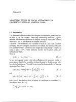
The hypericin-containing glands appear to the eye as almost black dots on stems, leaf
margins, and flower petal margins (Figure 10.3). There appear to be two general types of
glandular structures that produce the compound. H. perforatum L. has more flattened,
© 1999 by CRC Press LLC
“nodular” structures,
44
whereas other species such as H. hirsutum L.
43
and H. punctatum
Lam. display stalked nodules. We have found the pigment to be localized in spherical vac-
uolar inclusions of a group of relatively undifferentiated gland cells that are surrounded by
more flattened, unpigmented cells (unpublished data). Isolated vacuoles from the pig-
ment-containing cells are filled with hypericin. We speculate that hypericin is found
entirely within these “droplets” and that the oxygen concentration within the droplet is
insufficient for significant levels of singlet oxygen to be formed. One might expect oxida-
tive bleaching of the pigment if singlet oxygen were formed at the rate expected at atmo-
spheric oxygen levels.
Hypericin is a very expensive pharmaceutical at this time. We have not compared differ-
ent extraction methods for hypericin. However, tissue and cellular localization information
could be helpful in devising methods that produce higher yields.
10.2.2 Subepidermal Localization
In some cases, secondary products may be localized within a single cell layer within a leaf.
For example, the mung bean leaf has a subepidermal layer of cells with a higher concentra-
tion of phenolic compounds in the vacuoles than in the vacuoles of other cell layers. These
FIGURE 10.3
Leaf of Hypericum perforatum L. showing hypericin-containing glandular structures.
© 1999 by CRC Press LLC
compounds are paired with high polyphenol oxidase (PPO) activities compartmentalized
in the plastids
45,46
(K. C. Vaughn, unpublished). When the cell is broken, the vacuolar phe-
nolic compounds mix with the plastidic PPO, and highly reactive quinones are rapidly pro-
duced. These quinones could retard the activity of herbivores or pathogens.
A further example is that of the foliar localization of certain alkaloids. We have found tro-
pane alkaloids to be localized primarily in the adaxial subepidermal layer (palisade paren-
chyma), as well as spongy mesophyll and vascular parenchyma cells of the leaves of both
Erythroxylum coca var. coca and E. novogranatense var. novogranatense (Figure 10.4).
47
Other
plant species also concentrate alkaloids in similar subepidermal layers of the foliage.
e.g., 48
Furthermore, in E. coca, the alkaloids were found by immunocytochemistry to be localized
within the vacuoles of these subepidermal cells.
47
The alkaloids appear to be aggregated
around a core of phenolic compounds, making them perhaps immobile. Tropane alkaloids
could be extracted easily by dipping fresh, young leaves in chloroform with up to 75% of
the alkaloids of young leaves extracted with an 80 sec dip. This technique did not work
with dried leaves.
FIGURE 10.4
Cross section of Erythroxylum coca var. coca leaf stained with Dragendorf’s solution. Note intensely stained
subepidermal cells (→). (From Ferreira, J.F.S., Duke, S.O., and Vaughn, K.C., Intl. J. Plant Sci., 159, 492, 1999.
With permission.)
© 1999 by CRC Press LLC
What is the functional significance of the tissue and cellar location of these compounds?
Several functions are plausible. Subepidermal localization may be a good location for plant
defense by chemical toxicants. Plant pathogens and small herbivorous insects would
quickly come in contact with the subepidermal layer during infection or grazing. But why
are these compounds not found in all cell layers? We speculate that producing these com-
pounds in all cells of the leaf would reduce the productivity of the plant due to some level
of phytotoxicity and/or the high amount of resources required to produce the compounds.
Their vacuolar localization within the cells indicates that they play no role in normal cellu-
lar functions. Further, localization may result in higher concentrations of toxicants at stra-
tegic sites and, thus, a more effective deterrent. Secondary compounds so localized
probably have biological activity against one or more organisms with which the plant species
has coevolved.
10.3 Conclusions
If phytotoxic compounds produced by higher plants are autotoxic, they must be seques-
tered to avoid self-poisoning. Conversely, if a compound has been sequestered, one might
suspect that it could be a phytotoxin. Thus, the secondary compound contents of glands
and specialized cell layers should be the most lucrative sources of phytotoxic allelochemi-
cals. Plant-derived phytotoxins have been the sources of two commercial herbicides: a 1,8-
cineole-derived herbicide, cinmethylin,
49
and the newly commercialized triketones,
50
a
group of compounds derived from leptospermone, a constituent of the bottlebrush plant,
Calistemon spp. As discussed above, the glandular localization of 1,8-cineole provided a
clue to its activity. We are not aware of the tissue or cellular localization of leptospermone.
We have not discussed every mechanism or structure by which a plant can sequester a
secondary compound and only a few examples are provided of the discussed sequestration
mechanisms. Some highly potent phytotoxins, such as sorgoleone
51
and polyacetylenes,
52
are secreted or exuded by roots of producing plants rather than being sequestered. How
these plants avoid autotoxicity is not known. However, their molecular target site as a her-
bicide may not reside in the roots and they may not be translocated to potential foliar sites
of action.
A generally positive correlation between the degree of sequestration and the phytotoxic-
ity of sequestered compounds might be expected. If so, one would expect compounds from
peltate glands to generally be more phytotoxic than those from subepidermal layers. How-
ever, this and other aspects of the coevolution of anatomical features and secondary prod-
ucts will require considerably more study. Our understanding of the complex interactions
between the selection pressures of biotic stress and autotoxicity in the shaping of anatom-
ical structures and physiological mechanisms that aid in the safe and benign delivery of
cytotoxic compounds to appropriate sites for plant defense is quite limited. A more com-
plete understanding of these interactions will be useful in discovery of new uses for natural
products and in production of these compounds by biosynthesis.
© 1999 by CRC Press LLC
References
1. Williams, D.H., Stone, M.J., Hauck, P.R., and Rahman, S.K., J. Nat. Prod., 52, 1189, 1989.
2. Stone, M.J. and Williams, D.H., Molec. Microbiol., 6, 29, 1992.
3. Ferreira, J.F.S. and Duke, S.O., AgBiotech New Info., 9, 309N, 1997.
4. Dell, B. and McComb, A.J., Adv. Bot. Res., 6, 277, 1978.
5. Fahn, A., New Phytol., 108, 229, 1988.
6. Duke, S.O. and Paul, R.N., Intl. J. Plant Sci., 154, 107, 1993
7. Serrato-Valenti, G., Bisio, A., Cornara, L., and Ciarallo, G., Ann. Bot., 79, 1997, 329.
8. McLaughlin, G.A., in Pyrethrum, The Natural Insecticide, Casida, J.E., Ed., Academic Press, New
York, 1971, 3.
9. Simon, J.E., in Advances in New Crops, J. Janick and J.E. Simon, Eds., Timber Press, Inc., Portland,
OR, 1990, 472.
10. Martinez, B.C. and Staba, J., Adv. Cell Cult., 6, 69, 1988.
11. Fulzele, D.P., Sipahimalani, A.T., and Heble, M.R., Phytotherapy Res., 5, 149, 1991.
12. Neal, J.J., Tingey, W.M., and Steffens, J.C., J. Chem. Ecol., 16, 487, 1990.
13. Muller, W.H. and Muller, C.H., Bull. Torrey Bot. Club, 91, 327, 1964.
14. Croteau, R. and Johnson, A., in Biology and Chemistry of Plant Trichomes, Rodriguez, E., Healey,
P.L., and Mehta, I., Eds., Plenum, New York, 1984, 133.
15. Kelsey, R.G., Reynolds, G.W., and Rodriquez, E., in Biology and Chemistry of Plant Trichomes,
Rodriguez, E., Healey, P.L., and Mehta, I., Eds., Plenum, New York, 1984, 187.
16. Muller, W.H., Lorber, P., and Haley, B., Bull. Torrey Bot. Club, 95, 415, 1968.
17. Klayman, D.L., Science, 228, 1049, 1985.
18. Chen, P.K. and Leather, G.R., J. Chem. Ecol., 16, 1867, 1990.
19. Duke, S.O., Vaughn, K.C., Croom, E M., and Elsohly, H.N., Weed Sci., 35, 499, 1987.
20. Duke, S.O., Paul, R.N., and Lee, S.M., Amer. Chem. Soc. Symp. Ser., 380, 318, 1988.
21. Lydon, J., Teasdale, J.R., and Chen, P.K., Weed Sci., 45, 807, 1997.
22. Bryson, C.T. and Croom, E.M., Weed Technol., 5, 117, 1991.
23. Duke, M.V., Paul, R.N., Elsohly, H.K., Sturtz, G., and Duke, S.O., Intl. J. Plant Sci., 155, 365, 1994.
24. Ferreira, J.F.S. and Janick, J., Intl. J. Plant Sci., 156, 801, 1995.
25. Lin, Y. and Wagner, G.J., J. Chem. Ecol., 20, 1907, 1994.
26. Duke, S.O., in Handbook of Natural Toxins. vol. 6. Toxicology of Plant and Fungal Compounds,
Keeler, R.F. and Tu, A.T., Eds., Marcel Dekker, New York, 1991, 269.
27. Wagner, G.J., Plant Physiol., 96, 675, 1991.
28. Kowalski, S.P., Tingey, W.M., and Steffens, J.C., J. Hered., 81, 475, 1990.
29. Laughlin, J.C., Trans. Royal Soc. Trop. Med. Hyg., 88, Suppl. 1, 21, 1994.
30. White, N.J., Trans. Royal Soc. Trop. Med. Hyg., 88, Suppl. 1, 3, 1994.
31. Delabays, N., Benakis, A., and Collet, G., Acta Hortic., 330, 203, 1993.
32. Laughlin, J.C. Acta Hortic., 331, 53. 1993)
33. ElSohly, H.N., Croom Jr., E.M., El-Feraly, F.S., and El-Sherei, M.M., J. Nat. Prod., 53, 1560, 1990.
34. Vonwiller, S.C., Haynes, R.K., King, G., and Wang, H J., Planta Med., 59, 562, 1993.
35. Zhao, S.S. and Yeng, M.Y., Planta Med., 51, 233, 1985.
36. Mathis, C. and Ourisson, G., Phytochemistry, 2, 157, 1963.
37. Müller, W.E., Rolli, M., Schäfer, C., and Hafner, U., Pharmacopsychiatry, 30 (Suppl.), 102, 1997.
38. Hudson, J.B., Harris, L., and Towers, G.H.N., Antiviral Res., 20, 173, 1993.
39. Lavie, G., Mazur, Y., Prince, A.M., Pascual, D., Liebes, L., Levin, B., and Meruelo, D., Transfu-
sion, 35, 392, 1995.
40. Kimura, H., Harris, M.S., Sakamoto, T., Gopalakrishna, R., Gundimeda, U., Cui, J.Z., Spee, C.,
Hinton, D.R., and Ryan, S.J., Current Eye Res., 16, 967, 1997.
41. Zhang, W., Law, R.E., Hinton, D.R., and Couldwell, W.T., Cancer Lett., 120, 31, 1997.
42. Knox, J.P., Samuels, R.I., and Dodge, A.D., Amer. Chem. Soc. Symp. Ser., 339, 265, 1987.
© 1999 by CRC Press LLC
43. Knox, J.P. and Dodge, A.D., Plant Cell Environ., 8, 19, 1985.
44. Curtis J.D. and Lersten, N.R., New Phytol., 114, 571, 1990.
45. Hurkman, W.J. and Kennedy, G.S., Protoplasma, 89, 171, 1976.
46. Duke, S.O. and Vaughn, K.C., Physiol. Plant., 54, 381, 1982.
47. Ferreira, J.F.S., Duke, S.O., and Vaughn, K.C., Intl. J. Plant Sci., 159, 492, 1998.
48. White, H.A. and Spencer, M., Can. J. Bot., 42, 1481, 1964.
49. Grayson, B.T., Williams, K.S., Freehauf, P.A., Pease, R.R., Ziesel, W.T., Sereno, R.L., and Reins-
felder, R.E., Pestic. Sci., 21, 143, 1987.
50. Lee, D.L., Prisbylla, M.P., Cromartie, T.H., Dagarin, D.P., Howard, S.W., Provan, W.M., Ellis,
M.K., Fraser, T., and Mutter, L.C., Weed Sci., 45, 601, 1997.
51. Nimbal, C.I., Pederson, J.F., Yerkes, C.N., Weston, L.A., and Weller, S.C., J. Agric. Food Chem.,
44, 1343, 1996.
52. Tsao, R. and Eto, M., Chemosphere, 32, 1307, 1996.
© 1999 by CRC Press LLC