Burns Regenerative Medicine and Therapy - part 5 docx
Bạn đang xem bản rút gọn của tài liệu. Xem và tải ngay bản đầy đủ của tài liệu tại đây (201.6 KB, 16 trang )
Experimental and Clinical Study on Burns Regenerative Medicine and Therapy with MEBT/MEBO 59
This study was designed to investigate the effect of
MEBO on plasma ET/NO so as to further discover the
mechanisms of MEBT therapy. ET, separated from super-
natant fluid of cultured pig aorta endothelial cell by Ya-
nagisawa in 1988, is mainly synthesized in the vascular
endothelial cell and is known to be the strongest vasocon-
strictive peptide in addition to other biological actions.
However, the excessive release of ET may cause micro
blood vessels to contract and spasm for an extended peri-
od of time, resulting in microthrombus formation. NO is
a potent gaseous free radical and is synthesized in the
cytoplasm of vascular endothelial cells, vascular smooth
muscle cells, macrophages, platelets, etc. This reaction
occurs when nitrogen monoxide synthetase (NOS) cata-
lyzes the guanidin-end nitrogen of L-Arg to combine with
oxygen. There are two types of NOS: constitutional NOS
(cNOS) and inducible NOS (iNOS). NO has a very short
half-life as it may react with circulating oxyhemoglobin,
deoxyhemoglobin, superoxide anion or free oxygen to
create stable products such as nitrite and nitrate. These
are metabolized mainly through the kidney.
NO expresses its effects in two ways. On the one hand,
NO is antagonistic with ET in dilating micro blood ves-
sels, forming a protective layer in the tunica intima, pre-
venting platelets and neutrophils from adhering to the
vessel wall, and inhibiting the formation of microthrom-
bus. On the other hand, the excessive release of NO may
result in severe wound exudation and edema in the early
stages postburn, thus increasing damage to tissue and
cells. Under the normal situation, NO and ET maintain a
certain dynamic equilibrium as an increase of ET pro-
motes an elevation of NO synthesis while NO inhibits the
synthesis of ET. A physiologic ratio of ET/NO represents
a healthy balance.
According to the studies in recent years, the iNOS and
ET mRNA genes located in burns wounds, heart, lung,
kidney, liver and gastrointestinal tract express an increas-
ing activity postburn, which leads to a massive release of
ET and NO, especially from the wound. The significant
increase of ET and NO in the circulation and the imbal-
ance of the ET/NO ratio have a close correlation with
shock, acute renal failure, acute respiratory failure, stress
ulcer and cerebral edema after severe burns. Great care
must be paid to the role of ET/NO if one is to prevent
progressive damage of the wound tissues in the zone of
stasis at the early stages postburn [2]:
1 NO and ET, known as the most potent vasoconstric-
tion and vasodilatory factors, should maintain a
healthy balance during the critical postburn period.
2 The increment of NO and ET in blood plasma post-
burn has a positive relationship with the increase of the
capillary permeability, leading to massive exudation
and hyperedema [3, 4].
3 The increase of ET in plasma, the contraction and
spasm of the micro blood vessels, the thrombus forma-
tion around the wound and underlying tissue, and ede-
ma pressure may cause secondary ischemia and necro-
sis to the adjacent tissues.
4 The exudates, being rich in protein, provide a good cul-
ture medium for bacteria growth.
It was found that the application of a non-selective ET
receptor antagonist, AK-044, may alleviate the secondary
damage to burns wounds resulting from the increase of
ET [5]. In this study, the increment of ET and NO in the
BRT with MEBT/MEBO group was obviously lower than
that in the dry-exposed group. In the former group, the
ET/NO ratio in plasma decreased after 1 day toward a
normal level of 2.95, representing a timely improvement
in the tissue microcirculation status particularly in the
zone of stasis. This prevented the occurrence of progres-
sive microthrombosis and prevented the progressive ne-
crosis of tissue by optimizing the wound environment. In
the dry-exposed group, ET and NO increased significantly
at all phases and the ET/NO ratio in plasma remained
much higher than normal (2.95), usually at around 6. ET
might play a dominant role in this change by keeping the
micro blood vessel contracted and spasming for a long
time resulting in microthrombosis, low vitality of wound
tissues, and poorer healing process compared to that in
the BRT with MEBT/MEBO group. The results suggest
that the BRT with MEBT/MEBO group is superior to the
dry-exposed group in wound healing time and outcome.
However, further study is yet to be conducted for the spe-
cific mechanism on how MEBO controls the adequate
release of ET and NO from the burns wounds, and on how
to promote the return of the ET/NO ratio to normal as
rapidly as possible.
References
1 Bonaldi LA, Frank DH: Pathophysiology of the burn wound; in Achauer
BM (ed): Management of the Burned Patient. Norwalk, Appleton & Lange,
1987, pp 21–47.
2 Battal MN, Hata Y, Matsuka K, et al: Reduction of progressive burn injury
by using a new nonselective endothelin-A and endothelin-B receptor antag-
onist, Tak-044: An experimental study in rats. Plast Reconstr Surg 1997;
99:1610–1619.
3 Kowal VA, Walenga JM, Sharp PM, et al: Postburn edema and related
changes in interleukin-2, leukocytes, platelet activation, endothelin-1, and
C
1
esterase inhibitor. J Burn Care Rehab 1997;18:99–103.
4 Sozumi T: The role of nitric oxide in vascular permeability after a thermal
injury. Ann Plast Surg 1997;39:272–277.
5 Jia XM, Zhu ZM, Zeng Q, et al: The dynamic changes of endothelin-1 in
rat plasma, wound and organs at the early stages postburn. Med J PLA
1997;22:40–42.
60 Burns Regenerative Medicine and Therapy
Experimental Study of the Effect of BRT with
MEBT/MEBO on Hematological Parameters in
the Treatment of Burned Rabbits
Introduction
Many researchers found that treatment with BRT with
MEBT/MEBO improved the microcirculation in burns
wounds. However, the effect of BRT with MEBT/MEBO
on systemic postburn situations has not been established.
The authors used a rabbit model for the following experi-
mental study on this subject.
Materials and Methods
Seventy-two healthy adult rabbits of either sex eating a uniform
diet and weighing 2.0 B 0.5 kg were divided randomly into three
groups: group 1 (normal control, n = 12), group 2 (treated with
MEBO, n = 30), and group 3 (dry exposure, n = 30). Without anesthe-
sia, all animals were shaved on the waist with barium sulfide. Ani-
mals in group 1 received no treatment and cardiac blood was sam-
pled for measuring normal hematological parameters. Animals in
groups 2 and 3 sustained deep second-degree burns of 10% BSA via
100
°
C water applied for 5 s (confirmed by pathological examina-
tion). Animals in group 2 were then treated with BRT with MEBT/
MEBO renewed every 6 h. Animals in group 3 were offered no treat-
ment with wounds remaining dry and exposed. No fluid infusion was
administered. Blood samples were taken from the left ventricle of the
heart for determining hematological values at 4, 24, 48, 72 h and 6
days postinjury. The parameters tested included apparent blood vis-
cosity at different shear rates, plasma viscosity, hematocrit (HCT),
erythrocyte agglutinative index (EAI) and erythrocyte transforma-
tion kinetics (ETK). To standardize the experimental conditions and
instruments, blood sampling was performed in accordance with
requirements for determination of blood flow viscosity established
by the International Council for Standardization in Haematology
(ICSH) in 1988. These laboratory determinations were made by a
specially assigned technologist at room temperature using a Type N
E-1 viscometer (manufactured by Chengdu Instruments, China) [1].
Apparent blood viscosity at the shear rates of 230, 115, 46, 11.5,
and 5.75 s
–1
were determined, respectively, using 1.3 ml blood. Plas-
ma viscosity is the viscosity at the shear rate of 115 s
–1
. Hematocrit
(HCT) was determined by a Wintrope tube, type LXJ-64-01 centri-
fuge, at 3,000 rpm for 30 min [2]. Erythrocyte agglutinative index
(EAI) was the ratio of apparent blood viscosity obtained at low and
high shear rates (5.75 and 230 s
–1
). Erythrocyte transformation kinet-
ics (TK) was calculated according to the formula given in Chen [3].
Results
Owing to the minor difference of sex and strain in the
hematological parameters of the rabbits, it was feasible to
work out a uniform range [1–4]. Randomized division
kept animals in a normal control group, allowing the
MEBO-treated and the dry-exposed groups to live under
the same conditions. Therefore, no repeated blood sam-
pling was allowed and the data were significantly valu-
able. Apparent blood viscosity and plasma viscosity at
different shear rates in group 3 were higher than those in
group 2. Table 13 shows that the parameters in group 3
began to increase at 4 h post-injury, peaked at 24 h, and
remained higher at 48 h, 72 h and 6 days than those in
group 2. There were significant statistical differences (ta-
ble 14; p ! 0.01). Compared to the normal group, parame-
ters including apparent blood viscosity, plasma viscosity
and others in the MEBO-treated group began to increase
at 4 h postinjury, peaked at 24 h, decreased at 48 h, fur-
ther decreased at 72 h and nearly returned to normal at 6
days (table 15). But statistical analysis showed that hema-
tological parameters did not change significantly (p 1
0.05), except at 24 h postinjury when the blood viscosity
at shear rates of 11.5 s
–1
(t = 2.4696, p ! 0.05) and 5.75 s
–1
(t = 2.700, p ! 0.05) increased markedly (table 16).
Table 13. Changes of hemorrheological parameters in group 3 (dry group) (mean B SE)
Parameters 4 h
postinjury
24 h
postinjury
48 h
postinjury
72 h
postinjury
6 days
postinjury
Blood viscosity
230 s
–1
5.45B0.54 8.66B0.59 6.24B0.188 4.57B0.588 5.61B0.43
115 s
–1
5.92B0.66 8.86B1.37 6.55B0.28 5.27B1.13 6.57B0.46
46 s
–1
6.49B0.71 10.87B1.42 7.99B0.48 5.98B0.44 7.14B0.41
23 s
–1
7.26B0.43 12.71B1.43 9.32B0.33 8.03B0.46 8.84B1.59
11.5 s
–1
12.89B1.32 21.13B2.65 13.12B0.34 10.74B1.52 10.88B0.73
5.75 s
–1
18.12B3.42 25.22B0.96 19.99B4.32 14.39B1.74 12.39B0.12
Plasma viscosity (115 s
–1
) 2.71B0.08 2.64B0.32 2.33B0.123 2.15B0.116 2.30B0.095
HCT 0.42B0.051 0.43B00.4B0.026 0.36B0.04 0.036B0.015
EAI 3.29B0.27 2.86B0.12 3.17B0.68 3.14B0.023 2.21B0.255
TK 0.64B0.086 0.96B0.098 0.84B0.093 0.82B0.21 0.95B0.144
Experimental and Clinical Study on Burns Regenerative Medicine and Therapy with MEBT/MEBO 61
Table 14. Comparison of hemorrheological parameters between groups 2 and 3
Parameters 4 h postinjury
tp
24 h postinjury
tp
48 h postinjury
tp
72 h postinjury
tp
6 days postinjury
tp
Blood viscosity
230 s
–1
4.888 ! 0.001 10.74 ! 0.001 5.716 ! 0.001 1.412 1 0.05 3.558 ! 0.01
115 s
–1
3.776 ! 0.01 8.212 ! 0.001 3.364 ! 0.01 2.765 ! 0.05 3.289 ! 0.01
46 s
–1
1.838 1 0.05 8.031 ! 0.001 4.528 ! 0.001 4.125 ! 0.01 2.691 ! 0.05
23 s
–1
0.861 1 0.05 7.014 ! 0.001 4.435 ! 0.01 3.502 ! 0.01 3.909 ! 0.01
11.5 s
–1
4.611 ! 0.001 116.15 ! 0.001 6.572 ! 0.001 4.985 ! 0.001 3.261 ! 0.01
5.75 s
–1
4.564 ! 0.001 10.47 ! 0.001 23.12 ! 0.001 6.031 ! 0.001 2.043 1 0.05
Plasma viscosity 0.634 1 0.05 0.125 1 0.01 1.188 1 0.05 1.426 1 0.01 0.254 1 0.05
HCT 1.866 1 0.05 1.237 1 0.05 0.563 1 0.05 0.574 1 0.05 0.563 1 0.05
EAI 2.038 1 0.05 0.121 1 0.05 1.075 1 0.05 9.306 ! 0.001 0.547 1 0.05
TK 2.648 ! 0.05 1.748 1 0.05 6.106 ! 0.001 3.276 ! 0.01 5.048 1 0.001
Table 15. Hemorrheological parameters in group 1 and changes in group 2 (mean B SD)
Parameters Group 1
(normal
control)
Group 2
4 h
postinjury
24 h
postinjury
48 h
postinjury
72 h
postinjury
6 days
postinjury
Blood viscosity
230 s
–1
4.20B0.05 4.13B0.52 4.48B0.7 4.01B0.65 4.00B0.72 4.12B0.72
115 s
–1
0.69B0.71 4.54B0.68 5.67B0.91 4.58B0.98 4.27B0.8 4.42B1.10
46 s
–1
5.53B0.99 5.44B1.00 6.97B1.03 5.12B1.07 4.88B0.86 5.11B1.26
23 s
–1
6.79B1.32 6.59B1.30 8.49B1.20 6.00B1.18 5.59B1.17 6.04B1.40
11.5 s
–1
7.61B1.69 8.5B1.69 9.55B1.26 7.34B1.46 6.69B1.52 7.41B1.81
5.75 s
–1
9.33B2.58 12.21B2.7 13.45B1.9 9.92B1.86 8.33B1.88 9.57B2.28
Plasma-viscosity (115 s
–1
) 2.05B0.39 2.48B0.6 2.60B0.55 2.60B0.38 2.28B0.16 2.27B0.20
HCT 0.36B0.05 0.37B0.05 0.40B0.04 0.38B0.06 0.34B0.06 0.34B0.06
EAI 2.19B0.58 2.73B0.47 0.82B0.55 2.65B0.86 2.07B0.19 2.32B0.34
TK 0.73B0.08 0.75B0.08 0.82B0.34 0.75B0.05 0.64B0.04 0.72B0.09
Table 16. Comparison of hemorrheological parameters between groups 1 and 2
Parameters 4 h postinjury
tp
24 h postinjury
tp
48 h postinjury
tp
72 h postinjury
tp
6 days postinjury
tp
Blood viscosity
230 s
–1
0.1490 1 0.05 0.6514 1 0.05 0.4635 1 0.05 0.1826 1 0.05 0.1437 1 0.05
115 s
–1
0.3052 1 0.05 1.6984 1 0.05 0.1818 1 0.05 0.2855 1 0.05 0.4152 1 0.05
46 s
–1
0.0128 1 0.05 2.0160 1 0.05 0.5630 1 0.05 0.9918 1 0.05 0.5243 1 0.05
23 s
–1
0.3239 1 0.05 1.8600 1 0.05 0.3276 1 0.05 0.7940 1 0.05 0.2600 1 0.05
11.5 s
–1
0.7449 1 0.05 2.4696 ! 0.05 0.2430 1 0.05 0.8100 1 0.05 0.1632 1 0.05
5.75 s
–1
1.5470 1 0.05 2.7000 ! 0.05 0.3907 1 0.05 0.6635 1 0.05 0.1456 1 0.05
Plasma viscosity 1.2020 1 0.05 1.6320 1 0.05 2.0210 1 0.05 1.0440 1 0.05 1.0040 1 0.05
HCT 0.2916 1 0.05 1.2964 1 0.05 0.5249 1 0.05 0.5249 1 0.05 0.5249 1 0.05
EAI 1.4471 1 0.05 1.5768 1 0.05 0.8872 1 0.05 0.3239 1 0.05 0.4260 1 0.05
TK 0.3536 1 0.05 1.7166 1 0.05 2.4241 1 0.05 2.0142 1 0.05 0.1661 1 0.05
62 Burns Regenerative Medicine and Therapy
Conclusion
Treatment with MEBO after burns could ameliorate
total body stress reaction, reduce water evaporation from
the wound surface, lessen local and systemic capillary
exudation and thus improve the hemorrheological charac-
teristics of microcirculation. It also suggested that when
the minor or moderately burned patient was treated with
MEBO at an early stage, fluid infusion and/or blood trans-
fusion would not be necessary, whereas for severely
burned patients, the amount of fluid infusion could be
reduced as tolerated.
Discussion
Hemorrhagic change is considered to be one of the
pathophysiological changes following burns and serves as
a basis of microcirculation disorder. Subsequent to exten-
sive burns, microvascular permeability increases and co-
pious intravascular plasma exudes toward the wound sur-
face and tissue space leading to localized hemoconcentra-
tion, reduction of effective blood volume, decreased plas-
ticity of red blood cells and increased blood viscosity.
These hemorrhagic changes comprise the pathophysiolog-
ical basis of burn shock and contribute to the deleterious
stress reaction immediately following the trauma of
burns. For instance, adrenaline, 5-HT, and prostaglandin
may all increase the activation of platelets to erythrocytes,
thereby changing the localized electrical potential. The
increased secretion of catecholamines due to stress reac-
tion directly promotes platelet adhesiveness. The injured
sub-microstructure of the vascular wall elaborates an
adhesion protein on platelets and erythrocytes causing
significant intravascular platelet aggregation and contrib-
uting to adhesion and aggregation of platelets and erythro-
cytes. This, of course, precipitates thrombotic events [5].
In this study, we compared the blood viscosity of a
MEBO-treated group with controls and demonstrate that
the viscosity of the MEBO group approximated that of the
normal controls. As animals in the MEBO-treated group
were treated only with MEBO (they received neither fluid
replacement nor special feeding), such changes verified
that MEBO alleviated body stress reaction following
burns and ensured body recovery.
Despite becoming a systemic disease, burn injuries
begin with a wound on one region of the body surface. It
has been reported that BRT with MEBT/MEBO could
have both local and systemic therapeutic effects on burn
management [6]. We are pleased to report that rabbits in
the MEBO-treated group were as active as normal rabbits
and fed freely. Blood viscosity did not change significant-
ly (p 1 0.05), except at shear rates of 11.5 and 5.75 s
–1
24 h
postinjury.
The blood viscosity value of normal rabbits in this
study was slightly lower than the mean values as indicated
in many domestic and international reports. This differ-
ence is probably explained by the fact that venous blood
from rabbits (weighing 2.5 kg) was sampled in those
reports instead of cardiac blood from rabbits (weighing
2.0 B 0.5 kg) which we used in our study. Different
dietary factors as well as geographical differences may
contribute as well. Blood viscosity is a comprehensive
marker as it indicates aggregation, deformability and the
rheological properties of platelets, RBCs and WBCs. In
this study, blood viscosity and plasma viscosity of rabbits
were compared to and found to be lower than those of
human beings. It remains to be further discussed whether
this lower viscosity is associated with low HCT, difficulty
of RBC aggregation, or whether it is simply some biologi-
cal characteristic associated with ‘herbivores’.
References
1 Du ZY, Jiang XQ: Analysis on hemorrheology of healthy rabbits. Trans
Taishan Med Coll 1991;12:134–136.
2 Zhang YM, Jiang XQ, et al: Investigation and analysis on hemorrheological
parameters of human health. Chin J Hemat 1992;13:312–313.
3 Chen WJ (ed): Hemorrheology. Tianjin, Tianjin Science & Technology
Press, 1987, pp 50–53.
4 Guo ZR, et al: Experimental study on the effects of different resuscitation
regimens on hemorrheology during burn shock period. Chin J Plastic Surg
Burns 1988;4:130–132.
5 Xu RX: The medicine of burn and ulcer: A general introduction. Part 1.
Chin J Burns Wounds Surface Ulcers 1989;1:11–21.
6 Xu RX: A summary report of the first session of the editorial board of the
journal and to the national symposium on the moist exposed burn therapy.
Chin J Burns Wounds Surface Ulcers 1992;4:2–5.
Experimental and Clinical Study on Burns Regenerative Medicine and Therapy with MEBT/MEBO 63
OOOOOOOOOOOOOOO OOOOOOOOOOOOOOOOOOOOOOOOO OOOOOOOOOOOOOOOOOOOO OOOOOOOOOOOOOOO OOOOOOOOO OOOOOOOOO OOOOOOOOOO OOOOOOOOO OOOOOOO OOOO
Studies on the Anti-Infection Effect of BRT with
MEBT/MEBO
Effect of BRT with MEBT/MEBO on the
Immunity of Burns Patients
Introduction
The antibiotic and wound-healing properties of MEBO
have been proven in clinical practice. There are different
opinions about the mechanism of this antibiotic effect, so
research on this subject is very important. During the
period from January 1993 through December 1995, we
conducted clinical observations on the effect of MEBO on
burns patients’ immunity and demonstrated that MEBO
enhanced patients’ immunity as part of its antibiotic and
wound-healing effects.
Materials and Methods
Clinical Data
One hundred and twenty burns patients were divided randomly
into two groups. Sixty patients in the MEBO group, including 40
males and 20 females, aged 6–65 (35.5 B 14.8) years. Course of dis-
ease: 1–36 h (18.5 B 8.8 h) before administration of MEBO. Cause of
burns: direct flame, 30 cases; scald, 16 cases, and chemical burn, 14
cases. Burn position: craniofacial, 17 cases; neck, 17 cases; trunk, 18
cases, and limbs, 23 cases. Burns depth: superficial second-degree
burns, 26 cases; deep second-degree burns, 24 cases; third-degree
burns, 10 cases. Burn area: 1–25% (13 B 6%) total body surface area
(TBSA). By contrast, there were 60 cases in the control group includ-
ing 41 males and 19 females, aged from 7 to 65 years (36 B 14.5
years). Course of disease: 1–35 h (18 B 8.5 h) before administration.
Cause of burns: direct flame, 29 cases; scald, 17 cases, and chemical
burns, 14 cases. Burn position: craniofacial, 13 cases; neck, 7 cases;
trunk, 18 cases, and limbs, 22 cases. Burn depth: superficial second-
degree burns, 24 cases; deep second-degree burns, 26 cases, and
third-degree burns, 10 cases. Burn area: 1–26% (13.5 B 6.3%) TBSA.
The data of the two groups were similar and comparable (p 1 0.05).
Treatment and Examination
All patients were subjected to debridement before collecting sam-
ples of skin tissue. Patients in the MEBO group were treated with
burns regenerative therapy (MEBT/MEBO) [1, 2], and skin tissues
were taken twice from the original sites before the treatment began
and after the wounds healed, respectively. Patients in the control
group were treated with another traditional Chinese burns oint-
ment – Jing Wan Hong –, and skin tissue was taken at the same time
phase as in the MEBO group. Patients in both groups were observed
closely, and their wound healing time and incidence of wound infec-
tion were compared.
Five pieces of skin tissues taken from the edge of the burns
wounds using a pair of biopsy forceps were immersed in 10% forma-
lin and kept in separate ice bottles, respectively. Four pieces of skin
tissue were also taken from the normal (non-burned) skin of the same
patient and treated identically as the other tissue. In addition, normal
skin tissues from 60 surgical cases were taken during subdermal cyst
operations and treated identically to both burns tissues. The levels of
IgA-, IgG-, and IgM-producing cells and C
3
were determined using
the frozen section immunohistochemical method. The first antibody
was supplied from Vector and the second from DAKO. Venous
blood samples of burns patients and healthy persons were taken in
the morning before breakfast. Peripheral blood immunoglobulin (Ig)
levels were determined using the agar diffusion method.
Classification Standard of Antibody Producing Cell and C
3
The classification standard was as follows: – = no positive cells
or particles were found or only occasionally found in the whole
slide; + = positive cells accounted for less than 30% of the total
number of interstitial cells in the lamina propria; ++ = positive cells
accounted for 31–70% of the total number of interstitial cells in the
lamina propria; +++ = positive cells accounted for more than 71%
of the total number of interstitial cells in the lamina propria. Histo-
logical diagnosis was based on the criteria stipulated at the China
National Pathology Research Group Conference held in Zhengzhou
in 1978.
Results
Clinical Efficacy Assessment
In the MEBO group, burns wounds with different
depths had shorter healing time than those in the control
group (p ! 0.01; table 17). Only one case (1.7%) in the
MEBO group had wound infection, compared to 9 cases
(15%) in the control group. The difference between the
Table 17. Comparison of healing time of wounds with different depth in two groups (day, mean B SE)
Group Superficial
second-degree
Deep
second-degree
Third-degree Average
healing time
MEBO 9.5B2.3 (5–14) 25.5B2.3 (21–30) 36.5B2.8 (31–43) 23.5B9.3 (5–42)
Control 13B3 (7–19) 29.5B2.8 (24–35) 42B3.5 (35–49) 28B10.5 (7–49)**
t value 4.65 5.49 3.88 2.49
Compared with MEBO group: ** p ! 0.01.
64 Burns Regenerative Medicine and Therapy
Table 18. Comparison of staining intensity of immune factors in burns and non-burns areas of the patients (%)
Area Cases IgA
^ (+) 6 (++)
IgG
^ (+) 6 (++)
IgM
^ (+) 6 (++)
C
3
^ (+) 6 (++)
Burns area 120 60 (50.0) 60 (50.0) 59 (49.2) 61 (50.8) 57 (47.5) 63 (52.3) 58 (48.3) 62 (51.7)
Non-burns area 120 80 (66.7) 40 (33.3) 79 (65.8) 41 (34.2) 77 (64.2) 43 (35.8) 82 (68.3) 38 (31.7)
Normal person 60 44 (73.0) 16 (28.0) 42 (70.0) 18 (30.0) 39 (65.0) 21 (35.0) 42 (70.0) 18 (30.0)
Table 19. Staining intensity of local immune factors and depth of burns wound (%)
Wound Cases IgA
^ (+) 6 (++)
IgG
^ (+) 6 (++)
IgM
^ (+) 6 (++)
C
3
^ (+) 6 (++)
Superficial second-degree 50 28 (56.0) 22 (44.0) 29 (58.0) 21 (42.0) 27 (54.0) 23 (46.0) 28 (56.0) 22 (44.0)
Deep second-degree 50 26 (52.0) 24 (48.0) 24 (48.0) 26 (52.0) 25 (50.0) 25 (50.0) 24 (48.0) 26 (52.0)
Third-degree 50 6 (30.0) 14 (70.0) 6 (30.0) 14 (70.0) 5 (25.0) 15 (75.0) 6 (30.0) 14 (70.0)
Table 20. Staining intensity of local immune factors and burn area (%)
TBSA Cases IgA
^ (+) 6 (++)
IgG
^ (+) 6 (++)
IgM
^ (+) 6 (++)
C
3
^ (+) 6 (++)
^5% 52 29 (55.8) 23 (44.2) 29 (55.8) 23 (44.2) 30 (55.7) 22 (42.3) 30 (57.2) 22 (42.3)
6–15% 50 26 (52.0) 24 (48.0) 25 (50.0) 25 (50.0) 22 (44.0) 28 (56.0) 23 (46.0) 37 (54.0)
616% 20 5 (27.8) 13 (72.2) 5 (27.8) 13 (72.2) 5 (72.8) 13 (72.2) 5 (72.8) 13 (72.2)
two groups was significant (¯
2
= 5.35, p ! 0.05). MEBO
was proven to have infection-controlling and healing-pro-
moting effects.
Experimental Results
IgA-, IgG-, and IgM-producing cells and C
3
in the
burns area had higher staining intensity compared to
those of the non-burns area and to those of normal per-
sons (p ! 0.01). The immunity of the local area changed
postburn. Immunologic function and reaction were en-
hanced. In the non-burns area, the immunity was similar
to that of normal persons. The difference was not marked
(p 1 0.05; table 18).
Local immunity was closely related to burn depth.
IgA-, IgG-, and IgM-producing cells and C
3
in the local
area of third-degree burns wounds had stronger staining
intensity than those in superficial second-degree burns
wounds (p ! 0.05). The deeper the wound, the stronger the
staining intensity (table 19).
Burn area was positively related to local immune factor
staining intensity. IgA-, IgG-, and IgM-producing cells
and C
3
of patients with burn area 616% TBSA had stron-
ger staining intensity than patients with burn area ^5%
TBSA (p ! 0.05). The larger the burn area of TBSA, the
stronger the staining intensity of the immune factors (ta-
ble 20).
In burns wounds after treatment with MEBO, the local
IgA-, IgG-, and IgM-producing cells and C
3
had stronger
staining intensity than before treatment and than those in
the control group (p ! 0.05). MEBO significantly en-
hanced the staining intensity of local IgA-, IgG-, and IgM-
producing cells and C
3
, while the Jing Wan Hong oint-
ment treatment group did not show a significant effect
(p 1 0.05; table 21).
There was no significant correlation between staining
intensity of local IgA-, IgG-, and IgM-producing cells and
C
3
, and the duration of MEBO treatment (p 1 0.05). The
immunity of the patients was enhanced, irrespective of
the duration of the treatment (table 22).
There was no positive correlation between the Jing Wan
Hong ointment treatment and the staining intensity of local
IgA-, IgG-, and IgM-producing cells and C
3
(p 1 0.05).
Experimental and Clinical Study on Burns Regenerative Medicine and Therapy with MEBT/MEBO 65
Table 21. Staining intensity of local immune factors of patients in the two groups, before and after treatment (%)
Group Cases IgA
^ (+) 6 (++)
IgG
^ (+) 6 (++)
IgM
^ (+) 6 (++)
C
3
^ (+) 6 (++)
MEBO treatment
Before 60 31 (51.7) 29 (48.3) 29 (48.3) 31 (51.7) 28 (46.7) 32 (53.3) 30 (50.0) 30 (50.0)
After 60 18 (30.0) 42 (70.0) 17 (28.3) 43 (71.7) 16 (30.0) 44 (70.0) 17 (28.4) 43 (71.7)
Control treatment
Before 60 29 (48.3) 31 (51.7) 30 (50.0) 30 (50.0) 29 (48.3) 31 (51.7) 28 (46.7) 32 (53.3)
After 60 29 (48.3) 31 (51.7) 28 (46.7) 32 (53.3) 28 (46.7) 32 (53.2) 28 (46.7) 32 (53.3)
Table 22. Staining intensity of local immune factors and the course of MEBO treatment (%)
Course
days
Cases IgA
^ (+) 6 (++)
IgG
^ (+) 6 (++)
IgM
^ (+) 6 (++)
C
3
^ (+) 6 (++)
^10 19 6 (31.6) 13 (68.4) 6 (31.6) 13 (68.4) 5 (26.3) 14 (73.7) 6 (31.6) 13 (68.4)
10–20 10 3 (30.0) 7 (70.0) 3 (30.0) 7 (70.0) 3 (30.0) 7 (70.0) 3 (30.0) 7 (70.0)
21–30 13 4 (30.8) 9 (69.2) 3 (23.1) 10 (76.9) 4 (30.8) 9 (69.2) 4 (30.8) 9 (69.2)
631 18 5 (27.8) 13 (72.2) 5 (27.8) 13 (72.2) 4 (22.2) 14 (77.8) 4 (22.2) 14 (77.8)
Table 23. Staining intensity of local immune factors and the course of Jing Wan Hong ointment treatment (%)
Course
days
Cases IgA
^ (+) 6 (++)
IgG
^ (+) 6 (++)
IgM
^ (+) 6 (++)
C
3
^ (+) 6 (++)
^10 18 9 (50.0) 9 (50.0) 8 (44.4) 10 (55.6) 8 (44.4) 10 (55.6) 9 (50.0) 9 (50.0)
10–20 11 6 (54.5) 5 (45.5) 5 (45.5) 6 (54.5) 5 (45.5) 6 (54.5) 4 (36.4) 7 (63.6)
21–30 12 6 (50.0) 6 (50.0) 6 (50.0) 6 (50.0) 6 (50.0) 6 (50.0) 5 (41.7) 7 (58.3)
631 19 8 (42.1) 11 (57.9) 9 (47.4) 10 (52.6) 9 (47.4) 10 (52.6) 10 (52.6) 9 (47.4)
Table 24. Staining intensity of local immune factors and depth of burns wounds treated with MEBO (%)
Wound Cases IgA
^ (+) 6 (++)
IgG
^ (+) 6 (++)
IgM
^ (+) 6 (++)
C
3
^ (+) 6 (++)
Superficial second-degree 26 9 (34.6) 17 (65.4) 8 (30.8) 18 (69.2) 8 (30.8) 18 (69.2) 7 (26.9) 19 (73.1)
Deep second-degree 24 7 (29.2) 17 (71.8) 7 (29.2) 17 (71.8) 6 (25.0) 18 (75.0) 7 (29.2) 17 (71.8)
Third-degree 16 2 (20.0) 8 (80.0) 2 (20.0) 8 (80.0) 2 (20.0) 8 (80.0) 3 (30.0) 7 (70.0)
Changes in staining intensity of immune factors were not
attributed to Jing Wan Hong ointment (table 23).
After treatment with MEBO, the deeper the burn
wound, the stronger the staining intensity of local IgA-,
IgG-, and IgM-producing cells and C
3
, but no statistical
difference (p 1 0.05). MEBO enhanced the local immuni-
ty of different depths of burns wounds (table 24).
After treatment with MEBO, the larger the burn
wound, the stronger the staining intensity of local IgA-,
IgG-, and IgM-producing cells and C
3
, but no statistical
66 Burns Regenerative Medicine and Therapy
Table 25. Staining intensity of local immune factors and area of burns wounds treated with MEBO (%)
Area
(TBSA)
Cases IgA
^ (+) 6 (++)
IgG
^ (+) 6 (++)
IgM
^ (+) 6 (++)
C
3
^ (+) 6 (++)
^5% 24 9 (37.5) 15 (62.5) 8 (33.3) 16 (66.7) 8 (33.3) 16 (66.7) 8 (33.3) 16 (66.7)
6–15% 25 7 (28.0) 18 (72.0) 6 (24.0) 19 (76.0) 6 (24.0) 19 (76.0) 7 (28.0) 18 (72.0)
616% 11 2 (18.2) 9 (81.8) 3 (27.0) 8 (73.0) 2 (18.2) 9 (81.8) 2 (18.2) 9 (81.8)
Table 26. Changes in peripheral blood and
serum immunoglobulin level of patients
in two groups before and after treatment
(g/l, mean B SE)
Group Cases Blood IgA Blood IgG Blood IgM Serum IgA
MEBO treatment
Before 60 2.43B0.71 12.50B1.51 1.56B0.36 2.68B0.64
After 60 2.72B0.72 13.42B1.55 1.88B0.38 2.93B0.65
Control treatment
Before 60 2.41B0.70 12.48B1.50 1.64B0.35 2.68B0.63
After 60 2.42B0.72 12.51B1.27 1.66B0.35 2.67B0.62
Normal persons 60 2.41B0.70 12.50B1.50 1.65B0.25 2.65B0.65
Table 27. Peripheral blood and serum
immunoglobulin level and depth of
burns wounds treated with MEBO
(g/l, mean B SE)
Wound Cases Blood IgA Blood IgG Blood IgM Serum IgA
Superficial second-degree 26 2.57B0.70 12.76B1.48 1.76B0.35 2.82B0.62
Deep second-degree 24 2.67B0.72 13.49B1.56 1.84B0.40 2.89B0.65
Third-degree 10 2.92B0.74 14.01B1.58 2.04B0.40 3.08B0.68
Table 28. Peripheral blood and serum
immunoglobulin level and area of
burns wounds treated with MEBO
(g/l, mean B SE)
Area Cases Blood IgA Blood IgG Blood IgM Serum IgA
^5% 24 2.57B0.70 12.78B1.51 1.78B0.34 2.79B0.63
6–15% 25 2.69B0.71 13.50B1.56 1.86B0.37 2.80B0.64
616% 11 2.90B0.75 13.98B1.58 2.00B0.42 3.20B0.68
difference (p 1 0.05). MEBO enhanced the local immuni-
ty of burns wounds with different areas (table 25).
There was no significant difference in peripheral blood
IgA, IgG, and IgM and serum IgA levels between burns
patients and normal persons (p 1 0.05). After treatment
with MEBO, peripheral blood IgA, IgG, and IgM and
serum IgA levels were significantly higher than those
before treatment and those in the control group and nor-
mal persons (p ! 0.05), while in the control group, the
before and after treatment difference was not significant
(p 1 0.05). MEBO significantly raised the levels of periph-
eral blood and serum immunoglobulin (table 26).
The deeper the burns wounds treated with MEBO, the
higher the level of peripheral blood and serum Ig, but no
statistical difference (p 1 0.05). MEBO enhanced the
immunity of peripheral blood and serum of patients with
different burn depths (table 27).
The larger the burns wounds treated with MEBO, the
higher the level of peripheral blood and serum Ig, but no
statistical difference (p 1 0.05). MEBO enhanced the
immunity of peripheral blood and serum of patients with
different burns area (table 28). Statistical analysis is
shown in tables 29 and 30.
Experimental and Clinical Study on Burns Regenerative Medicine and Therapy with MEBT/MEBO 67
Table 29. Checklist of statistical analysis about the data of local immunity (¯
2
test)
Table Comparing parameters IgA
¯
2
p
IgG
¯
2
p
IgM
¯
2
p
C
3
¯
2
p
Table 18 Burns area:non-burns area 6.86 ! 0.01 6.82 ! 0.01 6.76 ! 0.01 9.87 ! 0.01
Burns area:normal person 8.93 ! 0.01 7.05 ! 0.01 4.92 ! 0.05 7.61 ! 0.01
Non-burns area:normal person 0.83 1 0.05 0.32 1 0.05 0.01 1 0.05 0.05 1 0.05
Table 19 Superficial second-degree:deep second-degree 0.16 1 0.05 1.00 1 0.05 0.16 1 0.05 0.64 1 0.05
Superficial second-degree:third-degree 3.87 ! 0.05 4.48 ! 0.05 4.84 ! 0.05 3.87 ! 0.05
Deep second-degree:third degree 2.79 1 0.05 1.87 1 0.05 3.65 1 0.05 1.89 1 0.05
Table 20 ^5%:6–15% 0.15 1 0.05 0.34 1 0.05 1.91 1 0.05 1.40 1 0.05
^5%: ^16% 4.19 ! 0.05 4.19 ! 0.05 4.79 ! 0.05 4.79 ! 0.05
6–15%: 616% 2.87 1 0.05 2.65 1 0.05 1.45 1 0.05 1.31 1 0.05
Table 21 Before:after treatment 5.84 ! 0.05 5.08 ! 0.05 5.16 ! 0.05 5.91 ! 0.05
Before control:after control 0 10.05 0.13 1 0.05 0.04 1 0.05 0 1 0.05
After MEBO treatment:after control treatment 4.23 ! 0.05 4.30 ! 0.05 5.16 ! 0.05 4.30 ! 0.05
Table 30. Checklist of statistical analysis about the data of blood and serum immunity (t test)
Table Comparing parameters Blood IgA
tp
Blood IgG
tp
Blood IgM
tp
Serum IgA
tp
Table 26 Before MEBO treatment:normal person 0.16 1 0.05 0 1 0.05 0.11 1 0.05 0.25 1 0.05
Before MEBO treatment:after MEBO treatment 2.22 ! 0.05 1.99 ! 0.05 2.11 ! 0.05 2.12 ! 0.05
After MEBO treatment:normal person 2.39 ! 0.05 2.00 ! 0.05 2.43 ! 0.05 2.37 ! 0.05
Before control treatment:after control treatment 0.08 1 0.05 0.07 1 0.05 0.20 1 0.05 0.18 1 0.05
After MEBO treatment:after control treatment 2.28 ! 0.05 2.06 ! 0.05 2.13 ! 0.05 2.24 ! 0.05
Conclusion
The results revealed:
1 Staining intensity of immune factors at the burn site
was significantly higher than at the non-burn site and
in healthy people (p ! 0.01).
2 Burns depth and area had a positive relationship with
the local immune factor staining intensity. The deeper
and the larger the burns wounds, the higher the stain-
ing intensity of the local immune factor and the higher
the immunity (p ! 0.05).
3 MEBO shortened the healing time of burns wounds of
different degrees. Compared with the non-MEBO con-
trol, the difference was very significant (p ! 0.01).
MEBO promoted healing.
4 Patients treated with MEBO had higher local immune
factor staining intensities than patients treated with
the non-MEBO method (p ! 0.05).
5 Patients treated with MEBO had higher peripheral
blood and serum immunoglobulin levels than before
MEBO treatment and also higher levels than patients
treated with the non-MEBO method (p ! 0.05). MEBO
significantly promoted immunity.
6 The wound infection rate of the patients treated with
MEBO was significantly lower than that of patients
treated with the non-MEBO method (p ! 0.05). MEBO
had an antibiotic effect.
The relationship between changes of local and systemic
immunity and the depth and the area of the burns was
also investigated. Through the enhancement of native
immunity of burns patients, MEBO was capable of con-
trolling burn wound infection by promoting the endoge-
nous defense to bacteria, virus and toxin invasion as well
as through the immune enhancement of burns patients.
MEBO shortened the healing time of the wounds.
Discussion
Burns is a severe injury which both destroys the skin
barrier and lowers the body’s native defense against bacte-
rial and viral invasions. At the ACCP and SCCM held in
August 1991 in the United States, a new definition of
infection was advanced [3]. Invasions of exogenous bacte-
ria and virus cause local infection of the burn wound
which can progress to systemic infection. Meanwhile, host
68 Burns Regenerative Medicine and Therapy
defenses also induce an inflammatory immunologic reac-
tion. Thus, systemic inflammatory reaction syndrome
(SIRS) may result [4]. MEBO itself does not have a direct
bactericidal effect in vitro. Some researchers considered
that MEBO serves as an immunologic barrier in burn
wound surface thereby protecting the injured skin. MEBO
may create an environment (temperature, humidity, nu-
trition, oxygen supply, metabolism, etc.) suitable for re-
sidual skin tissue repair. In effect, it creates an ideal iso-
lated ‘aseptic ward’. In addition, MEBO is capable of
altering the toxic potential of bacteria and virus in burns
wounds therefore lowering the infection rate [5].
This study investigated the effect of MEBO on the local
and systemic immunity of burns patients, and proved that
MEBO, through regulating human immunity, protected
burns wounds from infection at the same time as it pro-
moted wound healing. After MEBO treatment, the inci-
dence of wound infection was reduced to 1.7%, signifi-
cantly lower than that in the control group (p ! 0.05). The
average wound healing time in the MEBO treatment
group was 23.5 B 9.3 days, representing a significant
course of treatment as compared with the control group
(p ! 0.01). A vast amount of clinical data proved that
MEBO significantly lowered the infection rate of burns
wounds when compared with other methods [1]. MEBO is
applied directly onto the wound surface and is therefore
easily absorbed into the local tissue fluid circulation en
route to participation in systemic metabolism. MEBO
stimulates the immune system via enhancement of the
immunoglobulin level and strengthens body resistance
against infections. This paper reports the results of local
and peripheral blood and serum immunity of patients
with different depths and different areas of burns. It
proved that MEBO enhanced local and systemic immu-
nologic function as well as enhancing resistance to infec-
tion for burns patients.
Determination of local and peripheral blood and se-
rum immunoglobulin level of burns patients is an impor-
tant criterion for evaluating the effect of MEBO on hu-
man immunologic function. IgA, IgG, and IgM are im-
portant proteins with anti-bacterial, anti-viral and anti-
toxin activities. They also activate complement C
3
to
achieve bacteriolysis, phagocytosis and neutralization of
toxins. C
3
takes part in nonspecific and specific immune
reactions and is a factor of body defense. It helps produce
immunoglobulin IgA, IgG and IgM. With appropriate reg-
ulation of the neurohumoral system, the human body can
enjoy enhanced resistance to infectious factors. MEBO
contains polysaccharides, lipids and proteins, which in
combination and when applied to burns wounds can bind
with bacteria and toxins to form protein complexes.
These complexes, in turn, stimulate the human immune
system, and induce a variation of the bacteria which
reduces their toxicity. Therefore, MEBO should not be
used together with other topical drugs which may lessen
its efficacy. When applying MEBO to the burn wound, the
thickness of the ointment should be appropriate since the
combination of MEBO with the proteins will be hindered
if MEBO is smeared too thickly or too thinly. For the
same reason, the time interval of MEBO application
should be appropriate.
Before treatment, it was found that the local immunity
of the burns patients was higher than in the non-burn area
and normal persons. Burns patients with large area and
deep wounds had higher local immunity than small area
and superficial burns patients. However, the systemic
immunity of these burns patients was almost the same as
compared with that of normal persons. After MEBO treat-
ment, local and systemic immunity of the burns patients
increased significantly more than before treatment and in
controls. Therefore, we see that MEBO enhanced local
and systemic immunologic function of patients suffering
with burns of different depths, different areas and with
different courses of treatment. Our results may provide a
basis for further research and clinical application of
MEBO.
References
1 Xu RX: General introduction to medicine of burns, wounds and ulcers.
Chin J Burns Wounds Surface Ulcers 1989;1:11–21.
2 Xu RX: The principles of the treatment of burn wounds. Chin J Burns
Wounds Surface Ulcers 1992;4:7–21.
3 Bone RC, Balk RA, Cerra FB, Dellinger RP, Fein AM, Knaus WA, Schein
RM, Sibbald WJ: Definitions for sepsis and organ failure and guidelines for
the use of innovative therapies in sepsis. The ACCP/SCCM Consensus
Conference Committee. American College of Chest Physicians/Society of
Critical Care Medicine. Chest 1992;101:1644–1655.
4 Sinclair S, Singer M: Intensive care. Postgrad Med J 1993;69:340–358.
5 Qu YY, Qiu SC, Wang YP, et al: Experimental research on the mechanism
of the anti-infection effect of MEBO. Industr Med J 1995;8:1–3.
Study on the Bacterial Count of Viable
Tissue of Burns Wounds Treated with BRT
with MEBT/MEBO
Introduction
In order to verify the ability of BRT with MEBT/
MEBO to inhibit localized infections, we conducted a
study on the bacterial number on viable tissue of burns
wounds. The results showed that MEBO therapy effec-
tively controlled bacterial number to less than 10
4
per
gram viable tissue during the whole treatment procedure.
This result suggests a strong capacity for the prevention of
wound invasive infection.
Experimental and Clinical Study on Burns Regenerative Medicine and Therapy with MEBT/MEBO 69
Materials and Methods
The backs of 28 adult healthy guinea pigs of either sex were depi-
lated and scalded on both sides by a hot test tube to obtain full-thick-
ness necrotic wounds with a diameter of 3 cm. One wound on each
side of every animal randomly served as a blank exposed group (con-
trol group) while the other side served as the BRT with MEBT/
MEBO treatment group (test group). Wounds in the control group
were kept clean and allowed to heal spontaneously, while wounds in
the test group received BRT in the typical manner. Twenty-eight ani-
mals were harvested seven at a time at four different time intervals
(days 3, 6, 10 and 20 postinjury). Viable tissue underlying the
wounds was sampled for bacterial count and compared with normal
subcutaneous tissue.
Viable Tissue Sampling and Bacterial Counting
The wound surface was sterilized with iodine and alcohol after
each animal was sacrificed. Sterilized tissue scissors were used to cut
tissues from each wound without touching the deep fascia and mus-
cle tissue. The sampled wound tissues were spread flat in sterilized
cloth with the subcutaneous tissue exposed. Sterilized ophthalmic
scissors were then used to excise viable subcutaneous tissues (exclud-
ing necrotic tissues). The sampled viable tissues were weighed, tritu-
rated and diluted. Diluents were inoculated into agar culture me-
dium at a specific concentration and cultured for 48 h (at a constant
temperature of 37
°
C). Bacterial colony count was performed from
which total bacterial count and bacteria per gram of viable tissue
were obtained.
Results
The bacterial number in viable tissues of burns wounds
was less than 10
4
/g in both groups, indicating there was no
wound invasive infection during the whole test period.
However, the bacterial number in wound viable tissue of
the test group was significantly less than that in the con-
trol group at each observation time (table 31).
Discussion
There are several general clinical methods for de-
termining wound bacterial determination, including
(1) swab culture of wound surface; (2) quantifying bacte-
rial number of full-thickness burns wound; (3) bacterial
counting of sub-eschar viable tissues. The first method is
easy to perform, but fails to provide appropriate proof for
the existence of invasive infection on burns wounds. Clin-
ical application and lack of comparability limit the second
method. The third method is usually adopted in clinics to
predict the possible success or failure of skin grafting and
is regarded as an ideal method for detecting the existence
of wound invasive infection. Many authors used this
method for early diagnosis of wound sepsis.
In 1983, Bharadulj reported that more than 10
5
bacte-
ria per gram sub-eschar viable tissue is diagnostic for
wound sepsis. He also found that patients who died from
systemic infection bore more than 10
8
bacteria per gram
viable tissue. Other authors agree [Robson et al.]. There-
fore, we too adopted this method for the purposes of this
Table 31. Bacterial number per gram of viable tissue
Groups Test group Control group p
Day 3 2.61B1.14!10
3
4.43B2.09!10
3
! 0.05
Day 6 3.81B0.27!10
3
3.24B0.73!10
4
! 0.001
Day 10 3.57B0.64!10
3
1.08B0.10!10
5
! 0.001
Day 20 2.82B1.16!10
3
7.02B0.43!10
3
! 0.05
Normal skin 2.10B0.52!10
3
2.2B0.63!10
3
1 0.05
study. The results demonstrate that in different stages
postburn, no more than 10
4
/g was detected in sub-eschar
viable tissues taken from the BRT with MEBT/MEBO
group. We also noted that there was no obvious difference
throughout all stages although the peak occurred during
the period of wound rejection reaction. In the control
group, although no infection symptoms were observed on
wounds, bacterial number per gram sub-eschar viable tis-
sue increased progressively until a peak of 10
5
/g during
the wound-rejecting reaction period. A significant differ-
ence occurred between the two groups. Based upon bacte-
rial number, we see that MEBO had a unique effect in
controlling wound infection compared to normal tissue.
There are four dominant sources for burn wound infec-
tion: (1) parasitic bacteria in underlying wound viable
tissue; (2) burned tissues; (3) external contamination;
(4) hematogenous infection. It has been believed for a
long while that a dry, clean or sterilized wound environ-
ment may lead to a lower incidence of infection than
would a damp environment. Some experiments have
been conducted while maintaining the wound moist, but
the results suggested that a dry wound status was pref-
erable. Therefore, a therapeutic principle of drying the
wound was established to prevent infection and damp-
ness was itself regarded as a risk factor for infection. Inter-
estingly, our research reached a contrary conclusion.
A wet environment, according to the general rule, was
understood to provide a favorable surrounding for bacte-
rial growth despite the realization that tissue necrosis,
which results from a dry wound status, provides a much
more nutrient substrate for microbial proliferation. Ac-
cordingly, we advocated that a physiologically moist envi-
ronment, assuming appropriately regulated humidity,
was favorable for tissue recovery as well as enhancement
of endogenous resistance to infection with its resultant
reduction of infection. Furthermore, it is well understood
that burn wound infections are caused by a variety of fac-
tors and that each of these should be accounted for along
with a clean wound environment. For example, blocked
sub-eschar drainage resulted from dryness of wound and
eschar formation would support bacterial colonization,
thereby increasing the possibility of wound infection. If
70 Burns Regenerative Medicine and Therapy
wounds were kept wet without taking other effective mea-
sures, infection would be likely. We invented BRT with
MEBT/MEBO in order to maintain the wound in a physi-
ologically moist environment, which, while causing no
infection, also allowed for dramatically less bacterial
number in sub-eschar viable tissue than in wounds treated
by dry therapy. Why? According to the designing theory
of BRT with MEBT/MEBO, we had the following analy-
sis.
Moist Environment for Wound
In our study, a ‘moist’ instead of ‘wet’ environment
was emphasized. The special formulation of MEBO de-
veloped on the basis of Chinese traditional philosophy
ensured an appropriate ‘moist’ wound environment.
Composed of beeswax and non-water edible plant oil, the
dosage form of MEBO has a strong affinity to skin and
wound tissues. Its unique structure protects burn tissues
from direct immersion by exuded endogenous fluid, pre-
vents bacteria in water from contacting tissues and there-
fore keeps wounds moist, but not macerated. The physical
change of the ointment from semisolid to liquid allows a
circulation of ointment across the wound, ensuring effec-
tive drug concentration and addressing the requirements
of tissue repair. Prior to the invention of MEBO, exposure
therapy with the application of other topical drugs was
considered as the predominant measure to monitor
wound condition. To its credit, wound exposure allowed
better assessment of the need for drug renewal as well as
for timely drainage and manual discharge of exudation.
Unobstructed Drainage and Isolation
BRT with MEBT/MEBO features an automatic drain-
age system that enables the timely drainage and discharge
of exudation and liquefied products from the wound sur-
face. This mechanism of action destroys the bacterial
growth environment, interrupts bacterial nutriment sup-
ply, reduces bacterial concentration and therefore effec-
tively arrests bacterial proliferation and invasion. The
unique dosage form of MEBO effectively isolates the
wound from bacterial contamination originating from the
surrounding environment by forming a barrier that ac-
tually provides the wound with a clean and relatively
‘sterile’ environment.
Drug Ingredients and Other Claims
MEBO also claims to increase local blood flow, pro-
mote recovery of the microcirculation, and encourage
wound healing. These three factors all enhance the ability
of local tissue to resist infection.
Comparative Study of the Effects of
Moist-Exposed Burn Ointment, Silver
Sulfadiazine and Hot Dry-Exposed Therapy on
Controlling Burn Wound Infection with
Pseudomonas aeruginosa
Introduction
Since 1964 when Teplitz et al. [1] successfully estab-
lished a representative animal model, burn wound inva-
sive infection has been regarded as one of the main causes
of burns-related death. Therefore, topical use of antibac-
terial agents has played an important role in the control of
burn wound infection. In the 1960s, antibacterial agents
were developed including sulfamylon [2], silver nitrate [3]
and silver sulfadiazine (SD-Ag) [4]. Although SD-Ag has
been widely used historically because of its risk/benefit
ratio, we now are discouraged by its tendency to enhance
drug resistance [5, 6]. Researchers have addressed this
weakness in SD-Ag therapy by developing other antimi-
crobial agents to fight against burn wound infection
caused by Pseudomonas aeruginosa and other bacteria.
These newer agents include a variety of other topical
agents such as silver pipram [7, 8], silver norfloxacin [9,
10] and MEBO [11]. We designed a comparative study to
verify the effects of MEBO, SD-Ag and hot dry-exposed
therapy on controlling P. aeruginosa invasive infection of
burns wounds.
Materials and Methods
Pathogenic P. aeruginosa was collected from burns wounds of
invasive infection, cultured for 16–24 h, then produced into a 4 !
10
8
suspension using normal saline.
One hundred and twenty healthy adult Wistar rats of either sex
weighing 100–200 g were anesthetized intraperitoneally with sodium
pentobarbital (40 mg/kg), shaved of dorsum hair, and scalded on the
back with 100
°
C water for 10 s to each form a full-thickness burn
wound with 20% BSA (determined by pathological examination) [1].
A 1-ml suspension containing 4 ! 10
8
P. aeruginosa was smeared
evenly onto the wound surfaces to achieve contamination and infec-
tion. The animals were kept in separate cages, and divided randomly
into 4 groups as follows (30 in each). Group 1 (control group) re-
ceived no treatment. Group 2 (MEBO group) was treated with
MEBO according to the method of BRT with MEBT/MEBO which
kept the wound moisturized and covered by MEBO throughout the
duration of the study. Group 3 (SD-Ag group) was treated with 1%
SD-Ag cold cream, which was applied once a day. Prior to each
administration of the SD-Ag, the residual cream and necrotic tissue
was wiped off according to the SD-Ag protocol. Group 4 was treated
with continuous hot, dry-exposed therapy using a heat-controlled air
fan to keep the wound dry.
Six animals in each group were killed under aseptic manipulation
at the 1st, 3rd, 5th, 7th and 9th days after treatment. A sample of
heart blood was collected and cultured and a specimen of wound skin
tissue was taken by sterile scalpel, as deep as the muscular fascia [12],
and then cut into two parts. One part was used for bacterial count in
sub-eschar viable tissue. The other was fixed in formalin for patho-
Experimental and Clinical Study on Burns Regenerative Medicine and Therapy with MEBT/MEBO 71
logical examination. Sections were observed under a light microscope
and the extent of bacterial invasion was classified according to three
grades: ‘0’ referring to absence of bacterium, ‘I’ to invasion of bacteria
to necrotic tissues, and ‘II’ to invasion of bacteria to viable tissues.
Results
Bacterial Count of Sub-Eschar Viable Tissues
Table 32 shows the mean logarithmic values of bacte-
rial count of sub-eschar viable tissues in each group. The
results indicated the mean values in groups 2 and 3 were
significantly lower than those in groups 1 and 4 (p ! 0.01).
No marked difference of this value was noted between
groups 2 and 3 (p 1 0.05) or between groups 1 and 4 (p 1
0.05).
Correlation between Bacterial Count of Sub-Eschar
Viable Tissue and Clinical Course
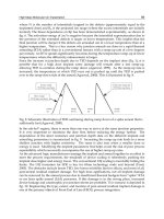
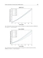
As figure 13 shows, the bacterial count of sub-eschar
viable tissues in groups 1 and 4 increased progressively
during the whole course of the disease. The bacterial
count in groups 2 and 3 remained at low levels, less than
10
5
/g throughout, and even declined, indicating that both
these topical drugs were effective in controlling the prolif-
eration of P. aeruginosa.
Results of Blood Culture
The incidence of positive blood cultures in groups 2
and 3 was markedly lower than in groups 1 and 4 (p !
0.005), as is shown in table 33. There was no significant
difference of positive rates between groups 1 and 4 (p 1
0.50), or between groups 2 and 3 (p 1 0.75).
Pathological Examination
In the study, grades of ‘0’ and ‘I’ in the pathological
examination were referred to as negative while grade ‘II’
was referred to as positive [13] (table 33).
As can be seen in table 33, positive rates of pathologi-
cal examination in groups 2 and 3 were significantly lower
than those in groups 1 and 4 (p ! 0.005). There was no
significant difference of positive rates either between
groups 1 and 4 (p 1 0.50), or between groups 2 and 3 (p 1
0.50).
Comparison of Incidence of Invasive Infection of Burns
Wounds
According to the data that bacterial invasion to viable
tissue of wound and/or bloodstream in the circulation
were indicative of invasive infection of burns wounds
[13], table 34 shows the incidence of invasive infection in
each group. It was found that the incidences of invasive
infection in groups 2 and 3 were dramatically lower than
those in groups 1 and 4 (p ! 0.005). There was no signifi-
cant difference of positive rates either between groups 1
and 4 (p 1 0.25) or between groups 2 and 3 (p 1 0.50).
Fig. 13. Illustration of the correlation between bacterial count in sub-
eschar viable tissues and the clinical course in each group.
Table 32. Mean logarithmic values of bacterial count of sub-eschar
viable tissues (mean B SE)
Group Bacterial count
(logarithmic value)
1 (control) 5.8B2.6
2 (MEBO) 3.8B2.0
3 (SD-Ag) 3.1B3.1
4 (hot dry-exposed) 5.4B2.0
Table 33. Results of blood culture and pathological examination
Group Positive
number
Negative
number
Positive
rate,%
Blood culture
1 (control) 25 5 88.33
2 (MEBO) 7 23 23.33
3 (SD-Ag) 8 22 26.67
4 (hot dry-exposed) 19 11 63.33
Total 59 61 49.17
Pathological examination
1 (control) 21 9 70.0
2 (MEBO) 11 19 36.67
3 (SD-Ag) 12 18 40.0
4 (hot dry-exposed) 20 10 66.67
Total 64 56 53.33
Table 34. Incidence of wound invasive infection
Group Positive
number
Negative
number
Positive
rate, %
1 (control) 26 4 86.67
2 (MEBO) 12 18 40.0
3 (SD-Ag) 11 19 36.67
4 (hot dry-exposed) 23 7 76.67
Total 72 48 60.0
72 Burns Regenerative Medicine and Therapy
Table 35. Results of bacterial count of sub-eschar viable tissues and
pathological examination
Bacterial
count
Pathological examination
positive
number
negative
number
total
! 10
2
3 19 22
10
2
3 12 15
10
3
10 4 14
10
4
253
10
5
8513
10
6
11 1 12
10
7
10 5 15
1 10
8
17 5 22
Total 72 48 60
Table 36. Results of positive bacterial count and positive pathologi-
cal examination
Bacterial count 610
5
Pathological examination
positive
number
negative
number
total
Positive number 46 16 62
Negative number 18 40 58
Total 64 56 120
Coincidence rate 86/120 (71.67%)
Non-coincidence rate 34/120 (28.33%)
Comparison of Bacterial Count of Sub-Eschar Viable
Tissue and Pathological Examination for Diagnosis of
Burn Wound Infection
According to table 35, there was a direct correlation
between the positive rates of the bacterial count of sub-
eschar viable tissue and the pathological examination (r =
0.808, p ! 0.005). The positive rate of pathological exami-
nation increased as did the bacterial count.
In further analysis, we took a bacterial number of 10
5
/g
sub-eschar viable tissue, the level defining invasive infec-
tion, as the boundary for positive and negative [14, 15]. It
was found by comparison that positive rates of pathologi-
cal examination of tissue specimens that yielded counts of
10
5
/g sub-eschar viable tissue or more reached 74.2%.
The negative rate of pathological examination of those
yielding counts lower than 10
5
/g reached 69%. The coinci-
dence and noncoincidence rates of two diagnostic meth-
ods were 71.67 and 28.33%, respectively (table 36). Sta-
tistical analysis showed an obvious relationship between
both methods (¯
2
= 17.62, p ! 0.005) and there was no
significant difference between both methods in the diag-
nosis of burn wound infection (¯
2
= 0.031, p 1 0.75).
Conclusion
1 MEBO has a similar effect to SD-Ag in controlling
burn wound invasive infection by P. aeruginosa.
2 Hot dry-exposed burns therapy has no effect on con-
trolling third-degree burn wound invasive infection by
P. aeruginosa.
3 The bacterial count of sub-eschar viable tissue can still
be used as one of the feasible methods in the early diag-
nosis of burn wound invasive infection.
Discussion
Evaluation of Antibacterial Effect of Hot Dry-Exposed
Therapy
In 1949, Wallace introduced the concept of dry-
exposed burns therapy [16, 17] in which the wound was
directly exposed to air at a certain temperature. He be-
lieved that direct exposure of the wound might allow the
formation of a layer of dry eschar/crust by exudation and
necrotic tissue on the wound surface, which served as
a barrier against bacterial contamination. Our study
showed that comparing the hot dry-exposed therapy
group to the untreated control group, there was no ob-
vious difference with respect to bacterial count of sub-
eschar viable tissue, positive rate of blood culture and
positive rate of pathological examination. We now suggest
that the hot, dry-exposed therapy has no significant effect
on controlling invasive infection with P. aeruginosa of
third-degree burns wounds.
Moist-Exposed Burn Ointment (MEBO) –
New Topical Drug for Burns
Infection is the leading cause of death due to burns
complications, and burn wound infection is of great clini-
cal concern as it can result in burn wound sepsis and septi-
cemia. An enormous amount of research has been con-
ducted in this field which has produced many advances in
burn infection treatment. However, the very existence of
necrotic tissue in deep burns provides culture medium
conducive to the growth of pathological micro-organisms.
Furthermore, the blocked local blood circulation hinders
the delivery of anti-bacterial and immune-enhancing pep-
tides which are integral to host-defense competency.
Mafenide (Sulfamylon), silver nitrate and SD-Ag were
developed in the 1960s [2–4]. Sulfamylon is a useful
antimicrobial agent which penetrates into eschar but has
the disadvantage of inhibiting carbonic anhydrase. There-
fore, absorption of topical Sulfamylon may result in meta-
bolic acidosis that limits its use in larger burn areas. Silver
nitrate was the initial topical agent but its tendency to
stain discouraged widespread use. SD-Ag has a strong
antimicrobial effect which, despite its poor penetration
into eschar, made it the topical agent of choice against
burn infection. Our study gave good proof for this.
Experimental and Clinical Study on Burns Regenerative Medicine and Therapy with MEBT/MEBO 73
In order to improve the antimicrobial effect of a topi-
cal agent while reducing deleterious side effects, re-
searchers developed other metal sulfonamides such as
zinc, ammonium, cerium and erbium for topical therapy
[18–21]. However, a comparison of relative antimicrobial
effects showed SD-Ag to be the best of the lot so it
remained the agent of choice against P. aeruginosa. This
remained the case despite its worrisome profile of creat-
ing multidrug resistance [5, 6]. Great efforts have been
made to deal with P. aeruginosa resistance to SD-Ag. In
the 1970s, on the basis of nalidixic acid, great improve-
ments were attained in the research of pyridine, pefloxa-
cin and its derivative in the prevention and treatment of
burn infection [7, 8, 22]. Recently, silver norfloxacin has
emerged, which was found to be effective in the treatment
of P. aeruginosa with drug resistance to SD-Ag [9, 10].
In 1988, a new topical drug for burns wounds was
invented, called moist-exposed burn ointment (MEBO)
[11]. This innovation has become widely accepted in clin-
ical use [23–25]. In this study, animals with infection of
third-degree burns wounds by P. aeruginosa were used,
and the comparison showed that MEBO was effective in
controlling burn wound P. aeruginosa infection. MEBO
had a similar effect to SD-Ag in reducing the bacterial
concentration of sub-eschar viable tissues, positive rate of
blood culture and incidence of invasion infection. In
addition to its ability to kill P. aeruginosa, the other
advantages of MEBO are as follows: easy to apply; non-
painful, no need for excruciating debridement between
applications, and easy assessment of healing progression.
It suggested that MEBO was a useful alternative topical
drug for burn treatment. Further investigation is needed
in order to find whether MEBO controls the infection of
other bacteria and micro-organisms as well as the mecha-
nism of MEBO against P. aeruginosa.
Roles of Bacterial Count of Sub-Eschar Viable Tissue
and Pathological Examination in the Diagnosis of
Burn Wound Infection
Infection has long been one of major life-threatening
causes of burn victims. The extent of infection depends on
the invasiveness of the pathogenic micro-organism and
the power of host resistance [12]. Micro-organism inva-
siveness has a close correlation to the strains, toxicity and
quantity. Therefore, a variety of methods for determining
the bacterial count on burns wounds have been devel-
oped.
As early as 1964, Teplitz et al. [1] put forward the con-
cept of burn wound invasive infection. They defined
wound invasive infection as occurring when bacterial
count exceeded 10
5
organisms per gram sub-eschar viable
tissue with bacteria penetrating into the underlying tissue
and blood vessels. Many researches agree that a bacterial
count of 10
5
/g sub-eschar viable tissue was a pivotal level
with wounds containing more than 10
5
/g being predis-
posed to invasive infection [14, 15]. Therefore, the value
of 10
5
/g of viable tissue is used as one indicator to predict
and diagnose burn wound invasive infection. However, in
a comparative study between bacterial count of sub-
eschar viable tissue and pathological examination of 200
cases, McManus and Kim [26] found that only 35.7% of
tissue specimen with 610
5
/g eschar viable tissue demon-
strated invasive infection by pathological examination.
They concluded that the bacterial density level of 10
5
or
more organisms per gram sub-eschar viable tissue was not
a sufficient indicator for the diagnosis of burn wound
invasion.
In our study, we made a bacterial count on the sub-
eschar viable tissues and performed pathological exami-
nation of 120 animals with the results showing a linear
correlation between bacterial count and positive rate of
pathological examination. The positive rate of pathologi-
cal examination increased as bacterial density did, and
there was a positive relationship (p ! 0.005). Among 62
specimens showing bacterial count 610
5
/g, 46 were
found positive in pathological examination with a rate of
74.2%. Of 58 specimens showing bacterial count ! 10
5
/g,
40 were found to be negative in pathological examination
with a rate of 69%. The coincidence rate of both diagnos-
tic methods was 71.67%. The statistical data demon-
strated that if a bacterial count 610
5
/g was used as the
critical level in the diagnosis of burn wound invasive
infection, there was a significant relationship between the
two methods. Therefore, the results of this study suggest-
ed that the bacterial count on sub-eschar viable tissue
remained one of the feasible methods for the prediction
and diagnosis of burn wound invasive infection. Although
it directly reveals the invasive extent of burn wound inva-
sive infection, pathological examination can give false-
negative results due to the impact of many factors includ-
ing sampling, section and staining techniques. Thus, we
must keep in mind that pathological examination for the
diagnosis of burn wound invasive infection has its limita-
tions and should not be the sole criterion. Similarly, blood
culture has its limitations in that it may result in a low
positive rate and delay appearance [27]. Therefore, we
conclude that bacterial count of sub-eschar viable tissue
can still be used as one of the feasible methods in the early
diagnosis of burn wound invasive infection.
References
1 Teplitz C, Davis D, et al: Pseudomonas burn wound sepsis. I. Pathogenesis
of experimental pseudomonas burn wound sepsis. J Surg Res 1964;4:200.
2 Lindberg RB, et al: The successful control of burn wound sepsis. J Trauma
1965;5:601.
3 Moyer CA, et al: Treatment of large human burns with 0.5% silver nitrite
solution. Arch Surg 1966;90:812.
4 Fox CL Jr: Silver sulfadiazine. Addendum to local therapy of burns. Ritten-
burg MS, et al. Mod Treat 1967;4:1259.
74 Burns Regenerative Medicine and Therapy
5 Bridges K, et al: Drug resistance in relation to use of silver sulphadiazine
cream in a burn unit. J Clin Pathol 1977;30:160.
6 Heggers JP, et al: The emergence of silver sulphadiazine resistant Pseudo-
monas aeruginosa. Burns 1978;5:184.
7 Ge SD, et al: Experimental study of topical chemotherapy in prevention
and treatment of burn infection. Acad J Sec Military Med Coll 1982;3:46.
8 Ge SD, et al: Experimental study of topical antimicrobial agent in burns.
Chin J Plast Surg Burns 1985;1:255.
9 Darrell RW, et al: Norfloxacin and silver norfloxacin in the treatment of
Pseudomonas corneal ulcer in the rabbit. Trans Am Ophthalmol Soc 1984;
82:75.
10 Ge SD, et al: The effect of pyridonic acid derivatives as topical antibiotics
on the prevention and treatment of pyocyaneous infection following burn.
Chin J Plast Surg Burns 1987;3:10.
11 Xu RX: The clinical application of moist exposed burn therapy. Chin J
Integr Tradition West Med 1988;8:204.
12 Krizek TJ, Robson MC: Evolution of quantitative bacteriology in wound
management. Am J Surg 1975;130:579.
13 Yang ZJ, Xu WS, Shi JX: Burn Management, ed 2. Beijing, People’s Health
Press, 1985.
14 Volence FJ, Clark GM, et al: Burn wound biopsy bacterial quantitation: A
statistical analysis. Am J Surg 1979;138:695.
15 Loebel EC, Marvin JA, et al: The method of quantitative burn wound biop-
sy cultures and its routine use in the care of the burned patient. Am J Clin
Pathol 1974;61:20.
16 Wallace AB: Treatment of burn, a return to basic principles. Br J Plast Surg
1949;1:232.
17 Wallace AB: The exposure treatment of burns. Lancet 1951;i: 501.
18 Fox CL Jr, et al: Metal sulfonamides as antibacterial agents in topical thera-
py. Scand J Plast Reconstr Surg 1977;13:89.
19 Monafo WW, et al: Control of infection in major burn wounds by cerium
nitrate/silver sulfadiazine. Burns 1977;3:104.
20 Fox CL Jr, et al: Topical chemotherapy for burns using cerium salts and
silver sulfadiazine. SGO 1977;104:668.
21 Ge SD, et al: N
1
-metal sulfa drugs and zinepalyanemine and its derivatives
in prevention of burn wound sepsis. Chin J Surg 1982;20:264.
22 Modak SM, et al: Control of burn wound infection by pefloxacin and deriv-
ative. Burns 1984;10:170.
23 Zhang LX, Yang KF: Clinical study report: 2076 cases treated with moist
exposed burn therapy. Chin J Burns Wounds Ulcers 1989;1:22.
24 Zhao RY, Tian LX, Zhao GX: Clinical analysis: 50 cases treated with moist
exposed burn therapy. Chin J Burns Wounds Ulcers 1989;1:32.
25 Ma EQ, Huang XY, et al: Clinical report of 69 cases treated with moist
exposed burn ointment. Chin J Burns Wounds Ulcers 1990;2:25.
26 McManus AT, Kim SH: Comparison of quantitative microbiology and his-
topathology in divided burn wound biopsy specimens. Arch Surg 1987;
122:74.
27 Baxter CR, Curreri PW, et al: The control of burn wound sepsis by the use
of quantitative bacteriologic studies and sub-eschar lysis with antibiotics.
Surg Clin North Am 1973;53:1509.
Experimental Research on the Mechanism
of the Anti-Infection Effect of BRT with
MEBT/MEBO
Introduction
Many basic studies and clinical research have proved
that BRT with MEBT/MEBO has beneficial effects of
anti-infection, promoting burn wound healing and reduc-
ing scar formation to name but a few of its attributes.
However, the mechanisms of these effects of MEBO
remain a bit mysterious. The following study was de-
signed to elucidate the anti-infection effect mechanism of
BRT with MEBT/MEBO.
Materials and Method
Apparatus and Reagents
Fully automatic enzyme labeling analyzer, Multiskan, MS; AC-
920 hemocyte analyzer; MEBO; RPMI-1640, Gibco, USA; Con A,
supplied from Guangzhou Medical Institute; MTT supplied from
Fluka; Pathogenic O
111
B
4
of Escherichia coli supplied from Binzhou
Medical College.
Animals and Methods
Thirty-two BALB/C mice, 8 weeks old, females, weighing 17–
19 g, were supplied from Beijing Medical University. Twenty-eight
Kunming mice of both sexes weighing 21–24 g, 18 rabbits weighing
2–2.5 kg, and 4 guinea pigs weighing 250–300 g were supplied from
Binzhou Medical College.
The animals were divided randomly into a control group and a
MEBO treatment group. In the MEBO treatment group, mice were
depilated (2 ! 2 cm) on their backs and smeared with MEBO. Guin-
ea pigs were depilated (3 ! 2.5 cm) on their backs and smeared with
MEBO. MEBO was applied 2 times a day. Biopsy of skin tissue was
done on day 9 to determine the activity of interleukin-1 (IL-1), and
was stained with hematoxylin and eosin (HE) for histopathological
observation.
Examination Indexes
Morphological Variation of Pathogenic E. coli (O
111
B
4
). The bac-
teria were cultivated on culture medium containing a given concen-
tration of MEBO [1]. Each generation of the bacteria was stained
using the G staining method. Staining reaction and morphological
variation of the bacteria were observed. The results were compared
with that of the E. coli communis cultured on the same kind of
medium containing MEBO.
Determination of IL-1 Level. MTT was used as a substrate. Since
living cells containing active succinate dehydrogenase can reduce
yellow-colored MTT to form violet or blue-colored formazam par-
ticles, the particles were dissolved by adding isopropyl alcohol hy-
drochloride [2]. The OD value was determined by colorimetry. The
amount of formazam was proportional to the number of living cells.
Determination was performed according to the modified methods
described previously [2]. Mice were killed under sterile conditions,
the thymus was removed, cut and screened. After being centrifuged,
a suspension with 10
7
/ml of cells containing 2 Ìg/ml of Con A was
prepared. On a 24-pore plate, we added to each pore 0.5 ml of this
suspension and then added mice skin tissue suspension and blood
plasma at 0.5 ml/pore. Two more control groups, one with 1640-
nutrient fluid (thymocytes plus nutrients) and the other with Con A
plus thymocytes were set. The 24-pore plate was placed into an incu-
bator at 37
°
C, 5% CO
2
for 68 h. From each pore, 0.5 ml of the
supernatant was drawn away, then 0.1 ml of 5 Ìg/ml MTT solution
was added to each pore and incubated again for 4 h. 0.5 ml of isopro-
pyl alcohol hydrochloride was added to each pore and then the plate
was shaken to dissolve the particles. Supernatant from each pore was
drawn and added to a 96-pore plate, 0.2 ml/pore. The plate was
placed into a fully automatic enzyme-labeling analyzer to determine
the OD value, reflecting the level of IL-1 in skin tissue cells and
blood plasma.
Effect of MEBO on Body Temperature of the Rabbit. The body
temperature of the rabbit was taken prior to application of MEBO.
Then, an area of 6.0 ! 6.5 cm was depilated on the back of the
animal and MEBO was applied twice a day while body temperature
was recorded simultaneously. This was done for a total of six mea-
surements. Changes in body temperature were recorded.
Effect of MEBO on the Classification of Mice Leukocytes. Every
mouse in the MEBO and control groups had a 0.2-ml blood sample
taken by enucleation of the eyeball. The blood was added to 10 ml of