Burns Regenerative Medicine and Therapy - part 6 potx
Bạn đang xem bản rút gọn của tài liệu. Xem và tải ngay bản đầy đủ của tài liệu tại đây (448.2 KB, 16 trang )
Experimental and Clinical Study on Burns Regenerative Medicine and Therapy with MEBT/MEBO 75
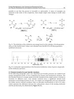
Fig. 14. a Bacillus proteus: Morphological variation of long rod. Light
microscope. !1,000. b Bacillus proteus: Morphological variation of
long rod. Electron microscope. !6,500. c The same as b but under
the electron microscope, nucleoplasm. !13,000.
the reagent solution and then examined using an AC-920 hemocyte
analyzer.
Assay of Humoral Immunologic Function. A quantitative hemo-
lysis spectrophotometry (QHS) method was used to determine the
amount of hemoglobin released after hemolysis of RBC mediated by
antibody-forming cells [3]. This amount (expressed as OD value)
reflected the amount of antibody-forming cells in mice, thus indicat-
ing the humoral immunologic function of the mice.
Table 37. Morphological variation of E. coli communis and patho-
genic E. coli
E. coli communis Pathogenic E. coli
Generation (50% MEBO) 1–6 7–9 10–12 1–3 4–6 7–10
Variation + ++ +++ + +++ ++++
Form of G
–
bacillus basically normal ‘+’; a little longer (like diplo-
bacillus) ‘++’; became larger like round ball ‘+++’; shape was normal,
but had deep particles in the bacteria ‘++++’.
Table 38. Effect of MEBO on classification of leukocytes (mean B
SE)
Group Animal
number
Lymphocytes, % Neutrophils, %
Control 8 69.94B3.35 30.06B3.35
MEBO 8 49.59B4.50 50.41B4.50
p value ! 0.01
Assay of Cellular Immunologic Function. Blood samples were tak-
en from mice tails in the MEBO and the control groups, respectively,
then smeared and stained according to the ·-naphthalene acetate
esterase (ANAE) method. These were then examined under the
microscope. 100 lymphocytes were observed randomly. The percent-
age of ANAE-positive lymphocytes reflects the cellular immunologic
function of the body.
Histological Changes of Mice Skin after Treatment with MEBO.
0.5-cm
2
skin tissue samples of mice taken from depilated areas of
normal skin and skin treated with MEBO were fixed, embedded,
stained with hematoxylin and eosin and then observed under the
light microscope.
Results
Anti-Infection Effect of MEBO
Morphological Variation of Bacteria. The results of
morphological variation of E. coli communis and patho-
genic E. coli cultured in medium with MEBO are shown
in figure 14 and table 37. These reveal that MEBO acts to
induce the variation of both E. coli communis (which is
common in burns wounds), and pathogenic E. coli. How-
ever, the variation of different bacteria might occur at dif-
ferent times.
Effect of MEBO on the Classification of Circulating
Leukocytes. Table 38 shows that the amount of neutrophil
in blood increased after MEBO treatment.
76 Burns Regenerative Medicine and Therapy
Table 39. Effect of MEBO on rabbit body temperature (
°
C, n = 6,
average)
Days after MEBO treatment
01357
Body temperature,
°
C 38.63 39.05 39.31 39.43 39.47
Average elevation of body
temperature,
°
C 0.84
Table 40. Effect of MEBO on the production of IL-1 in mouse skin
tissue cells (mean B SE)
Group IL-1 in skin tissue cells
animals OD
IL-1 in blood plasma
animals OD
Control 8 0.043B0.019 4 0.429B0.171
MEBO 8 0.142B0.039 4 0.733B0.105
p value ! 0.01 ! 0.05
Table 41. Effect of MEBO on specific immunologic function of mice
(mean B SE)
Group ANAE positive rate, %
(n = 6)
QHS
(n = 8)
Control 58.83B10.61 1.296B0.021
MEBO 54.17B10.23 1.317B0.027
p value 1 0.05 1 0.05
Effect of MEBO on Rabbit Body Temperature. 75% of
the rabbits had an increase in body temperature after
MEBO treatment. On day 7, the average elevation of body
temperature was 0.84
°
C (table 39).
Effects of MEBO on Wound Healing and Scar
Formation
Effect of MEBO on the Production of IL-1 in Mouse
Skin Tissue Cells. It was found that MEBO was effective
in inducing synthesis of IL-1 from IL-1-delivering cells of
the skin. IL-1 is capable of promoting the proliferation of
thymocytes and has a synergistic action with Con A. The
IL-1 levels in both skin tissue and blood plasma of the
MEBO treatment group were significantly higher than
those of the control group (table 40).
MEBO Promoted Proliferation of Skin Cells and Cells
at the Margin of the Sebaceous Gland. In this study, we
found that the number of skin basal cells in the division
phase was increased, and the number of juvenile flat cells
at the margin of the sebaceous gland was also increased in
the MEBO group. This finding indicates that the metabo-
lism of the cells was vigorous.
Effect of MEBO on the Specific Immunologic Function
of Mice
Table 41 shows that MEBO did not affect the cellular
and humoral immunologic functions.
Conclusion
The results suggested that: (1) MEBO can prevent
infection; (2) the wound-healing benefit and the reduction
of scar formation in the MEBO group were related to its
effect of increasing the production of IL-1 by skin cells.
Discussion
Clinical practice has strongly proved that MEBO
has anti-infection, pain-killing, wound-healing-promot-
ing and scar-forming-reducing effects. Based on our
study, we discussed the mechanisms of its actions as the
following.
Anti-Infection Effect of MEBO
MEBO has potent ability to control wound infection
and keep the wound moist but not macerated. The active
ingredients in MEBO ointment and its unique dosage
delivery system create an environment hostile to bacterial
growth. In culture medium containing MEBO, morpho-
logical structural and physiological variations of the bac-
teria occurred. MEBO affected the synthesis of the com-
ponents for formation of a bacterial wall and inhibited
related enzymes. Also, the synthesis of DNA was inhibit-
ed and the bacteria proliferation rate was decreased. Deep
pigments were found in the bacteria, this indicated that
the bacteria were in a stable phase of proliferation, during
which a high level of glycogen, lipid, etc. was stored in the
bacteria.
MEBO induced morphological and physiological vari-
ations in bacteria while influencing the production of
plasma coagulase of Staphylococcus aureus [4]. Bacterial
pathogenicity is related to the bacterial wall component
and thus MEBO reduced the pathogenicity of the bacte-
ria. The bacterial variation characteristics were varied in
different species of bacteria and at different MEBO con-
centrations. The initiation time of the variation was not
standard.
In clinical care, we found that after treatment with
MEBO, the body temperature of burn patients might rise
Experimental and Clinical Study on Burns Regenerative Medicine and Therapy with MEBT/MEBO 77
by 1–1.5
°
C during the initial stage. At 3 h after applica-
tion of MEBO, the temperature of burn patients with
superficial second-degree wounds rose. In this study, the
body temperature of rabbits increased after application of
MEBO, and rose by 0.84
°
C on day 7. Furthermore,
MEBO induced the production of IL-1, from IL-1-deliver-
ing skin cells, such as epidemic cells, keratinocyte, and
Langerhans cells. In the early 1940s, people recognized
that certain extracts from acute inflammatory foci could
cause fever after injection into the body. This type of
inflammatory substance is called endogenous pyrogen
(EP). In 1979, purified EP was first proved to have IL-1
activity, and EP and IL-1 were considered the same mole-
cule [5]. MEBO stimulates local skin cells to produce IL-1,
which in turn was absorbed into the systemic circulation
thereby affecting the temperature-regulating center lead-
ing to elevated body temperature.
The effect of fever on body resistance of mammals is
not clear yet. Some researchers think that fever may pro-
mote the immunity of the host. When body temperature is
raised, but still ! 41
°
C, the phagocytic power of most
phagocytes is enhanced. We also found that MEBO can
promote phagocytic power of abdominal cavity macro-
phages in mice [6]. In this study, the quantity of neutro-
phils in blood circulation was significantly increased after
treated with MEBO. Bone marrow stimulated by IL-1
may account for this interesting finding.
MEBO Promotes Wound Healing and Reduces Scar
Formation
Clinical data revealed that after treatment with MEBO,
patients with superficial or deep second-degree burns
wounds healed with full epithelization; superficial third-
degree burns wounds healed with a mild scarring that
appeared smooth and soft. In this study, we observed that
the production of IL-1 was increased in mice skin and sub-
dermal tissues after treatment with MEBO. The difference
between the MEBO group and the control group was very
significant. Besides macrophages, many other tissue cells
when stimulated can produce IL-1 in 1 h [9]. IL-1, IL-8 and
tumor necrosis factor (TNF) are cellular factors which are
capable of activating and inducing differentiation of T and
B lymphocytes, enhancing the activities of monocytes, NK
cells and killer cells, thus stimulating lysosomal enzyme
activity and phagocytic activity of neutrophils.
Recently, both animal experiments and clinical prac-
tice proved that IL-1 does induce a series of pathophysio-
logical changes. These changes are similar to the host’s
response to infection [6], indicating that IL-1 is an impor-
tant regulatory factor of the body as regards inflammation
and immunologic reaction. We must differentiate be-
tween the local effect of IL-1 and the effect of high lev-
els of IL-1 systemically. These are two totally different
concepts [6]. In this study, the level of IL-1 in mouse skin
and extracellular subdermal tissues in the MEBO treat-
ment group were significantly different from those of the
control group, as well as the IL-1 level in blood plasma.
IL-1 is closely related to wound healing and we know that
wound exudate typically contains IL-1. IL-1 promotes
proliferation of fibroblast and secretion of collagenase [6].
The effect of IL-1 is complicated. It induces inflammation
and fever while at the same time promoting wound heal-
ing. Inflammation induced by IL-1 is understood to be a
kind of host defense reaction [7]. After mice were treated
with MEBO, their skin basal cell in division stage in-
creased. Juvenile cells observed around the sebaceous
gland were very metabolically active. This proves that
MEBO promotes wound healing.
Effect of MEBO on Specific Immune Function
In this study, MEBO could increase the quantity of
neutrophils in blood, while relatively decreasing the num-
ber of lymphocytes. In addition, MEBO did not affect the
cellular and humoral immunologic function.
References
1 Qu YY, et al: Experimental research on the anti-infective mechanism of
MEBO. Chin J Burns Wounds Surface Ulcers 1996;40:19–23.
2 Mosmann T: Rapid colorimetric assay for cellular growth and survival:
Application to proliferation and cytotoxicity assays. J Immunol Methods
1983;65:55.
3 Bi AH (ed): Medical Immunology. Tongji, Tongji Medical University,
1986, p 7.
4 Yu H: Medical Microbiology. Beijing, People’s Health Publishing House,
1983, p 54.
5 Yang GZ (ed): Outline and Technology of Immunology and Bioengineer-
ing. Jilind, Jilin Science & Technology Press, 1991, pp 2–15.
6 Chen WJ (ed): Molecular and Cell Biology of Blood. Beijing, Chinese Med-
ical Science & Technology Press, 1993, vol 126–127, p 362.
7 Yang GZ, et al: Progress in genetic engineering and clinical immunology;
in: Domestic and International Progress in Medical Sciences. Shanghai,
Shanghai Institute of Medical Science & Technology Information, 1987,
p 124.
Primary Exploration on the Mechanism of the
Anti-Infection Effect of BRT with MEBT/MEBO
Introduction
Clinical practice has revealed that MEBO has a re-
markable anti-infection effect [1]. During the period May
to June 1992, fourteen burns cases were treated in the
Burns Department, Affiliated Hospital of this College,
and bacteria isolated from the burns wounds were exam-
ined. We found that Bacillus proteus had the morphologi-
cal Hauch-ohne Hauch (H-O) variation, and the plasma
coagulation ability of S. aureus decreased. In order to
investigate the mechanism of the anti-infection effect of
MEBO, we studied the biological variability of some com-
mon bacteria, such as Bacillus proteus, P. aeruginosa,
78 Burns Regenerative Medicine and Therapy
E. coli and S. aureus, cultured in medium containing a
certain amount of MEBO. The effect of MEBO on non-
specific immunity in vivo was also observed.
Materials and Methods
Clinical Data
During the period from May to June 1992, 14 cases of burns were
treated with MEBO (hospitalized for 4–20 days). Swab samples were
taken from the upper and lower (contact with wound) layers of the
MEBO ointment, before changing the dressing. Bacteria were iso-
lated and cultured.
Reagents, Bacterium Species and Culture
Antibiotic sensitivity test paper and nutrient agar were purchased
from Shanghai Medical Chemistry Institute. B. proteus, P. aerugino-
sa, E. coli and S. aureus were prepared in our department.
The four above bacteria were cultured on ordinary culture me-
dium and medium containing different concentrations of MEBO,
respectively, and continuously transferred to 10–15 generations.
Each generation of the bacteria was checked. The biological charac-
teristic and drug sensitivity of bacteria were examined.
Animal Experiment
Forty healthy adult mice of both sexes weighing 20–24 g were
randomly divided into 3 groups, i.e. blank control group (group 1),
liquid paraffin control group (group 2) and MEBO group (group 3).
Animals in groups 2 and 3 were depilated (2.5 ! 2.5 cm) on their
backs and liquid paraffin or MEBO was applied on the depilated
area. The frequency was twice a day for 10 successive days. On the
11th day, the mice were sacrificed and abdominal cavity fluids were
sampled 30 min after intraperitoneal injection of 0.5 ml of 2% sheep
erythrocytes.
Observation Indexes
Bacteria Variation. The four bacteria were cultured and trans-
formed, 18–24 h as one generation. The dynamics of every genera-
tion of the bacteria was observed under a dark-field microscope. The
bacteria were stained with the G method to observe the staining reac-
tion and morphological characteristic. The colony features, biochem-
ical reactions, and the ability of plasma coagulation were also exam-
ined.
Nucleoplasm Staining. The 7th generation of B. proteus was
smeared, fixed with the vapor of 1% molybdic acid solution, hydro-
lyzed DNA with 1% hydrochloric acid for 15 min, then stained with
Löffler’s methylene blue and observed.
Antibiotic Drug Sensitivity Test. The four bacteria were cultured
on ordinary agar medium and medium containing 20% of MEBO,
respectively. Drug sensitivity test paper of eight kinds of antibiotics,
i.e. gentamicin, neomycin, erythromycin, carbenicillin, ampicillin,
kanamycin, chloromycetin and polymyxin, were stuck on the me-
dium, respectively, and observed after 24 h. The diameter of the bac-
terial inhibition zone was measured [2].
Determination of Phagocytic Function of the Intraperitoneal
Phagocytes. The abdominal cavity fluids were smeared and stained
with Wright’s method. The percentage of phagocytes was determined
[3].
Determination of Lysozyme Activity in Abdominal Cavity Fluid
and Serum. The agar plate method was used: 2 ml of Micrococcus
solution (9 ! 10
10
/ml) were added to 1% agar at 70
°
C, then after
mixing well, this was poured into a Petri dish. After cooling, holes
(3 mm in diameter) were bored into the substance. Mouse serum
from 3 groups was added to the holes in one set of Petri dishes and
abdominal cavity fluid was added to the holes in another set of Petri
dishes. These were incubated at 37
°
C for 24 h. The diameter of the
bacteriolytic ring was measured [4].
Total Number and Classification of Leukocytes in Peripheral
Blood. Blood samples from the tails were taken from 3 groups of
mice. Leukocytes were counted, stained with Wright’s method and
classification was determined.
Results
Species and Variation of Bacteria Isolated from Burns
Wounds
Table 42 shows that after treatment with MEBO, B.
proteus in the wounds had H-O morphological variation.
S. aureus in the wounds turned from positive plasma co-
Table 42. Species and variation of bacteria
isolated from burns wounds
1
Case No. Bacteria from upper layer of MEBO Bacteria from lower layer of MEBO
1 S. aureus; P. aeruginosa P. aeruginosa
2 Staphylococcus albus; E. coli E. coli
3 S. albus (negative coagulase) S. albus (negative coagulase)
4 S. aureus (positive coagulase) S. aureus (coagulating ability decreased)
5 B. proteus B. proteus (H-O morphological variation)
6 S. aureus (positive coagulase) S. aureus (negative coagulase)
7 S. aureus; E. coli E. coli
8 S. aureus (positive coagulase) S. aureus (positive coagulase)
9 B. amotile B. amotile
10 B. amotile B. amotile
11 P. aeruginosa P. aeruginosa
12 S. albus S. albus
13 P. aeruginosa P. aeruginosa
14 G
+
diplococcus G
+
diplococcus
1
Samples collected on days 4–20 after MEBO treatment.
Experimental and Clinical Study on Burns Regenerative Medicine and Therapy with MEBT/MEBO 79
agulase to negative, or the plasma coagulating ability de-
creased.
Bacteriostasis of MEBO
Eight species of bacteria, i.e. S. aureus, S. albus, E. coli,
B. proteus, P. aeruginosa, B. typhosus, B. paratyphoid A
and B. dysenteriae, were cultured in simple agar dishes.
The scraps of MEBO filter paper were pasted on the bac-
terial surface of streak plating. The results showed that
MEBO had no direct bacteriostasis or bactericidal ac-
tion.
Effect of MEBO on Bacterial Biological
Characteristics
Effect of MEBO on B. proteus Biological Features. B.
proteus was cultured for several generations on medium
containing certain amounts of MEBO. We noted that the
motility of the bacteria gradually decreased before finally
vanishing. Also, we noted that H-O variation occurred.
The 7th generation of the bacteria became long and fila-
mentous. In culture medium containing 25% MEBO,
90% of the bacteria became long filamentous or long rod,
and then became small bacillus. Dark pigments ap-
peared, colonies became small and the bacteria grew very
slowly. The decomposition activity of the bacteria to glu-
cose and lactic acid was retarded (table 43; fig. 15). The
effect of MEBO on H antigen of B. proteus is shown in
table 44.
Fig. 15. a Normal appearance of E. coli. b Appearance of variant
E. coli cultured in medium containing MEBO for 6 generations.
Table 43. Effect of MEBO on B. proteus biological features
Medium Motility
G 1 G 2 G 3 G 4–10
Morphology
G 1–2 G 3–4 G 5–8 G 9–10
H-O variation
G 1 G 2 G 3–4 G 5–10
H
2
S test
G 1–7 G 8-10
50% MEBO + B – – + + ++ +++ + + – – + retarded
decomposition
25% MEBO + BB– +++++*+++ +++– +retarded
decomposition
+ = Motile; B = weak motility; – = no motility.
+ = Typical G
–
bacillus; ++ = long rod or filament; +++ = G
–
with dark pigment.
+ Colonial migration: + = 1–3 cm; – = no migration.
G = Generation; * 90% of the 7th generation of the bacteria became long filamentous.
Table 44. Serological test of B. proteus
cultured in medium containing MEBO
Original
bacteria
Medium containing MEBO, 12th generation
10% MEBO 25% MEBO 50% MEBO
B. proteus 1:1,280 1:640 1:640 1:320
H antiserum ++ ++ ++ ++
80 Burns Regenerative Medicine and Therapy
Effect of MEBO on the Biological Characteristic of
P. aeruginosa. It was found that P. aeruginosa cultured in
MEBO-containing medium started to decrease its motili-
ty from the 5th generation, and motility vanished in the
10th generation. Variations of morphology and coloniza-
tion features of the bacteria were also found (table 45).
Effect of MEBO on the Biological Characteristics of
E. coli. From table 46, we can see that after proliferation
to the 10th generation while cultured in MEBO contain-
ing medium, E. coli changed as follows: it lost motility,
became sphere shaped, colonies became smaller, dry and
flat (fig. 16). The decomposition activity of the bacteria to
glucose and lactose was retarded (after 32 h).
Table 45. The effect of MEBO on the biological characteristic of P. aeruginosa
Medium Motility
G 1–4 G 5–9 G 10–12
Morphology
G 1–4 G 5–9 G 10–12
Colonial feature
G 1–5 G 6–9 G 10–12
Oxidase test
G 1–12
MEBO 50% + B – + ++ +++ + ++ ++
x
+
MEBO 25% + B – + ++ +++ + + ++ +
+ = Typical G
–
bacillus; ++ = a few became long rod or diplococcus; +++ = deep pigment appeared.
+ = Colony moderate size and smooth; ++ = colony smaller and dry; ++
x
= pigment formed but not good.
G = Generation.
Table 46. Effect of MEBO on the biological characteristic of E. coli
Medium Motility
G 1–3 G 4–9 G 10–12
Morphology
G 1–6 G 7–9 G 10–12
Colony feature
G 1–3 G 4–8 G 9–12
Fermentation test
glucose
G 1–9 G 10–12
lactose
G 1–9 G 10–12
MEBO 50% + B – + ++ +++ + ++ +++ + retarded
decomposition
+ retarded
decomposition
MEBO 25% + B – + ++ +++ + ++ +++ + retarded
decomposition
+ retarded
decomposition
+ = Typical G
–
bacillus; ++ = long rod (like diplococcus); +++ = bacteria swelling (sphere shaped).
+ = Colony medium size and smooth; ++ = colony medium size, a little flat; +++ = colony small, rough and flat.
G = Generation.
Table 47. Effect of MEBO on the biological characteristic of S. aureus
Medium Morphology
G 1–7 G 8–11 G 12–15
Colony feature
G 1–7 G 8–11 G 12–15
Plasma coagulase test
G 1–7 G 8–12 G 13–15
Manicol test
G 1–11 G 12–15
MEBO 50% + ++ +++ + ++ +++ ++ + B + retarded
decomposition
MEBO 25% + ++ ++ + ++ ++ ++ + + + +
+ = G
+
arranged in grape shape; ++ = part of the bacteria became diplococcus-like or arranged in short chain;
+++ = piled up in grape shape and had scattered diplococcus-like and short chain arrangement.
+ = Colony medium size and smooth; ++ = colony small and slightly flat; +++ = colony smaller, flat and dry.
Plasma coagulase test: ++ = Fluid was clear and obviously coagulated; + = fluid turbid and small coagulate;
B = fluid turbid, small and few coagulate.
G = Generation.
Experimental and Clinical Study on Burns Regenerative Medicine and Therapy with MEBT/MEBO 81
Effect of MEBO on the Biological Characteristics of
S. aureus. We found that after 10 generations of S. aureus
cultured in medium containing MEBO, bacteria were
piled up in grape shape and had scattered diplococcus-like
and short chains. Colonies became smaller, flat and dry.
The decomposition activity of the bacteria to mannitol
was retarded (after 32 h), and the variation of plasma
coagulation ability was very significant (table 47) [3].
Nucleoplasm Staining of B. proteus. Cultured in me-
dium containing 25% MEBO and proliferated to the 7th
generation, B. proteus appeared as a long filamentous
variant. Nucleoplasm staining was done and examined.
As the RNA in cytoplasm was hydrolyzed, the nucleo-
plasm was stained blue. The bacteria became long rod or
long filament in the course of binary division, because the
formation of cell wall was slower than the division of the
nucleoplasm. This result proved that bacteria prolifera-
tion was retarded when cultured in medium containing
MEBO.
Synergistic Effect of MEBO and Antibiotics on Bacte-
riostasis. When cultured in medium containing 20%
MEBO, P. aeruginosa resistant to carbenicillin and chlo-
romycetin became moderately sensitive and B. proteus
resistant to chloromycetin and ampicillin became sensi-
tive. Both carbenicillin and kanamycin had a synergistic
effect with MEBO against E. coli. Carbenicillin, ampicil-
lin, kanamycin and erythromycin had a synergistic effect
with MEBO against S. aureus (table 48).
Fig. 16. a Normal appearance of B. proteus. b Appearance of variant
B. proteus cultured in medium containing MEBO for 2 generations.
Table 48. Synergistic bacteriostasis of MEBO and antibiotics
Culture
medium
Carbenicillin
DR MS S
Ampicillin
DR S
Kanamycin
MS S
Erythromycin
S
Chloramphenicol
DR MS S
Ordinary
S. aureus 30211720
P. aeruginosa 16 12
E. coli 12 21
B. proteus 512
Containing 20% MEBO
S. aureus 38302025
P. aeruginosa 20 16
E. coli 22 25
B. proteus 20 20
DR = Drug-resistant; MS = moderate sensitivity; S = sensitivity.
82 Burns Regenerative Medicine and Therapy
Table 49. Effect of MEBO on peripheral
blood leukocytes (mean B SE)
Group Animals WRC, 10
9
/l PMN, %
Blank control 8 6.87B0.85 26.0B1.53
Vaseline control 8 6.93B1.22 28.3B3.86
MEBO 8 8.75B0.91 48.5B2.56
p value ! 0.01 ! 0.01
Table 50. Effect of MEBO on phagocytic
function and lysozyme activity
(mean B SE)
Group Animals Phagocytosis
%
Lysozyme diameter, cm
abdominal cavity fluid serum
Blank control 8 54.23B6.20 1.70B0.16 2.08B0.17
MEBO 8 65.50B4.18 2.33B0.38 2.10B0.11
p value !0.05 ! 0.05 1 0.05
Effect of MEBO on Nonspecific Immunity
Effect on Peripheral Blood Leukocytes. MEBO signifi-
cantly increased the peripheral blood count of white blood
cells and polymorphonuclear leukocytes (PMN%) in mice
(table 49).
Effect on Phagocytic Function and Lysozyme Activity.
MEBO significantly promoted the phagocytic function of
phagocytes and increased the lysozyme activity in the
abdominal cavity fluid (table 50).
Conclusion
MEBO can increase the number of WBC in the periph-
eral blood of mice. It can also enhance the function of
phagocytes in the abdominal cavity of mice. MEBO can
induce a variation of bacteria and improve nonspecific
immunity.
Discussion
Bacterial inhibitory test proved that MEBO does not
have a direct bacteriostatic or bacteriocidal effect. This
finding may be understood in part due to the oily texture
of MEBO making it very difficult to infiltrate and diffuse
into a watery culture medium. After continuous culture
in medium containing MEBO, many species of bacteria
showed variations in morphological structure and biolog-
ical characteristics that are closely related with the
growth environment. The morphological variation of
bacteria may cause changes in its biochemical character-
istics, antigenicity and toxicity. B. proteus and P. aerugi-
nosa had deep pigmented particles and E. coli became
sphere-shaped after culture in medium containing
MEBO.
These variations are non-genetic. MEBO had a syner-
gistic bacteriostatic effect with antibiotics. This is benefi-
cial to the control of local and systemic infections second-
ary to severe burns. MEBO promoted the phagocytic
function of abdominal cavity phagocytes and release of
lysozymes, and increased the leukocyte and neutrophil
counts in the peripheral blood. This is very important for
clearing out the bacteria and toxins both locally and sys-
temically. In summary, the mechanism of the anti-infec-
tive effect of MEBO includes inducing variation of the
bacteria, decreasing their proliferation rate, reducing bac-
terial pathogenicity and promoting nonspecific immunity
of the body.
References
1 Zhang LX, Yang KF: Clinical report of 2076 burn cases treated with moist
exposed burn therapy. Chin J Burns Wounds Surface Ulcers 1989;1:22–
26.
2 Shanghai Medical Laboratory: Test of Sensitivity to Antibiotics. 1983.
3 Yu H (ed): Medical Microbiology. Beijing, People’s Health Publishing
House, 1983, vol 13–28, p 239.
4 Wang MX (ed): Medical Microbiology and Immunology. Beijing, People’s
Health Publishing House, 1989, p 116.
Experimental Research on the Anti-Anaerobic
and Anti-Fungal Effect of MEBO
Introduction
Clinical research data of BRT with MEBT/MEBO re-
vealed that MEBO has a strong ability to retard wound
infection. Its mechanism is myriad [1]. This paper reports
the effect of MEBO on the morphological structure, colo-
Experimental and Clinical Study on Burns Regenerative Medicine and Therapy with MEBT/MEBO 83
ny character and pathogenicity of anaerobic spore bearing
bacillus (Bacillus tetani), anaerobic non-spore-bearing ba-
cillus (Bacteroides fragilis, Propionibacterium acnes) and
fungi (Candida albicans). MEBO has been proven to pos-
sess strong broad-spectrum antibacterial effects. MEBO
also creates an environment for preserving the residual
surviving cells in the burns area and to promote their pro-
liferation [2]. Thus, MEBO offers a dual regulatory ef-
fect.
Materials and Method
Materials
Aquarium Type B224 was designed by the laboratory of the Affil-
iated Hospital of Binzhou Medical College. Culture medium was
supplied from Shanghai Biological Preparation Institute. Bacteroides
fragilis and Propionibacterium acnes were purchased from Shanghai
Medical University; Bacillus tetani and Candida albicans from the
laboratory of the Affiliated Hospital of Binzhou Medical College.
These bacteria were cultured separately in anaerobic agar medium
for use.
Method
MEBO Group. The above-stated four species of bacteria were
inoculated separately into the medium containing a certain amount
of MEBO. Anaerobic bacteria were incubated at 37
°
C for 48–72 h as
one generation. Candida albicans was incubated at 37
°
C for 24–48 h
as one generation. After 4–6 successive generations, they were
treated with Gram stain and their staining reaction, morphology and
colony characteristics were observed.
Control Group. The above-stated original bacteria were observed
before being inoculated into the medium containing MEBO.
Examination Indexes
Variation of the Bacteria. (1) Bacteroids fragilis, Propionibacte-
rium acnes and Bacillus tetani were inoculated separately into the
medium containing MEBO and cultured for multiple generations
during which morphological and colony variations at each generation
were observed. (2) Variations of Candida albicans were observed
after culture in MEBO-containing media.
Spore Tube Test. Original Candida albicans and the 1st, 2nd, 5th
and 6th generations after being cultured in MEBO-containing media
were inoculated into 0.5 ml human serum medium and cultured at
37
°
C for 3 h, the fungi were smeared, stained and 500 counts of the
fungi were observed to determine the spore tube producing rates.
Effect of MEBO on Bacterial Growth
Equal amounts of the colonies of the 10th generation of Staphylo-
coccus aureus, and Bacillus pyocyaneus cultured in MEBO-contain-
ing media and the original bacteria of the two species were ground
and placed into 0.1 ml of saline, respectively, then 1 ml of saline was
added and the counts of the bacteria were compared.
Effect of MEBO on the Invasive Power of Bacillus pyocyaneus
The 10th generation of B. pyocyaneus cultured in MEBO-con-
taining media and the original B. pyocyaneus cultured in ordinary
media were taken and diluted separately to 3 ! 10
6
/ml. 0.1 ml of the
bacteria solutions were injected into mice intracutaneously. After
20 h the mice were killed. A block of rectangular skin tissue to the
muscular layer was taken from the injection site of each mouse. The
tissue blocks were made into sections and stained with HE and
observed.
Results
The effect of MEBO on anaerobic bacteria is given in
table 51 and depicted in figures 17–19. The effect of
MEBO on Candida albicans is given in table 52 and
depicted in figure 20. The effect of MEBO on the prolifer-
ation rates of Staphylococcus aureus and Pseudomonas
aeruginosa is given in table 53. The effect of MEBO on
the invasiveness of Pseudomonas aeruginosa is shown in
table 54.
Table 51. Effect of MEBO on anaerobic bacteria
(1) Bacillus tetani
Control group: G positive, slender bacillus, spores could be seen
occasionally on the top, bacteria in the shape of a
group of drumsticks, colonies grown in films
(fig. 17a)
MEBO group: 1, 2 generations: most of the bacteria were in a shape
of long rod or long filament
1
, a few had spores, colo-
nies were flat, rough and dry, none grown in films
(fig. 17b)
3, 4 generations: most of the bacteria were bacillus of
various length, many of them had spores. The bacte-
ria were in a shape of drumstick, a few were long
rods or long filaments, colonies were flat, rough and
dry (fig. 17c)
(2) Propionibacterium acne
Control group: G positive, non-spore-bearing bacillus, straight or
slightly crooked, colonies were small and round with
smooth surface (fig. 18b)
MEBO group: 1, 2 generations: basically the same as the control
3, 4 generations: bacillus of different length ap-
peared (like diplobacillus) with aggregation and con-
fluence, colonies were small, slightly flat, rough and
dry (fig. 18c)
5, 6 generations: most of them were small coccoba-
cillus with deep colored particulates, colonies were
small, slightly flat, rough and dry (fig. 18d)
(3 )Bacteroides fragilis
Control group: G positive, non-spore-bearing moderate bacillus,
with obtuse ends, colonies were a little convex with
smooth surface (fig. 18e)
MEBO group: 1, 2 generations: basically the same as the control
3, 4 generations: long bacillus (like diplobacillus)
and some coccobacillus appeared, colonies a little
flat with dry and rough surface (fig. 18f)
5, 6 generations: small coccus and coccobacillus
appeared; colonies aggregated to confluence to form
irregular round bodies; colonies were flat, dry and
rough (fig. 18g)
1
Cultured in MEBO-containing media, when MEBO concentra-
tion was 25%, the variation percentage of Bacillus tetani to form long
filaments was higher than that cultured in media containing higher
concentration of MEBO.
84 Burns Regenerative Medicine and Therapy
Fig. 17. a The normal Bacillus tetani showing slender rod-like shape.
b The 1–2 generations of Bacillus tetani in culture medium with
MEBO showing as a shape of long rod or long filament. c The 3–4
generations of Bacillus tetani in culture medium with MEBO show-
ing in various lengths, many of them having spores. The bacteria
were drumstick shaped, a few were long rods or long filaments.
Fig. 18. a The normal form of Bacteroides fragilis showing moderate
size. b The 3–4 generations of Bacteroides fragilis varied in length
(look like diplobacillus) and bacterial colonies fused together. c The
5–6 generations of Bacteroides fragilis in culture medium with
MEBO were sphere or egg shaped. Many bacterial colonies fused to
form irregular spheres.
Experimental and Clinical Study on Burns Regenerative Medicine and Therapy with MEBT/MEBO 85
Fig. 19. a The normal shape of Propionibacterium acne showing G
+
(some straight or slightly crooked). b The 3–4 generations of Propio-
nibacterium acne in culture medium with MEBO showing varied rod
length. c The 5–6 generations of Propionibacterium acne were small
coccobacilli with deep-colored particulates.
Table 52. Effect of MEBO on C. albicans
Control
The fungi were oval with spores and pseudohypha, colonies were mil-
ky with smooth and moisturized surface (fig. 19a)
Spore tube test: spore tube producing rate was 90% (fig. 19b)
MEBO
1, 2 generations: the same as the control, colonies were a little more
flat and small, compared to the control; spore tube producing rate
was about 85% (fig. 19c)
3, 4 generations: some of the fungi were sphere or oval of different
sizes, pseudohypha appeared; colonies were flat, dry and rough
(fig. 19d)
5, 6 generations: the fungi were sphere or oval, filaments with stick or
long rod shape and different length appeared; only few spores;
colonies were flat, dry and hard (fig. 19e)
Spore tube rate was about 0.5–2%
1
(fig. 19f)
1
Cultured in media with different concentrations of MEBO, their
spore-producing rates were different. In media containing 25% of
MEBO, the spore tube-producing rate was 2%, a little higher than in
that containing higher concentration of MEBO.
Table 53. Effect of MEBO on the proliferation of S. aureus and P.
aeruginosa
Bacteria Primary bacteria
cultured
counts/ml
10th generation after
cultured in MEBO
containing media
counts/ml
Staphylococcus aureus
1.4!10
8
1.5!10
6
Pseudomonas aeruginosa 2!10
8
6.5!10
6
Table 54. Effect of MEBO on the invasiveness of P. aeruginosa –
pathological examination
Control group
In subcutaneous tissue, there was congestion and edema, infiltration
of inflammatory cells and a suppurative zone
MEBO group
In subcutaneous tissue and striated muscles, there was infiltration of
a few inflammatory cells without suppurative phenomenon
86 Burns Regenerative Medicine and Therapy
Fig. 20. a The normal shape of Candida albicans showing egg shape
with many blastospores. b The germ tube tests on normal Candida
albicans gave a producing rate of 90%. c The germ tube producing
rate of 1–2 generations of Candida albicans in culture medium with
MEBO was 85%. d The 3–4 generations of Candida albicans in cul-
ture medium with 25% MEBO showing large oral or few sphere-
shaped and stick- or filament-shaped ones being observed occasion-
ally. e The 5–6 generations of Candida albicans showing in shapes of
stick or long rod. Bacterial filaments in various length and few blas-
tospores being observed. f The germ tube producing rate of 5–6 gen-
erations of Candida albicans in cultured medium with MEBO was
0.5–2%.
Experimental and Clinical Study on Burns Regenerative Medicine and Therapy with MEBT/MEBO 87
Discussion
Micro-organisms proliferate rapidly and are according-
ly susceptible to unfavorable factors in their growing envi-
ronment. These changes in their environment may result
in variations of their characteristics including morpholog-
ical structure, culture feature, toxicity, antigenicity and
drug resistance, etc. Morphological structure variations
are directly related to cell division. Cell division is a com-
plicated process and is more susceptible to unfavorable
factors in the environment than to protein and DNA syn-
theses. For example, Bacillus tetani and Bactorides fragilis
may have polymorphic variations such as polynuclear
filaments or spheres because, in an unfavorable environ-
ment, cell membrane synthesis is delayed and cell divi-
sion cannot proceed in a timely manner, while metabo-
lism proceeds normally. Variation is closely related to the
effect of the strength and the duration of action of the
unfavorable factors. In our experiment, we found that the
same bacteria demonstrated variation in morphological
features when cultured in media containing different con-
centrations of MEBO (data not shown).
In this experiment, we observed the antibacterial effect
of MEBO on Bacillus tetani, Propionibacterium acnes and
Candida albicans. These micro-organisms are normal flo-
ra and opportunistic pathogens in the human body. Un-
der certain conditions they become pathogenic and, in
most clinical cases, the infections are endogenous in etiol-
ogy. Burns patients often receive large doses of antibiot-
ics, which always create an imbalance in native flora
thereby facilitating the overgrowth of opportunistic
pathogens such as Candida albicans. Aerobic bacteria
infections such as Staphylococcus aureus, E. coli and
B. pyogeneus predominate in burns wounds though anaer-
obic bacteria infections may also occur since burns
wounds may become ischemic and necrotic. Therefore,
bacterial examination of wounds should always consider
both aerobic and anaerobic bacteria. In 1983, Wang
Dewang reported a positive anaerobic bacteria detection
rate of 23.9% in 34 cases of burns and most of them had
mixed infections.
Bacillus tetani is an anaerobic spore-bearing bacteria.
After being cultured in MEBO-containing media, long
filament variants appeared, and in 3 generations many of
them formed only one spore. This spore is not the vegeta-
tive form of the bacteria as it cannot proliferate under
normal conditions. Spore keeps the bacteria alive though
dormant. Only under favorable conditions can the spore
proliferate and produce exotoxin to cause disease. After
culture in MEBO-containing media for 6 generations, in
Candida albicans the number of blastospores was signifi-
cantly reduced, the spore tube producing rate was only
0.5–2%, while the normal fungi had a spore tube produc-
ing rate of 90%. The above facts prove that MEBO has an
inhibitory effect on the proliferation and pathogenicity of
Bacillus tetani and Candida albicans.
From table 53, we can see the effect on Bacillus pyocya-
neus. After this baceria was cultured in MEBO-containing
media for 10 generations, we again cultured the 10th gen-
eration for another 20 h and discovered that the bacteria
count/ml was greatly reduced to only 1/30 of that of the
original. The invasiveness of this organism to mice was
also greatly decreased. This proved that under an unfa-
vorable environment, not only morphological structures
but also physiological features of the bacteria are subject
to variations.
Our data as well as many experimental and clinical
data reported by other workers proved that MEBO has a
broad-spectrum antibacterial effect and can also promote
wound healing. Therefore, MEBO is a drug with a dual
regulatory effect.
References
1 Yang K, Ma J, Yang Q, et al: A comprehensive report on the therapeutic
effect of MEBO in treating 4954 cases of various wounds and ulcers. Chin J
Burns Wounds Surface Ulcers 1997;4:15–21.
2 Xu R: China Burns and Wounds Medicine. Burns Wounds Surface Ulcers,
1997, pp 137–140.
88 Burns Regenerative Medicine and Therapy
OOOOOOOOOOOOOOO OOOOOOOOOOOOOOOOOOOOOOOOOOOOOO OOOOOOOOOOOOOOOOOOOOOOOOOOO OOOOOOOOOOO OOOOOOOOOO O OOOOO O OOOO O OOOOO O OOOOOO OOOOOO
Studies on the Effects of BRT with MEBT/MEBO on
Regeneration and Healing of Burns Wounds
A Comparative Study of Fibronectin and
Moist-Exposed Burns Ointment (MEBO) in the
Treatment of Experimental Corneal Alkali Burns
in Rabbits
Introduction
Worldwide reports regarding the basic science and
clinical applications of fibronectin’s (FN) contribution to
the healing of trauma and burns wounds have shown that
FN can enhance the epithelial healing rate by promoting
the adhesion and migration of epithelial cells at the
wound site [1–3]. Clinical application of eye drops made
of FN in treating burned cornea has obtained satisfactory
results. However, we are still years from the widespread
use of FN because the extraction and production of FN
eye drops are time-consuming, expensive and offer spe-
cific preservation challenges. We have used MEBO since
1990 in the treatment of burned cornea and we have
achieved satisfactory results in terms of improving cor-
neal nutrition, promoting wound healing, ameliorating
pain, relieving irritation and reducing the incidence of
corneal ulceration. Therefore, we embarked on an experi-
mental study to verify the effect of MEBO as compared
with FN in the management of corneal burns in rabbits.
Materials and Methods
MEBO, developed by Beijing Guangming Chinese Medicine
Institute for Burns, Wounds and Ulcers, is an ointment containing
sesame oil, beeswax and other active ingredients derived from plants
such as Cortex phellodendri and Radix scutellariae. FN produced by
the Shanghai Institute of Biological Products was diluted to a solu-
tion containing 400 Ìg/ml with sterile normal saline.
Five healthy New Zealand white rabbits (body weight 2–3 kg)
without eye disease were anesthetized intramuscularly with 20 mg/kg
sodium thiopental and eye solution of 0.4% Novesine two drops in
each eye. 8-mm filter papers, previously soaked completely in 0.5 N
NaOH, were laid on the middle of both corneas, respectively. One
minute later, the paper was removed and the eyes were rinsed imme-
diately with sterile normal saline. Then eye drops made of FN were
administered to the right eyes, four times daily and MEBO was
applied on the left eyes, three times daily. Both courses lasted 2
weeks.
At 6, 24, 36 and 48 h postinjury, the corneal fluorescent staining
zones were photographed with a DF Haiou camera (China) at fixed
focus. After developing the film, we drew the fluorescent staining
zones, analyzed and measured the areas at different hours using a
computer image pattern analyzer, and then calculated the epithelial
healing rate (mm
2
/h) by linear regression. Observations were made
twice daily on days 3–14 postinjury and fluorescent staining zones
were regarded as a positive indicator. Epithelial damage rate = (fluo-
rescent staining positive number)/(number of eyes examined)
!100%.
Besides corneal fluorescent staining zone, conjunctival conges-
tion and corneal transparency were also observed. (1) Conjunctival
congestion: + = palpebral conjunctival congestion; ++ = palpebral
conjunctival and partial bulbar conjunctival congestion; +++ = whole
palpebral and bulbar conjunctival congestion. (2) Corneal transpar-
ency: + = slightly opacity with distinct structure of underlying iridial
texture; ++ = moderate opacity with blurred structure of iridial tex-
ture; +++ = severe corneal opacity with indistinct structure of iridial
texture.
Results
In the early stage (at 6 h postinjury) epithelial healing
was slow and at 6–48 h the healing rate remained con-
stant. The use of FN on eyes shows an epithelial heal-
ing rate of 1.279 B 0.317 mm
2
/h compared to 1.285 B
0.128 mm
2
/h with the MEBO treatment. There is no sig-
nificant difference between the two groups although in the
MEBO group treatment efficacy is a little faster.
Repeated corneal damage occurred from the early
healing stage until 2 weeks postinjury and there is a signif-
icant difference in damage rate in both groups: 58.3% in
the FN group and 33.3% in the MEBO group (u = 5.56,
p ! 0.01).
The cornea showed disk opacity a few minutes follow-
ing burn. Conjunctival congestion with white secretion
was observed at 3 h postinjury. On day 2 postinjury, eye
irritation became obvious with increased secretion and
corneal edema was noted until 1 week postinjury when
edema was still observed in epithelium and matrix. At 2
weeks postinjury, the above symptoms were further re-
lieved. The advantages of MEBO treatment as compared
to FN in the management of burns eyes were demon-
strated both in terms of corneal epithelial exfoliation and
regarding conjunctival congestion and opacity.
Conclusion
Some advantages of MEBO include inexpensive cost,
convenient application, safety and product stability. The
effect of MEBO in the treatment of alkali-burned corneas
compared favorably to that achieved with FN. MEBO
proved more effective than FN in reducing the epithelial
damage rate and was far easier to use given a widespread
application. We suggest that MEBO treatment is remark-
ably advantageous in reducing the repeated epithelial
damage rate of alkali burns eyes compared to FN treat-
ment.
Experimental and Clinical Study on Burns Regenerative Medicine and Therapy with MEBT/MEBO 89
Discussion
It has been reported that FN has the effect of promot-
ing epithelial healing rate on corneal defects [4]. This
study verified no significant difference in promoting cor-
neal epithelial healing between the two groups. However,
MEBO treatment is remarkably advantageous in reducing
the repeated epithelial damage rate of alkali burns eyes
compared to FN treatment. The repair of relative integri-
ty of the corneal epithelium is beneficial to the stability of
the corneal parenchyma and endothelium. The cornea in
the MEBO group showed slight transparency with slight
congestion. Subsequent to alkali burns, the integrity of
basement membrane underlying corneal epithelium was
damaged. The appropriate replacement of exogenous FN
may reduce the possibility of epithelium damage, as FN is
needed for the repair of basement membrane. This study
demonstrated that MEBO is superior to FN with regard to
firm adherence of corneal epithelium and maintenance
and basement membrane integrity. In addition, MEBO
supplies rich nutrients necessary for repairing and regen-
eration of alkali burns corneas.
FN is a kind of macromolecular glucoprotein which
operates at the wound surface. The surge of enzymes
released subsequent to alkali burn causes the degradation
of FN at the wound surface. The paucity of FN resulting
from enzymatic degradation can be compensated with
exogenous FN, which reduces the epithelial damage rate
by promoting epithelium adhesion. Many combining sites
in FN molecular structure served as a bridge between epi-
thelial cells and basement membrane, thereby improving
epithelium adhesion. Affinity between FN and intracellu-
lar actin may cause the change of epithelial intracellular
actin from sphere to fibriform and then induce cell migra-
tion.
As avascular tissue, the cornea receives the nutrition
and oxygen necessary for metabolism mainly from diffu-
sion of the vascular net in the corneal limbus, from tears
and from aqueous humor. Glucose is the main source of
energy for the oxygenic metabolism of corneal epithelium
and for intraparenchymatous anaerobic metabolism [5].
MEBO contains abundant glucose, organic acid, a variety
of vitamins, proteins and enzymes as well, all of which
directly provide energy and nutrition for alkali-burned
corneal tissue. In this manner, MEBO serves to promote
metabolism of the cornea, to accelerate the prompt re-
moval of necrotic tissue and to facilitate swift growth of
new epithelium. MEBO also has anti-inflammatory prop-
erties, bacterial inhibition, repercussive and analgesic ef-
fects [6]. By relieving local congestion and corneal irrita-
tion, blepharospasm due to pains and nictitation were
alleviated, which reduced the friction to corneal surface
and also improved local resistance.
References
1 Fujikawa LS, Foster CS, Harrist TJ, Lanigan JM, Colvin RB, et al: Fibro-
nectin in healing rabbit corneal wounds. Lab Invest 1981;45:120–129.
2 Nishida T, Nakagawa S, Awata T, Nishibayashi C, Manabe R: Rapid prep-
aration of purified autologous fibronectin eyedrops from patient’s plasma.
Jpn J Ophthalmol 1982;26:410.
3 Shi SM, et al: Comparative physical properties of ophthalmic viscoelastic
materials. J Pract Ophthalmol 1989;7:9.
4 Nishida T, et al: Fibronectin enhancement of corneal epithelial wound
healing of rabbits in vivo. Arch Ophthalmol 1984;102:455.
5 Liu JQ, et al (eds): Practical Ophthalmology. Beijing, People’s Health
Press, 1984, p 43.
6 Xu RX: The medicine of burn and ulcer: A general introduction. Chin J
Burns Wounds Surface Ulcers 1989;1:11.
A Comparative Study of the Effects of
Moist-Exposed Burns Ointment (MEBO) and
Other Drugs on the Healing Rate of Corneal
Epithelial Defect in Rabbits
Introduction
Corneal epithelium consists of three types of cells at
three different levels. They are basal cells in the deep layer,
wing cells in the middle layer and squamous cells at the cor-
neal surface. Microvillus in the superficial layer absorbs
tears to form a tear membrane. Basement membrane be-
hind the deep layer is located in the corneal Bowman mem-
brane (anterior limiting lamina). Corneal epithelial layer is
a natural barrier against micro-organism invasion. Once
injured, it is prone to infection and shedding. Some corneal
diseases such as dendritic keratitis, neuroparalytic kerati-
tis, exfoliation of recurrent corneal epithelium, alkali
burns, vesicular keratitis, etc., and surgeries such as scrap-
ing the corneal epithelium for intraocular examination and
operation when performing cutting of the vitreous and re-
pair for retinal detachment, may injure corneal epithelium.
Rapid and complete repair of corneal epithelial defects
plays a very crucial role for the recovery of corneal physio-
logical function and for good eyesight. Corneal physiologi-
cal function contributes greatly to the promotion of deep
corneal wound healing and formation of collagenous fiber
[1]. In this study, rabbits with corneal epithelial defects of
the same size were used and treated with MEBO, homolog-
ous serum, 0.5% dexamethasone, 25,000 U/ml vitamin A.
In addition, the rabbits wore soft corneal contact lenses to
observe the regenerating rate of corneal epithelia and to
assess the therapeutic effects of different measures.
Materials and Methods
Eighty-two healthy adult rabbits of either sex weighting 2–3 kg
were anesthetized by 35% urethane 2.5 ml/kg intravenously and, if
required, by inhalation of ether. Then 1% dicaine was dropped twice
for surface anesthesia into the palpebral fissure which was expanded
with eyelid retractor. The cornea center was constrictively marked at
90 Burns Regenerative Medicine and Therapy
Table 55. Average healing rate of corneal epithelium after injury
(mm
2
/h, mean B SE)
Groups Number
of eyes
Average healing rate
Group 1 (MEBO) 10 1.184B0.106
Group 2 (dexamethasone) 11 1.087B0.087
a
Group 3 (vitamin A) 11 1.065B0.066
b
Group 4 (homologous serum) 10 1.114B0.038
e
Group 5 (saline) 10 1.037B0.059
b, c, f
Group 6 (contact lens) 10 0.770B0.016
b, d, e, f, g
a
p ! 0.05,
b
p ! 0.01 compared with group 1;
c
p ! 0.05,
d
p ! 0.01
compared with group 2;
e
p ! 0.05 compared with group 3;
f
p ! 0.01
compared with group 4;
g
p ! 0.05 compared with group 5.
surface using an 8-mm corneal trephine and was stained with 1%
fluorescein followed by rinsing with normal saline. Subsequent to the
appearance of a circular mark, an 8-mm stained disk area of epithe-
lial defect was created with a lancet scraping off the whole epithelium
within cycle and by fluorescein staining.
The animals were then divided randomly into 6 groups, 12 in
each. In group 1, the animals were treated with MEBO every 6 h. In
group 2 to group 5, the animals were given eye drops with 0.5% dexa-
methasone, 25,000 U/ml vitamin A, homologous serum and normal
saline, respectively, every 2 h. In group 6, the animals wore soft cor-
neal contact lens without any eye drops, whereby 6 eyes was removed
of lens every 6 h for fluorescein staining and other 6 eyes stained 24 h
later. In addition, 10 rabbits had their secretions cleaned away with
cotton sticks soaked with normal saline and no eye drops were
applied. Corneas were removed at 0, 2, 4, 6, 12, 18, 24, 30, 36 and
48 h for microscopic observation on corneal epithelial healing [2].
Results
The observation record showed the following mean
healing time: 48.6 h in the normal saline group, 47.36 h in
the vitamin A group, 45.18 h in the homologous serum
group, 42.78 h in the MEBO group, 46.5 h in the dexa-
methasone group and 65.3 h in contact lens group. The
results revealed that there was no significant difference of
average healing rate between groups 1 and 4, as well as
groups 2 and 4, groups 2 and 3, groups 3 and 5. However,
there were statistical differences between other each two
groups. MEBO, homologous serum, vitamin A, and dexa-
methasone had superior healing effects compared with
normal saline, while wearing of contact lens retarded the
healing of the defect. Beside homologous serum, MEBO is
remarkably superior to other drugs in promoting corneal
epithelial healing rate (table 55).
Discussion
Cornea has sources of nutrition from the vascular net
in corneal limbus, tears and aqueous humor. Corneal epi-
thelium is a functional barrier between tear membrane
and intraocular tissues through which the fluid output
from the stroma is regulated in order to keep the stroma in
a normal hydration. A corneal epithelial defect caused by
injury or scraping off can repair rapidly in two stages. The
latent period comes first, with a mean time of 5.5 B 0.3 h
during which extensive cellular and subcellular changes at
the wound edges are expressed by desquamation of super-
ficial cells, by loss of columnar appearance of basal cells,
by damage on hemidesmosome link of the basement
membrane as well as by formation of a cell process, indi-
cating that residual viable epithelial cells are transforming
into functional cells. A healing period is followed after-
wards when epithelial cells around the wound migrate
towards the center at a constant pace without mitochysis.
This process starts from the uninjured epithelial cells
adjacent to wound edge. We see basal cells in particular
being enlarged and flatted with pseudopodium migrating
towards the center and becoming the migratory edge of
monolayer cells, which is followed by two or more layers
of epithelial cells. The migration stops as wounds com-
pletely close and a firm hemidesmosome adhesion to the
basement membrane is re-formed. At this point, mitochy-
sis begins and mature corneal stratified epithelium is
finalized.
The experiment revealed us the two periods for epithe-
lial healing. At 2 h postsurgery, the defects did not de-
crease, instead, some increased from 8 to 8.1–8.5 mm,
which may have resulted from the contraction of tissues at
the wound edge and the shedding of injured cells. It was
not until 6 h postsurgery that epithelial defects began to
diminish progressively until complete wound healing was
achieved. Systemic or local administration and either
intra- or extra-ocular agents may be factors contributive
to the successful repairing process of corneal epithelial
defects from the latent period till the end of the healing
period. By means of smearing, eye drops or wearing of
corneal contact lenses, the study investigated various fac-
tors relative to epithelial healing in order to verify the
therapeutic effects of methods and drugs.
MEBO is an ointment containing sesame oil, beeswax
and other active ingredients extracted from plants such as
Cortex phellodendri or Radix scutellariae. It is highly
effective in the treatment of burns, wounds and ulcers and
its advantages include superior analgesic effects, anti-
inflammation, infection prevention and minimal scar for-
mation post healing [3]. Although recent reports prove
MEBO is significantly superior to other therapies in treat-
ing various burns, wounds and ulcers [4], there is no com-
parative study on the management of eye injury reported.
Therefore, we conducted a preliminary comparative
study on fibronectin (FN) and MEBO in the treatment of
experimental corneal alkali burns in rabbits, which veri-
fied that MEBO was remarkably effective in the treat-
ment of burns on avascular corneal tissue [2, 5]. Further,