Cleft Lip and Palate - part 3 potx
Bạn đang xem bản rút gọn của tài liệu. Xem và tải ngay bản đầy đủ của tài liệu tại đây (18.17 MB, 79 trang )
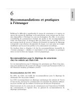
Chapter 6C
Complete Bilateral Cleft Lip and Palate 135
Fig. 6C.28. (continued) By 8-3 the anterior overjet is markedly reduced by growth. 13-11 Conventional orthodontics were eventu-
ally used to reduce the Class II occlusion and align the anterior teeth into an ideal overbite-overjet relationship
136
S. Berkowitz
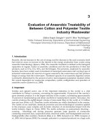
Fig. 6C.29. Case PM (KK-22). Computer-generated outlines of
the serial casts were performed using an electromechanical dig-
itizer.All casts are drawn to scale. The casts range from 12days
to 17years, 4 months of age. This series demonstrates the spon-
taneous closure of the anterior and posterior cleft spaces after
“molding action” brought on by uniting the lip and then by
gradual palatal growth at the border of the cleft space. The pre-
maxilla was initially aligned forward of the lateral palatal seg-
ments but was satisfactorily incorporated within the arch at a
later age
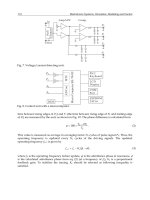
Table 6C.3. Surface Area of CBCLP.Case PM (KK-22).The palatal surface area increased by 4096 after 1 year,4 months and by 7696
at 2 years,1 month.By 8 years,2 months,the palatal surface area had increased two and a half times rohen the cleft space was closed.
Skeletal Area Cleft Space Total
Age Premax RLS LLS Tot Ant Post Tot SA + CS
0-0-12 145.5 335.5 282.7 763.7 127.4 417.0 544.4 1308.1
0-3 150.2 397.4 377.6 925.2 65.4 331.5 396.9 1322.1
1-4 154.0 469.5 464.3 1087.8 36.8 265.5 302.3 1390.1
1-10 211.8 502.6 506.2 1220.6 70.4 216.3 286.7 1507.3
2-1 217.6 589.0 549.5 1356.1 79.8 195.3 275.1 1631.2
2-10 220.7 603.9 551.3 1375.9 95.8 193.2 289.0 1664.9
3-10 271.6 660.4 616.5 1548.5 122.6 206.3 328.9 1877.4
5-8 273.3 673.0 675.9 1622.2 123.6 201.2 324.8 1947.0
6-7 273.6 811.0 820.5 1905.1 115.0 206.5 321.5 2226.6
7-4 277.3 813.0 839.5 1929.8 106.7 185.2 291.9 2221.7
8-2 306.5 844.6 890.8 2041.9 101.1 155.4 256.5 2298.4
12-3 346.8 1087.1 1116.1 2550.0 2550.0
14-0 348.7 1161.8 1226.4 2736.9 2736.9
14-5 351.1 1198.8 1237.0 2786.9 2786.9
17-4 353.5 1241.0 1246.3 2840.8 2840.8
Note: Premax = Premysxilla; RLS = Right Lateral Segment; LLS = Left Lateral Segment; Tot = Total Surface Area; Ant = Anterior
Cleft Space; Post = Posterior Cleft Space; Tot = Ant + Post; SA + SC = Bony Surface Area + Cleft Space Area; #: Changing teeth.
Chapter 6C
Complete Bilateral Cleft Lip and Palate 137
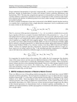
Fig. 6C.30. Case PM (KK-22). Palatal outlines were superim-
posed using the rugae for registration. This series shows that
the premaxilla’s position within the maxillary complex at
17 years of age is similar to that seen at birth. Excellent growth
occurs in all dimensions and is similar to the growth pattern
seen in noncleft palates. Increased posterior palatal growth is
necessary to accommodate the developing molars. Alveolar
bone growth with tooth eruption increases midfacial height.
Comments: The position of the anterior premaxilla relative to
the anterior cranial base (Nasion) to the anterior position of
pogonion of the mandibular symposium shows the same rela-
tive position from birth to 17years of age. These 2 studies con-
firm that midfacial growth is retarded
Fig. 6C.31. Case DK (AI-31). Premaxillary ventroflexion with
medial movement of the lateral palatal segments caused a great
reduction in the anterior and posterior cleft spaces by 5months
of age. Thereafter, for the next 19months, the anterior cleft
space gradually reduced,while the posterior cleft space showed
some increase due to the increase in palatal length. Both lateral
palatal segments showed a similar,gradually increasing growth
rate
138
S. Berkowitz
Fig. 6C.32. Case DK (AI-31). After the initial change in cleft
size brought on by medial movement of the lateral palatal seg-
ments and ventroflexion of the premaxilla, the greatest acceler-
ation of cleft space closure occurred between 2-10 and 3-5. The
premaxilla reached its largest size by 3 years, which is associat-
ed with eruption of the teeth. Palatal growth acceleration oc-
curred between 1 to 3 months and 12 to 14months and then
gradually tapered off. The palatal segments had increased 37%
in size by 1year and 74% by 2years. Palatal growth at its medi-
al borders still occurs; even though it narrows the cleft space,
the total cleft space is increasing in size due to the increase in
palatal length
Chapter 6C
Complete Bilateral Cleft Lip and Palate 139
Fig. 6C.33. Case PM (KK-22) Time sequence analysis of serial
palatal growth shows that both palatal segments are growing at
the same rate and to the same degree. The premaxilla is also in-
creasing in size with tooth eruption but at a lesser rate. The
greatest palatal growth acceleration occurs the first 2 years and
then tapers off. The anterior cleft space is initially reduced as a
result of premaxillary ventroflexion, but thereafter it remains
the same dimensions until the palatal cleft is closed. The poste-
rior cleft space initially is reduced with palatal medial move-
ment.The resulting posterior cleft space remains approximate-
ly the same size for the next 8years.It must be remembered that
the cleft length is increasing while the cleft width is decreasing.
The net cleft area is gradually reducing with growth.All fistulae
are closed by 12–3 years of age
140
S. Berkowitz
Fig. 6C.34. Serial midpalatal cross-sections showing vault
height and palatal width changes using stereophotogrammetry.
The left lateral palatal segment is attached to the vomer – while
the right lateral palatal segment is displaced laterally.A lip mus-
cle adhesion causes the displaced palatal segment to move me-
dially, narrowing the cleft space. The appositional growth of
the alveolar segments is continuous and becomes more obtuse.
Closure of the palatal cleft space with a modified vomer flap
maintains a normal vault height, and vault space flattening of
the vault occurs in almost all cases in the absence of a vomer
flap. Comments: A vomer flap with a von Langenbeck proce-
dure seems to create a minimum scarring. Vomer flap alone
performed early (6 months to 1year) created excessive scarring
Chapter 6C
Complete Bilateral Cleft Lip and Palate 141
Fig. 6C.35 a,b. Case PM (KK-22) a Cephalometric serial trac-
ings of the skeletal and soft tissue profile show marked reduc-
tion of the midfacial protrusion.
b Superimposed serial tracings
using Coben’s Basion Horizontal method show an excellent
facial growth pattern which straightens the skeletal profile.
There is very little forward midfacial growth between 11 and
20 years of age. During the same time period, growth at the
anterior cranial base and the mandible contributed to flattening
of the facial profile
a
b
Fig. 6C.36.
Case PM (KK-22). Cephaloradiographs taken at
5 years of age. Top : At rest with teeth together. Middle:Taken
while vocalizing “Youu . . .” Bottom: Taken while vocalizing
“Sss ”Comments:When vocalizing both sounds,the velum el-
evates and makes contact with the adenoids. The pharyngeal
depth is relatively small. The adenoids are of moderate size, the
velum is of good length and shows good elevation. This is not a
functional test to evaluate velopharyngeal closure but it does
show the well-proportioned oral and nasal pharyngeal spaces
which are conducive to good velopharyngeal closure. A gap
space more than 5mm may indicate VPI exist with inadequate
lateral pharyngeal wall movements
142
S. Berkowitz
ab
dc
e
Fig. 6C.37 a–e.
Various palatal expansion appliances. a “W”
appliance with finger springs designed to move the central in-
cisors forward while correcting the posterior crossbite.
b “W”
appliance.
c Arnold expander: a .040 wire is inserted into a
.040 tube; the compressed open coil spring exerts a gentle lat-
eral force moving the two segments apart. A larger diameter
(.045) tube wire allows the cuspids to be moved laterally more
than the molars.
d A Hyrax expander, which needs a lever
and parent involvement to activate the very strong expansion
force. This appliance is rarely necessary with meager
transpalatal scarring.
e A three-part removable expansion
plate used to simultaneously advance and expand the anterior
and buccal segments in a BCLP. Appliances that attach to the
teeth are more reliable and efficient than removable ones
Chapter 6C
Complete Bilateral Cleft Lip and Palate 143
Fig. 6C.38 a–s. Case ML (KK-56) demonstrates severe premax-
illary protrusion at birth in IBCLP. “Whisker” forked flap was
performed at 2months, definitive lip surgery at 6months, and
palatal cleft closure at 18 months. Secondary alveolar cranial
bone grafting was placed at 8years,3months.Maxillary surgery
with chin augmentation was performed at 15 years, 7 months.
a–g Facial and intraoral photographs show progressive facial
and occlusal changes
abc
d
fg
e
144
S. Berkowitz
Fig. 6C.38 a–s. (continued) h, i,and j Facial photographs at 8years of age. k After Lefort I advancement, the lateral incisor pontics
were attached to the arch wire for aesthetics.
l and m Occlusal photographs showing missing incisor spaces
hi j
k
l
mn
Chapter 6C
Complete Bilateral Cleft Lip and Palate 145
Fig. 6C.38 a–s. (continued) n, o,and p Intraoral photographs
showing retainer with lateral incisor pontics in place.
q, r,and s
Facial photographs at 17 years of age.Case ML (KK-56) demon-
strates severe premaxillary protrusion at birth in IBCLP.
h, i,
and
j Facial photographs at 8years of age. k After Lefort I ad-
vancement, the lateral incisor pontics were attached to the arch
wire for aesthetics.
l and m Occlusal photographs showing
missing incisor spaces.
n A partial upper retainer in place with
pontics for missing lateral incisors. Case ML (KK-56) demon-
strates severe premaxillary protrusion at birth in IBCLP.
o,and
p Intraoral photographs showing a fixed bridge to replace both
upper lateral incisors.
q, r,and s Facial photographs at 17years
of age. A prominent symphysis is noted. Comment: This case
shows the need to keep the lateral incisors spaces open in order
to obtain good anterior overbite and overjet in the presence of
strong mandibular growth
o p
qr s
146
S. Berkowitz
Fig. 6C.39. Case ML (KK-56). Serial casts from 0-0-2 to 4-3:
With the establishment of an intact lip musculature, the pre-
maxilla and lateral palatal segments molded into a good arch
form. The premaxilla, although latero- and ventroflexed, still
caused the upper lip to be pushed forward. The left buccal
crossbite was corrected by 8years of age and a fixed palatal
retainer placed
Chapter 6C
Complete Bilateral Cleft Lip and Palate 147
Fig. 6C.39. (continued) 15-7 Orthodontic treatment was de-
signed not to correct the slight Class II occlusion of the left seg-
ment. 15-9 After Lefort I osteotomy and final teeth alignment.
Because the premaxilla was positioned slightly to the right and
could not be centered orthodontically, it was decided to leave
the left occlusion in Class II and the right occlusion in Class I,
thereby equalizing the space for the lateral incisors. 17-0 A cus-
pid-to-cuspid fixed bridge replaced the missing lateral incisors
and stabilized the relationship of all segments
148
S. Berkowitz
Fig. 6C.40. Case ML (KK-56).Serial cephalo-
metric tracings showing well-proportioned
facial growth with a flattening of the facial
profile. The protrusive premaxilla was pres-
ent at 7-10. Orthodontia at 11years of age
improved the axial inclination of the maxil-
lary incisors. The profile at 15-3 is more at-
tractive than that at 17-3 as a result of the
chin augmentation at 15-7. The chin point is
too protrusive,resulting in a prominent sub-
labial fold
Fig. 6C.41. Case ML (KK-56). Superimposed polygons show an
excellent facial growth pattern flattened the profile by 15-3.
Midfacial osteotomy corrected the maxillary asymmetry.A chin
augmentation was performed at 15-7 years which created a
too prominent chin. The mandible continued to grow until
17-3 years of age,creating a slightly concave skeletal profile.The
patient is considering having the chin prominence reduced.
Comment: Rarely should a chin augmentation be performed
with a LeFort I advancement to avoid creating a “dished in”face
if the midfacial advancement relapses. Note the small forward
growth increments at the anterior cranial base and midface.The
midfacial changes did show good vertical growth to maintain
normal facial proportions
Chapter 6C
Complete Bilateral Cleft Lip and Palate 149
Fig. 6C.42. Case CW (BG-71).Serial casts demonstrate forward
advancement of the buccal segments to reduce a very large an-
terior palatal cleft space.There are instances when a marked os-
teogenic deficiency exists in both lateral incisor areas where the
premaxilla is in a slight overjet-overbite relationship and the
buccal segments in a good Class I relationship. In these cases,
there may be insufficient contiguous soft tissue to close the an-
terior palatal cleft space when performing a secondary alveolar
bone graft and create a normal site for tooth replacement. The
treatment of choice is to advance both buccal segments, simul-
taneously placing secondary alveolar bone grafts, yet leaving
space for the lateral incisors. After surgery the cuspids were to
return to their original Class I position.It was believed that with
early premaxillary setback there would be inadequate soft tis-
sue to obtain adequate cleft closure, but worst of all it would
have created a severely retruded midface which would have
required midfacial advancement
150
S. Berkowitz
Fig. 6C.42. (continued) 15-5 Good premaxillary relationship
with a large anterior palatal cleft space. Class I posterior occlu-
sion.Sectioned plaster casts with the posterior segments placed
in a Class II relationship. 16-3 and 17-2 Both buccal segments
relapsed into Class I. The main objective of closing the anterior
palatal cleft spaces was achieved. Final casts show a good Class
I occlusion with a satisfactory overjet-overbite relationship.The
anterior palatal cleft space was closed. However, the alveolar
bone graft did not take. In most cases the advanced lateral
palatal segments will remain forward with the cuspids in the
lateral space
Chapter 6C
Complete Bilateral Cleft Lip and Palate 151
Fig. 6C.43 a–z. Case CS (AF-48) demonstrates severe premaxil-
lary protrusion in a child with CBCLP at birth,resulting in max-
illary retrusion in adolescence with the eventual loss of the pre-
maxillary incisors. Lip adhesion was performed at 3months
with forked flap and posterior palate cleft closure at 3 years. No
secondary alveolar bone grafts were utilized. Premaxillary sur-
gical advancement at 15years to correct its retrusion. The pre-
maxillary incisors were extracted due to severe periodontal
bone loss, and the anterior palatal oronasal opening was closed
at 16years of age.
a and b Newborn. c–i Even with premaxillary
ventroflexion, the upper lip was still pushed forward
a bc d
e
ghi
f
152
S. Berkowitz
Fig. 6C.43 a–z. (continued) i–l A plastic obturator was utilized
at 6years of age to close the very large anterior palatal cleft
space to aid speech development and feeding.
m–o At 14years,
a retrusive looking midface with an extremely tight upper lip
and depressed nasal tip masked a good premaxillary overjet
j
m
pq r
no
kl
Chapter 6C
Complete Bilateral Cleft Lip and Palate 153
Fig. 6C.43 a–z. (continued) u At 15years following an unsuc-
cessful attempt to surgically center the premaxilla and close the
anterior cleft space. The maxillary incisor roots began to show
severe external root absorption.A very large anterior cleft space
remains.
v–y After lip and nose revision, the anterior teeth were
extracted and the oronasal opening closed with adjoining soft
tissue.
z A removable maxillary prosthesis replaced missing
teeth and bumpered the upper lip forward.Comments: It would
have been better treatment to have surgically advanced both
palatal segments to close the very large anterior palatal cleft
space. The root absorption was secondary to traumatic ortho-
dontics utilized to maintain the incisor overjet with a protective
facial mask and cross elastics
s tu
vw x
yz
154
S. Berkowitz
Fig. 6C.44. Case CS (AF-48). Serial casts show that the extreme premaxillary protrusion with large anterior cleft space at birth is
still present at 6-1 years of age
Chapter 6C
Complete Bilateral Cleft Lip and Palate 155
Fig. 6C.44. (continued) 11-6 With increased facial growth, the
increasing tonicity of the buccal muscle forces collapsed the
maxillary arch placing the posterior teeth in crossbite.The pre-
maxilla is now upright and in an acceptable overbite-overjet
relationship. 12-3 Maxillary expansion has been initiated. The
maxillary central incisors are in tip-to-tip relationship.14 Con-
tinued orthodontic treatment to advance the premaxilla and
position the incisor teeth in proper overjet-overbite relation-
ship
156
S. Berkowitz
Fig. 6C.44. (continued) 15-2, 16,and 16-6 Premaxillary reposi-
tioning with soft tissue closure of the anterior cleft space was
unsuccessful. As a result of the premaxillary central incisors
showing external root absorption and loss of periodontal sup-
port, they were extracted and the oronasal opening closed with
adjacent soft tissue. A removable maxillary prosthesis replaces
the missing teeth and bumpers the upper lip. Comment: As
already suggested, the treatment of choice is to reposition the
lateral palatal segments anteriorly while leaving the premaxilla
as is
Fig. 6C.45. Case CS (AF-48). Computerized tracings of serial
casts drawn to scale. This shows the lack of palatal growth and
reduction in cleft space over 2 years prior to surgical closure
of the palatal cleft. The anterior cleft space remains large up to
15-2 years. 16-9 and 17-8 The premaxillary incisors were ex-
tracted. Comment: This case clearly demonstrates the severe
degree of osteogenic deficiency that can exist in bilateral clefts
of the lip and palate. The once protruding premaxilla can
become retrusive with growth (time) and may eventually need
to be brought forward. Although palatal growth does occur, it
may not be sufficient to appreciably reduce the posterior cleft
space
Chapter 6C
Complete Bilateral Cleft Lip and Palate 157
Fig. 6C.46. Case CS (AF-48). The palatal segments show a very gradual growth acceleration curve, while the posterior cleft space
gradually reduces in size
158
S. Berkowitz
a
Fig. 6C.47 a,b.
Case CS (AF-48).a Serial cephalo-
metric tracings. This analysis show a protrusive
midface at 4-2 becoming recessive at 14-4.
b Seri-
al facial polygons superimposed according to
Basion Horizontal method (Coben). The mid-
face advanced only slightly after 6-4, while the
mandible showed progressive downward and for-
ward growth until 14-6, flattening the facial pro-
file. Comments: This case clearly shows (1) that,
even with a severely protruding premaxilla at
birth, the premaxillary incisors can be in anterior
crossbite after the pubertal growth spurt; (2)
traumatic orthodontic advancement of the pre-
maxillary incisors can lead to external root ab-
sorption and loss of alveolar support; and (3) sur-
gical advancement of one or both lateral palatal
segments, placing the cuspids in the lateral inci-
sor spaces,with secondary alveolar bone grafting
is the treatment of choice in cases when the pre-
maxilla is in good overjet-overbite relationship
and a large anterior cleft space exists. Only in
very rare instances should the premaxilla be sur-
gically set back to the lateral palatal segments. (4)
Premaxillary surgical setback is contraindicated
prior to the postpubertal facial growth spurt
b
Chapter 6C
Complete Bilateral Cleft Lip and Palate 159
Fig. 6C.48. Case ML demonstrates poor facial growth pattern
in a BCLP leading to a retruded midface with a severe anterior
open bite. This patient came to the clinic at 5years, 7months of
age with an anterior open bite.The upper lip was long and tight
with the lateral elements brought together below the prolabi-
um. The palatal cleft was closed at 12 months of age. At 13-10,
a removable plate replaced the right central incisor. At 15-11,
orthodontic preparation for Lefort I posterior-impaction. At
16-4, after maxillary surgery.At 19-9, after anterior fixed bridge
used to stabilize the palatal segments and replace missing teeth.
The changes in occlusion and total facial height led to a more
relaxed soft tissue profile. With reduction in lower vertical
facial height, the upper to lower lip position became more aes-
thetic