Recurrent Hernia Prevention and Treatment - part 9 ppt
Bạn đang xem bản rút gọn của tài liệu. Xem và tải ngay bản đầy đủ của tài liệu tại đây (770.92 KB, 41 trang )
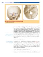
340 Treatment of Recurrent Inguinal Hernia
32
In the previous operation, the mesh had been placed
anterior to the posterior wall of the inguinal canal in
55 cases (59.8%) and in a preperitoneal position using
a posterior approach in 37 cases (40.2%). A Lichten-
stein repair as anterior onlay mesh had been carried
out in the majority of previous operations (56.5% or
n = 52). A previous endoscopic technique (13 TEP and
19 TAPP procedures) had been used in 34.8% of the
patients (n = 32).
There was a wide variety of reasons why a particular
operative procedure was chosen for the repair of a re-
current hernia. The ultimate decision as to which tech-
nique to use was made as late as during surgery in 71
cases (77.2%). In a mere 21 cases (22.8%), the surgeons
decided before surgery to perform either an endoscopic
procedure or a conventional ( Stoppa, Wantz) approach
(
⊡
Table 32.1).
After a previous anterior approach (Lichtenstein:
n = 52, plug and patch: n = 3), an anterior repair tech-
nique was again chosen in 24 cases. In 12 of these cases,
the surgeons used a Shouldice procedure or a direct
suture for the closure of small defects. The mesh was
removed in 8 of these cases. A Lichtenstein repair was
performed for the repair of both the previous and re-
current hernias in 10 cases (a larger medial overlap was
created in the majority of these cases). In one case, the
Lichtenstein technique was chosen after a previous
plug and patch repair. A total of 31 of the 55 patients
who had undergone a previous anterior repair had a
preperitoneal repair for a recurrent hernia. An endo-
scopic (TEP) approach was used in 7 of these cases
and a conventional TIPP repair was chosen in 15 cases
(6 meshes were removed). Last but not least, a Wantz
repair was performed in 5 cases and a Stoppa repair in
the remaining 4 cases
(
⊡
Table 32.1).
After a previous preperitoneal repair (32 endoscopic
TAPP or TEP procedures, 5 conventional Stoppa, Wantz
or TIPP procedures), the technique was changed and
an anterior placement of the mesh was chosen in 30
patients. A Lichtenstein repair (TAPP or TEP) was per-
formed in 15 of these cases, a Tipp repair in one case
and a direct suture or a Shouldice repair in another 15
cases. In six cases with a previous posterior repair, a
preperitoneal mesh was implanted again using a Stoppa
repair after a Wantz procedure in two cases, a TIPP
repair after a TEP procedure in one case, a Wantz repair
after a TAPP procedure in another case, a TAPP repair
after a Stoppa procedure in one case and a TAPP repair
was repeated in one case (
⊡
Table 32.1).
An analysis of the records showed that the decision
as to which repair technique to use was mostly made
on the basis of each individual case. In the majority of
cases, it is not possible to identify a definitive algorithm
for the selection of a technique. The following state-
ments can be made:
▬ There is a huge variety of previous techniques per-
formed for inguinal hernia repair.
▬ A transinguinal repair technique was usually used
for revision in patients presenting with pain and a
recurrent hernia.
▬ Where multiple recurrences could not be managed
using the commonly employed technique, a mini-
mal direct suture repair (either with or without the
placement of an additional small mesh) was used
0
year
10
8
14
18
2004
2000
2002
operation for recurrence [%]
0
20
40
100
mesh repair [%]
70
60
50
30
10
16
12
6
4
2
1998
1996
1994
1992
80
90
2006
2008
2010
operations for recurrent hernia (D)
to be expected
hernia repair with mesh
⊡
Fig. 32.1. In Germany, the mesh did
not eliminate recurrence as should be
expected
Schumpelick.indd 340Schumpelick.indd 340 05.04.2007 8:53:20 Uhr05.04.2007 8:53:20 Uhr
341
IX
Principle Actions for Re-Recurrences
for small defects or a preperitoneal (Wantz, Stoppa
or TAPP) approach was used for inserting a new
large mesh.
To follow up the patients, telephone interviews were
performed on the basis of a questionnaire in order to as-
sess the outcome of revision operations for recurrences
after previous mesh repairs (
⊡
Table 32.2). The mean
follow-up was 36.3 months (13 to 68 months; median:
33) or, in other words, slightly more than 3 years. It
was possible to conduct interviews with 87 of the 92
patients. One patient had died of another cause, but
had had no recurrence. Another 4 patients could not be
contacted. Accordingly, a follow-up rate of 94.6% was
achieved. Whereas 9 patients (10.3%) had undergone
surgery for a re-recurrence by the time of follow-up, all
other patients had had no recurrence. The re-operations
had been performed after an average of 19.9 months
(9–38 months) after the last repair. Only patients with
previous multiple recurrences were affected. Of the 26
patients who had undergone a non-mesh repair, 6 had
a recurrence. This group of patients showed the highest
re-operation rate (23.1%).
The surgical management of recurrent inguinal
hernia after a previous mesh repair is a technically
demanding challenge for a surgeon. Compared with
a suture repair, the mesh technique leads to consider-
ably more scarring, thereby making it usually much
⊡
Table 32.1. Repair techniques used in the previous and revision operations
Previous
operation
Revision operation Total
Shouldice
or suture
Lichten-
stein
TIPP TAPP TEP Wantz Stoppa
Lichten-
stein
12 10150744 52
TEP 14 18 110000 13
TAPP 10 17 101010 19
Wantz 11 10 100002 13
Stoppa 10 10 101000 11
Plug and
patch
11 11 100010 13
TIPP 10 11 100000 11
Total 28 27 16 2766 92
⊡
Table 32.2. High rate of re-recurrences following non-mesh repair after previous mesh repair
a
Re-recur-
rence
Suture Lichten-
stein
TEP TIPP TAPP Wantz Stoppa
No 20 25 7 14 2 5 5
Yes 16 (23%) 110 11001
a
Follow-up of 87 Patients (94.6%) after 36.3 months (13–68); re-recurrence rate 10.3% (n = 9)
Schumpelick.indd 341Schumpelick.indd 341 05.04.2007 8:53:21 Uhr05.04.2007 8:53:21 Uhr
342 Treatment of Recurrent Inguinal Hernia
32
more difficult for the surgeon to identify anatomical
landmarks and in most cases impossible to preserve
selective nerves (
⊡
Fig. 32.2). Especially the traditional
heavyweight small-pore meshes are often associated
with the formation of massive scar and fibrous tissue
[1, 6, 12].
Altogether 22 of 92 meshes were explanted in our
patient population. All Rutkow plugs were removed
during the revision operation. In the absence of pain
or signs of infection, meshes were left in situ during the
revision operation. Meshes were removed only if the
patient reported a relevant foreign-body sensation or
pain. Our approach is according to the literature. The
removal of a previously introduced mesh is indicated
if the patient complains of chronic pain that cannot be
managed by neurolysis, if the foreign material causes
discomfort or if a massive infection with abscess for-
mation develops around the mesh [1]. In addition, it is
postulated that there should be very strict indications
for the removal of mesh material and that the surgeon
must have extensive experience in hernia surgery and
experience in vascular surgery. Especially in the pres-
ence of massive adhesions in the region of the major
vessels, it is better to leave mesh material in situ than to
risk vascular or spermatic cord damage. A mesh that is
not causing a problem can usually be left in place.
There are no generally accepted guidelines and only
a paucity of data on the choice of repair technique for
recurrences after a previous mesh repair. Whereas some
authors recommend repeating the primary procedure
and the placement of an additional mesh [4, 9], others
advocate changing the procedure and using an anterior
approach after a posterior procedure and vice versa [3,
7, 8, 11].
In our experience, the choice of technique depends
on the previous repair technique and on the need for
removing the foreign material that was inserted before-
hand (
⊡
Fig. 32.3). The mesh must be removed if there
⊡
Fig. 32.2. Difficult dissection in scarry tissue with increased
risk for spermatic cord and nerves
⊡
Fig. 32.3. Algorithm for selecting the most appropriate type of revision operation for the management of recurrent hernia
after a previous mesh repair
recurrence
following
mesh repair
yes
access for
revision
transingiunal
anterior or posterior
posterior
open: Wantz, Stoppa
previous
approach
anteriour
Lichtenstein
posterior
Tapp, TEP
posterior
endoscopic or open
anterior
Lichtenstein
anterior
Lichtenstein
posterior
TAPP, TEP
mesh-related
complications?
no
Schumpelick.indd 342Schumpelick.indd 342 05.04.2007 8:53:21 Uhr05.04.2007 8:53:21 Uhr
343
IX
Principle Actions for Re-Recurrences
are complications such as a foreign-body sensation and
pain. The presence of these symptoms appears to re-
quire a conventional transinguinal approach for the
revision operation. The use of a posterior technique
for reconstructing the posterior wall of the inguinal
canal after a previous anterior procedure or vice versa
makes it easier for the surgeon to perform the operation
since mesh is placed in a non-operated area. Likewise, a
change of surgical approach in patients where the mesh
causes no complications has the main advantages that
the trauma of access is minimized and the surgeon can
operate through intact tissue.
An algorithm (
⊡
Fig. 32.3) is provided to advice on
the selection of the most appropriate repair technique.
Depending on local expertise, it is also possible to repeat
previous TAPP or TEP procedures, which, however, are
highly demanding and much more difficult to perform
than a repair after a change in technique. Patients with
multiple recurrences after a previous Stoppa repair
(GPRVS) present a particular challenge for the surgeon.
In our opinion, the best repair approach in these cases
appears to be a transabdominal reinforcement of the
abdominal wall using a TAPP approach. Both a lapa-
roscopic and an open repair are possible.
Conclusions
There is currently neither an algorithm for selecting
the most appropriate type of revision operation in the
management of recurrent hernia after a previous mesh
repair, nor is there general agreement on how to choose
a technique. The increasing use of mesh techniques
requires that we address this problem in a construc-
tive and effective way. As a general rule, re-operations
after mesh repairs are technically more demanding than
re-operations after previous Shouldice repairs and re-
quire a high level of professional skill on the part of the
surgeon. A change of technique from an anterior to a
posterior approach and vice versa enables the surgeon
to operate through intact tissue. The mesh should be
removed in patients presenting with complications
such as pain and a foreign-body sensation. Multiple
recurrences require a mesh repair and a preperitoneal
placement of the new mesh. This is emphasized by
our follow-up data, suggesting a high rate of failure for
the suture repair of recurrent hernias after a previous
mesh repair. The best way to minimize the number of
revision operations after mesh placement is a thorough
knowledge of potential weaknesses and limitations of
the primary operations and thus to avoid recurrences
due to technical failures.
References
1. Arlt G (2004) Explantation of meshes as a routine in future?
In: Schumpelick V, Nyhus LM (eds) Meshes: benefits and risks.
Springer, Berlin Heidelberg New York, pp 413–426
2. Atkinson H, Nicol S, Purkayastha S, Paterson-Brown S (2004)
Surgical management of inguinal hernia: retrospective co-
hort study in southeastern Scotland, 1985–2001. BMJ 329
(7478): 1315–1316
3. Barrat C, Surlin V, Bordea A, Champault G (2003) Manage-
ment of recurrent inguinal hernias: a prospective study of
163 cases. Hernia 7(3): 125–129
4. Ferzli GS, Shapiro K, DeTurris SV, Sayad P, Patel S, Graham A,
Chaudry G (2004) Totally extraperitoneal (TEP) hernia repair
after an original TEP. Is it safe, and is it even possible? Surg
Endosc 18(3): 526–528
5. Hermanek P (2004) Qualitätssicherung der Leistenhernien-
operation. Viszeralchirurgie 39:8–12
6. Klinge U, Zheng H, Si Z, Schumpelick V, Bhardwaj RS, Muys L,
Klosterhalfen B (1999) Expression of the extracellular matrix
proteins collagen I, collagen III and fibronectin and matric
metalloproteinase-1 and -13 in the skin of patients with in-
guinal hernia. Eur Surg Res 31: 480–490
7. Kurzer M, Kark AE, Belsham PA (2005) Open preperitoneal
mesh repair for recurrent inguinal hernias. Hernia 9(1): 105
8. Kurzer M, Belsham PA, Kark AE (2002) Prospective study of
open preperitoneal mesh repair for recurrent inguinal hernia.
Br J Surg 89(1): 90–93
9. Leibl BJ, Schmedt CG, Kraft K, Ulrich M, Bittner R (2000) Recur-
rence after endoscopic transperitoneal hernia repair (TAPP):
causes, reparative techniques, and results of the reoperation.
J Am Coll Surg 190(6): 651–655
10. Mohr D, Bauer J, Döbler K, Fischer B, Woldenga C (2003)
BQS-Qualitätsbericht 2002- Modul 12/3: Hernienoperation.
Bundesgeschäftsstelle Qualitätssicherung gGmbH, Düssel-
dorf
11. Richards SK, Vipond MN, Earnshaw JJ (2004) Review of the
management of recurrent inguinal hernia. Hernia 8(2):
144–148
12. Schumpelick V, Klinge U (2003) Prosthetic implants for hernia
repair. Br J Surg 90(12): 1457–1458
Discussion
Miserez: I agree with the lower parts of your slide com-
pletely. With the upper parts I would just like to say what
has been stressed by the previous speakers. If you have to
take out the mesh which is probably the case in infection,
even if it’s difficult then there is no problem in taking
out the mesh entirely and doing an endoscopical repair
posteriorly to place a new mesh if necessary, so there is,
I think, definitely place in those difficult cases for a com-
bined approach.
Schwab: Combined approach was exactly what I also
made possible and it also depends on the skill of the sur-
geon who performs it. If you are an absolute expert in
TEP or in TAPP, you will have probably an easier ap-
Schumpelick.indd 343Schumpelick.indd 343 05.04.2007 8:53:23 Uhr05.04.2007 8:53:23 Uhr
344 Treatment of Recurrent Inguinal Hernia
32
proach to the posterial wall than Prof. Flament with his
anterior method and his expertise, so it’s not a question
of this council here, it’s a question for the surgeons out
on the field performing 99.9% of the hernia repairs not
the 0.1% we perform here.
Amid:
Many surgeons are afraid of doing anterior repair
after an original mesh repair because it’s more scar tissue.
If I’m given a choice of doing a recurrent hernia repair I
will pick a patient who had a previous mesh repair and
this is, at least in my mind, for a very logical reason.
When there is mesh in the groin, that mesh for me is a
point of reference. I can stay on the mesh, shave off every-
thing else the mesh and then do the rest of the operation.
Whereas when there is no mesh in the inguinal canal it
is all scar tissue. My reference point is gone. If I go too
deep I may end up in the bladder. If I go too superficially
I may end up in the spermatic cord and cause testicular
problems. But when the mesh is there at least in one direc-
tion I’m safe and I have repeatedly mentioned that, but
it seems that it is only my preference. Nobody else agrees
with me. People are afraid of that extra scar tissue when
there is a mesh there, but the presence of mesh, as I said,
is good for me, it is a point of reference for me that makes
my operation safer at least in one direction.
Schwab: While writing the paper on our patients and on
our results I looked in the literature and find that most
surgeons suggest doing the redo in an untouched layer. It’s
easier for most surgeons, but might not be true for you.
Amid:
I know. As I said, this is surgeon-dependent. I’m
more comfortable with the anterior approach and I men-
tioned the reason, but recurrent hernias are difficult, no
matter what you do.
Young:
Dr Amid, I would agree with you. However, there
are many situations where I do refer these patients to
laparoscopists, even though I don’t do this procedure
myself.
Schumpelick.indd 344Schumpelick.indd 344 05.04.2007 8:53:24 Uhr05.04.2007 8:53:24 Uhr
X
Treatment of the Other Hernia
33 Laparoscopic Repair of Recurrent Childhood
Inguinal Hernias After Open Herniotomy 347
34 The Femoral Hernia – the Bête Noire of Hernias!
353
35 The Umbilical Hernia
359
36 Parastomal Hernia: Prevention and Treatment 365
37 Central Mesh Rupture – Myth or Real Concern? 371
Schumpelick.indd 345Schumpelick.indd 345 05.04.2007 8:53:24 Uhr05.04.2007 8:53:24 Uhr
X
33 Laparoscopic Repair of Recurrent Childhood
Inguinal Hernias After Open Herniotomy
K.L. C
Introduction
Repair of inguinal hernia (IH) is one of the most common
operations in paediatric surgical practice [1]. The incidence
of IH ranges from 0.8 to 4.4% in children of all ages. It is
particularly common in the first year of life.
Open repair is still the popular method of treatment for
paediatric IH [2, 3] which is the result of a patent processus
vaginalis only. There is no need for muscle strengthening
procedure after the division and ligation of the hernia
sac. However, the recurrence rate still ranged from 1.76
to 6.3% [4–6]. The high recurrence rate was attributed to
the setting of a general department, where several sur-
geons and residents operated upon a limited number of
paediatric patients [6], the other reasons suggested being
junior surgeons or surgeons without specific paediatric
surgical training performing the operations.
In boys, re-operations are difficult and required tedious
and careful dissection of dense fibrous tissue resulting
from the previous surgery. There is a definite risk of dam-
aging the vas deferens and testicular vessels, which are
situated in the midst of the dense fibrous tissue.
Our centre reported a safe laparoscopic method for
paediatric IH repair [7–9]. The operative site is above the
previous operative field if it is a recurrent hernia after an
open operation. The laparoscopic method should have
less chance of damaging the vas deferens and testicular
vessels.
The present study was to evaluate our laparoscopic
repair for paediatric recurrent inguinal hernia after open
repair. The results were also compared with the historic
data of the same laparoscopic method used as the first
attempt at IH repair
Materials and Methods
The medical records of all paediatric patients who were
treated laparoscopically in our institution for recurrent
IH after open surgery were reviewed retrospectively.
The parameters of sex, age, follow-up duration, opera-
tion time, success rate and complications of the patients
were noted. The data were compared with the historic
data from our previously reported IH patients who
were treated laparoscopically as the first initial hernia
operation [9].
Continuous data were expressed as mean +/- stan-
dard deviation (SD) and statistical significance with
two-tail t test or Mann-Whitney test. For proportion
data, Chi-square or Fisher’s exact test was used. Statisti-
cal significance was set at p < 0.05.
Surgical Technique
The detailed technique has been reported elsewhere
previously [7–9]. Briefly, after the induction of general
endotracheal anaesthesia, the patient was placed in the
Trendelenburg position. A 5-mm port was then inserted
through the umbilicus. Pneumoperitoneum of pres-
Schumpelick.indd 347Schumpelick.indd 347 05.04.2007 8:53:24 Uhr05.04.2007 8:53:24 Uhr
348 Treatment of the Other Hernia
33
sure between 8 and 10 mmHg was created with carbon
dioxide. The internal opening of the hernia was first
confirmed and then the opposite side was inspected.
Two more 3-mm ports were placed under telescopic
vision via the abdominal wall medial to the anterior
superior iliac spine. Contents of the hernia, such as
omentum or bowel loop were gently dissected from
the hernia sac (
⊡
Fig. 33.1). For girls, 3/0 prolene stitch
was placed into the peritoneal cavity through the ab-
dominal wall. A purse-string suture was placed around
the internal hernia opening and tied using intraperi-
toneal knotting. The ends of the stitches were then cut
after the needle was passed out through the abdominal
wall.
For boys, to separate the important structures of vas
deferens and testicular vessels from the peritoneum,
normal saline injection was given at the extraperitoneal
space with the injector (6F, 155 mm, NM-3k injector,
Olympus, Tokyo, Japan) which was guided by a metal
cannula (Stryker, Santa Clara,LA) (
⊡
Fig. 33.2). On plac-
ing the needle for the purse-string stitch, “needle sign”
was emphasized. “ Needle sign” is the sign in which the
⊡
Fig. 33.1. Laparoscopic photo showing the right internal in-
guinal opening of the recurrent hernia. O omentum; TV testicular
vessels; VAS Vas deferens
⊡
Fig. 33.2. a Appearance of the internal inguinal opening after the portion of omentum dissected from the opening. There was
not much fibrous tissue around the opening. b Extraperitoneal saline injection easily separated the testicular vessels and vas
deferens from the peritoneum. c Purse string stitch was put around the internal inguinal opening. d An intracorporal knot tightly
closed the internal inguinal opening.
ab
cd
VAS
O
TV
Schumpelick.indd 348Schumpelick.indd 348 05.04.2007 8:53:25 Uhr05.04.2007 8:53:25 Uhr
349
X
Laparoscopic Repair of Recurrent Childhood Inguinal Hernias After Open Herniotomy
needle could be seen clearly underneath the peritoneum
without the vas and the testicular vessel in between. The
sign further protected these important structures to be
included in the stitch.
The stitch ends were pulled and tightened slightly
before they were tied together. A complete ring of peri-
toneum without the presence of visible significant por-
tion of raw stitch was named the complete ring sign.
Only then were the ends tied and the opening closed
completely. The complete ring sign was used to prevent
recurrence.
After the pneumoperitoneum was released, the ports
were removed. The umbilical wound was closed with
absorbable stitches and the lateral ones with sterile
strips.
Results
From September, 2002, to October, 2005, four boys and
one girl were treated in our institution for recurrent IH
after open operation. Their mean age was 58.8 months
(
⊡
Table 33.1). One patient had bilateral hernias after an
open operation on one side in another institution. Both
hernias of the patient were treated laparoscopically in
one operative setting.
All patients were treated successfully with our lapa-
roscopic technique. There was no recurrence detected
in the group of patients with the mean follow-up period
of 21 months. There was no testicular atrophy nor other
possible complications detected on follow up.
The present data such as operative time, complica-
tions, when compared with our previous reported data
from a series of patients who had laparoscopic hernia
repair as the first operation and their data were collected
prospectively [9] and showed no statistical significance
(
⊡
Table 33.1).
Discussion
After reviewing 71 recurrent IH after open repair in 62
children, Grosfeld et al. [10] suggested adequate high
ligation at the internal ring, snugging of a large internal
ring, avoidance of injury to the canal floor and closure
of the internal ring in girls to prevent indirect hernia
recurrence. From the above technical considerations,
the laparoscopic method theoretically can avoid recur-
rence. However, the recurrence rate was reported to
be 3.4% in a three-centre experience with 933 repairs
[11]. The main reason may be due to the presence of
testicular vessels and vas deferens in close proximity to
the peritoneum at the expected site of closure near the
internal ring (see
⊡
Fig. 33.1
). Our technical refinement
in the use of saline injection to separate these structures
from the peritoneum (see
⊡
Fig. 33.2) and the emphasis
of the complete ring sign during surgery has reduced
the recurrence rate to 1% [8].
⊡
Table 33.1. Comparison between laparoscopic repair of recurrent childhood hernias with historic data for first laparo-
scopic attempt repair of childhood hernias
Recurrent lap hernias (n = 5) Historic lap hernias (n = 41) P value
a
Sex (male:female) 4:1 34:7 0.634
Age [months] 58.8 +/– 68 56+/– 45.67 0.91
Follow-up [months] 21 +/– 13 12.2 +/– 2.83 0.121
OT time(unilateral) 25 +/– 5.58 min 23.25 +/– 6.26 min 0.842
OT time(bilateral) 35 min 34.0 +/– 6.26 min 0.642
Successful rate [%] 100 100 > 0.05
Testis atrophy [%] 0 0 > 0.05
Recurrence [%] 0 0 > 0.05
a
Statistic significance is p < 0.05; data expressed as mean +/– SD
Schumpelick.indd 349Schumpelick.indd 349 05.04.2007 8:53:27 Uhr05.04.2007 8:53:27 Uhr
350 Treatment of the Other Hernia
33
In a first initial operation for IH, laparoscopic repair
is also found to be superior to open operation with
regard to postoperative pain, recovery and cosmesis.
It can also allow detection of contralateral hernias and
have them repaired at the same operation [9]. The
findings were based on our prospective randomized
single-blinded control study to compare the two forms
of operation for paediatric IH.
For recurrent hernias after open operation, re-op-
eration with the open method needs to go through the
old operation site which in boys almost always has the
vas deferens and testicular vessels embedded in dense
fibrous tissue. The operation is always tedious and pos-
sesses the danger of damaging these important struc-
tures. From the present retrospective study, the laparo-
scopic method is the preferred operation for recurrent
hernias after open hernia repair. It has all the superior
aspects of laparoscopic method and can also avoid the
previous operation site. Thus, it can avoid damaging the
vas deferens and testicular vessels. Further, it is as simple
as a fresh hernia repair because the time taken for the
repair of recurrent hernia laparoscopically was the same
as the fresh laparoscopic repair (see
⊡
Table 33.1). There
was no added complication nor was it less successful as
compared with the initial laparoscopic operations. There
was no recurrence in the present group of patients after
a mean follow-up of 21 months.
In conclusion, laparoscopic repair is the preferred
operation for recurrent childhood IH after open opera-
tion. With refinements in the technique in laparoscopic
repair, recurrence can be prevented even in this group
of patients.
References
1. Cheung TT, Chan KL (2003) Laparoscopic inguinal hernia
repair in children. Ann Coll Surg HK 7: 94–96
2. Levitt MA, Ferraraccio D, Arbesman MC, Brisseau GF, Caty
MG, Glick PL (2002) Variability of inguinal hernia surgical
technique: A survey of North American pediatric surgeons.
J Pediatr Surg 37: 745–751
3. Antonoff MB, Kreykes NS, Saltzman DA, Acton RD. (2005)
American academy of pediatric section on surgery hernia
survey revisited. J Pediatr Surg 40: 1009–1014
4. Carneiro PM (1990) Inguinal herniotomy in children. East Afr
Med J 67: 359–364
5. Harvey MH, Johnstone MJ, Fossard DP. (1985) Inguinal herni-
otomy in children: a five-year survey. Br J Surg 72: 485–487
6. Nazir M, Saebo A (1996) Contralateral inguinal hernial devel-
opment and ipsilateral recurrence following unilateral hernia
repair in infants and children. Acta Chir Belg 96:28–30
7. Chan KL, Tam PK (2003) A safe laparoscopic technique for
the repair of inguinal hernias in boys. J Am Coll Surg 196:
987–989
8. Chan KL, Tam PK. (2004) Technical refinements in laparo-
scopic repair of childhood inguinal hernias. Surg Endosc
18: 957–960
9.
Chan KL, Hui WC, Tam PK (2005) Prospective randomized
single-center, single-blinded comparison of laparoscopic vs
repair of pediatric inguinal hernia. Surg Endosc 19: 927–932
10. Grosfeld JL, Minnick K, Shedd F, West KW, Rescorla FJ, Vane
DW. (1991) Inguinal hernia in children: factors affecting re-
currence in 62 cases. J Pediatr Surg 26: 283–287
11. Schier F, Montupet P, Espostito C (2002) Laparoscopic ingui-
nal herniorrhaphy in children: A three-center experience
with 933 repairs. J Pediatr Surg 37: 395–397
Discussion
Ceydeli:
Thanks, Dr. Chan, for this great presentation
and I think that as pediatric surgeon I have to say that
this is really a revolution in how we’re doing hernia sur-
gery on children. I just have one quick comment and
then a couple of questions for you. Firstly I’m doing this
operation laparoscopically as well but I do not put the
sutures in place intracorporally. I find that managing a
suture, especially in a premature infant, and a needle is
not necessarily an easy task and so what we’re doing is
replacing a 2-mm incision – just a stab incision – over
the internal ring and then passing the suture circumfer-
entially around the neck of the hernia sac and tying it
down in the subcutaneous tissues. This we find is faster
than trying to place the suture inside. I agree with you
that the recurrent hernia – I’ve had one recurrent hernia
in a child who was constipated in straining and the suture
released – and the recurrent hernia is as easy as doing the
initial hernia operation. A couple of questions: How do
you decide whether you should close the opposite side or
not, given the high chance of spontaneous closure of the
pin processes? The next question is how young are these
patients and also what about patients who have ascites,
or are you using laparoscopy for these patients?
Chan:
Thank you for the comment and also for your ques-
tions. There are a number of ways to kill a cat and you
have mentioned one and then I mention mine. I think I
can do the knotting. I find no problem. You found that
there is a problem in diagnosis. I think you just continue
the operation and there is a contralateral repair. I think if
we are doing a laparoscopic method we find holes in the
other side because is a sign to put stitches with minimal
or no chance of damaging anything. So whenever we see
something, we close it if we are doing the laparoscopic
repair; for closure I think there is no prospective study
proof that the patent process will definitely close. So there
is no evidence of this kind. So I think at operation you
have to close the other side as well if you find the holes
open on the other side.
Schumpelick.indd 350Schumpelick.indd 350 05.04.2007 8:53:28 Uhr05.04.2007 8:53:28 Uhr
351
X
Laparoscopic Repair of Recurrent Childhood Inguinal Hernias After Open Herniotomy
Ceydeli: The patients that may have ascites – are you
using laparoscopy on them?
Chan:
At the present moment we do not. Maybe later
we’ll try, but the thing is that we don’t know the cause
of ascites.
Read:
Dr. Chan, in your first statement regarding the
cause of these hernias in infants you mentioned a patent
processus vaginalis. We know that the patent processus
vaginalis can persist through life without any hernia de-
velopment. My own son, who is now 54 years of age, as
a neonate had a communicating hydrocele of the cord.
That went away. He has never had a hernia. But we do
know he did have a real patent processus vaginalis. I’d
like you to comment on that.
Chan:
If it’s a hernia it means there is a big patent
processus vaginalis, then I think at the present mo-
ment there is no definition as to how or why the patent
processus vaginalis is a hernia. I suppose if it is more
than half a centimetre, then the bowel can get in and
it can become a hernia. There was a paper published
in the Asian Journal of Surgery in the recent issue.
They will close a patent processus vaginalis that is half
a centimetre in diameter – but the thing is that is no
data.
Schumpelick.indd 351Schumpelick.indd 351 05.04.2007 8:53:28 Uhr05.04.2007 8:53:28 Uhr
X
34 The Femoral Hernia –
the Bête Noire of Hernias!
R. B
Introduction
“An error made on your own is safer than ten truths accepted
on faith” (Ayn Rand, Atlas Shrugged 1957). Rand’s aphorism
summarizes all the fears one must experience to become
familiar with the difficult clinical diagnosis and surgical
treatment of femoral hernias. And more than one error
it will be! The cause of this all too common fear is the
lack of familiarity with the problem. Femoral hernias are
less frequently seen than inguinal hernias and make up
only 2 to 5% of all groin hernia series. If the average gen-
eral surgeon treats 50 hernias a year, this means that he
may handle from one to perhaps two femoral hernias a
year [1].
Femoral Laws
At the risk of sounding repetitive and trite and to
hammer a point home (is it not what Madison Avenue
advertising agencies do with publicity spots?), some
platitudes about femoral hernias must be enshrined as
“ Femoral Laws”.
First Law of Femoral Hernias. Remember that the first
operation is the best chance of a cure. All subsequent
attempts will be attended by danger, fear, failure and
complications [2].
Second Law of Femoral Hernias.
You must search for
and exclude femoral hernias during all surgeries in the
groin. These hernias account for more than 8% of all
recurrences and can be especially difficult [3].
Third Law of Femoral Hernias. Whether surgery is car-
ried out through an open or a laparoscopic technique,
never disturb any fat pad or lymph node present at or
within the femoral ring [4].
Fourth Law of Femoral Hernias.
All femoral hernias must
be repaired with a mesh from, or within, the preperi-
toneal space. Suture repairs, however small the defect,
can no longer be trusted [5].
Fifth Law of Femoral Hernias. Surgery for femoral her-
nias must be done at the earliest convenience if elective.
In emergencies, whether incarcerated of strangulated,
never delay. In strangulation, complications and mortal-
ity vary directly and proportionally with the duration
of the delay [6].
Femoral hernias have been described as the most
treacherous of all hernias and when incarcerated, they
outnumber all other forms of incarcerated abdominal
wall hernias combined [6]. The diagnosis is missed in
25% of cases [7]. Incarceration and strangulation have
been reported in 2–25% [6, 7].
Schumpelick.indd 353Schumpelick.indd 353 05.04.2007 8:53:29 Uhr05.04.2007 8:53:29 Uhr
354 Treatment of the Other Hernia
34
The incidence of recurrence is often quoted as be-
ing between 0 and 1.1% after mesh repairs and from
0–6.5% after sutured repair [8]. I have long suspected
these figures to be low. The suspicion was confirmed
when one of the largest series ever reported and with
which I was associated (508 cases) revealed that 50% of
femoral hernias admitted to the hospital were already
recurrences. This pattern had been noted in previous
years. That same series which reflected a careful follow-
up of the patients (84.7% after 4 years), revealed that
recurrences ranged from 11.8–75% depending on the
number of previous operations (
⊡
Table 34.1) [9].
In the selection of the patients to be followed, those
who were included were patients who had had a femoral
hernia confirmed at surgery. When a recurrence took
place, only those patients who had a femoral hernia
recur, in other words a true recurrence of the original
pathology, were included in the follow-up study. If the
recurrence after a femoral repair was an inguinal her-
nia or if a femoral hernia followed an inguinal repair,
these patients were not included in the study. The aim
of the study was to identify and confirm a pure femoral
hernia and document the recurrence of a pure femoral
hernia. Interesting additional facts which emerged was
that women made up 52.5% of 251 primary femoral
hernias while they made up only 18% of 257 recurrent
femoral hernias. All these patients underwent elective
surgery. However, when patients present in emergen-
cies with incarceration or strangulation, sometimes
requiring a bowel resection, 76.7% turn out to be
females [10].
Three significant factors have accounted for the
complexity of femoral hernia as a clinical entity. These
factors are: the intricacy of the anatomy, the flimsy na-
ture of the tissues available for repair, and tension.
Intricacy of the Anatomy
True understanding of the femoral canal was the major
contribution of Chester McVay [11] and Fruchaud [12].
In simplest terms, the femoral canal is formed by the
development of the femoral vessels which drag along
with them, the true
into the thigh. This transversalis fascia is that part of
the endopelvic fascia, flimsy as it is. It is not to be con-
fused with what is commonly called the transversalis
fascia but is, in fact, the transversus abdominis apo-
neurosis. The latter on its deepest surface is adjacent
to the true transversalis fascia and both are referred to,
erroneously, as the “transversalis fascia”. The femoral
canal is therefore lined with true transversalis fascia
which comes to lie and fit against nearby elements.
These surrounding elements create a funnel shaped
structure with an inlet and a body.
The inlet is rigid and its limits are:
▬ Posteriorly: the pubic crest and Cooper’s ligament.
▬ Anteriorly: the inguinal and Thomson’s ligaments.
▬
Medially: the lateral edge of the lacunar ligament of
Gimbernat.
▬ Laterally: the femoral vein.
The body of the funnel, however, is walled by:
▬ Anteriorly, the anterior leaf of the fascia lata.
▬ Posteriorly: the pectineus fascia (medially) and the
posterior leaf of the fascia lata (laterally).
▬ Medially: the lacunar ligament of Gimbernat.
▬ Laterally: the femoral vein.
It is important to distinguish, as pointed out by
Fruchaud, that the crural canal is that which houses
the femoral artery, femoral vein and the lymphatic canal
as they descend from the abdominal cavity into the
thigh, while the femoral canal is the most medial part
of the crural canal, covered superiorly by a fat pad and
or a lymph node. It is the canal into which a femoral
hernia will descend and enlarge in the direction of the
fossa ovalis where the latter makes room for the hook
of the saphenous vein.
Nature of the Tissues
It becomes readily apparent that the tissue forming the
femoral canal is of no substance. Laterally, where it is
called the femoral sheath and is adjacent to the femoral
vein, it is so thin that the naked eye can rarely identify it.
Certainly, it is of no surgical value in terms of retaining
a suture. Whence, the tenuous nature of suture repairs
resulting in frequent failures.
⊡
Table 34.1. Re-recurrence rate of femoral hernias
1x recurrent femoral hernia 11.8%
2x recurrent femoral hernia 34.7%
3x recurrent femoral hernia 34.6%
4x recurrent femoral hernia 30.0%
5x recurrent femoral hernia 75.0%
Average 22.0%
Mean 37.2%
Schumpelick.indd 354Schumpelick.indd 354 05.04.2007 8:53:29 Uhr05.04.2007 8:53:29 Uhr
355
X
The Femoral Hernia – the Bête Noire of Hernias!
Tension
All suture repairs of femoral hernias imply tension. This
tension is generated by the architecture of the groin.
The area in question is triangular with the base formed
by the femoral vein, the rounded apex of this triangle,
by the lacunar ligament of Gimbernat, the posterior
side of the triangle being the pubic ramus and pectineal
ligament while the anterior side is the iliopubic tract of
Thomson and inguinal ligament. These structures are
fixed rather rigidly and attempts to approximate them
effectively will either cause a tear in tissues or impinge
on the femoral vein. The latter is a constant if low occur-
rence in McVay repairs [13, 14]. It is in femoral hernias
that meshes have found their most efficient expression.
The literature detailing this definite advance in hernia
surgery is abundant and is the subject of an entirely
different interest.
Conclusion
There is little doubt that femoral hernias are among
the most difficult hernias to repair. Certainly the most
stressful! How does one go about “creating a femoral
hernia”. One sure way is to be unfamiliar with anatomy.
The other is to insist on a suture repair. No matter how
tension-free a suture repair may look and feel, it is only
an appearance without substance. One must not suc-
cumb to that illusion. The mesh repairs of femoral her-
nias must avoid the use of gadgets for which there is “no
need to know anatomy”! A simple sheet of mesh 6 to
8 cm in diameter (with a suture threaded at its centre if
need be) can be inserted by any method that one is most
familiar with: infrainguinal, transinguinal, suprapubic
or laparoscopic. The net result of the repair should be
a preperitoneal position of the mesh.
References
1. Bendavid R. Femoral hernias: why do they recur? Probl Gen
Surg 12 (1995) 147–149
2. Koontz A. The disaster of recurrent hernia. Curr Med Digest
29, 1962
3. Obney N, Chan CK. Repair of multiple time recurrent inguinal
hernias with reference to common causes of recurrence.
Contemp Surg 25 (1984) 25–32
4. Georgievski A. Surgeon-in-chief, Shouldice Hospital (1995–
2000). Personal communication (1990)
5. Bendavid R. A femoral “umbrella” for femoral hernia repair.
Surg Gynecol Obstetr 165 (1987) 153–156
6. David T. Strangulated femoral hernia. Med J Aust 1 (1967)
256–261
7. Ponka JL, Brush BE. The problem of femoral hernia. Arch Surg
102 (1971) 417–423
8. Bendavid R. The need for mesh. In: Bendavid R (ed) Prosthesis
and abdominal wall hernia. Landes Biomedical Publishers,
Austin, 1994, pp 116–122
9. Bendavid R. Femoral hernias: primary vs. recurrence. Int Surg
74 (1989) 99–100
10. Xavier H, Bouras-Kara T. Should prostheses be used in emer-
gency hernia surgery? In: Bendavid R (ed) Abdominal wall
hernia: principles and management. Springer, New York,
2001, pp 557–559
11. McVay CB. Hernia. The pathologic anatomy and their ana-
tomic repair of the more common hernias. Charles C Thomas,
Sprinfield, IL, 1954
12. Fruchaud H. Surgical anatomy of inguinal hernias in the
adult, translated and edited by Bendavid R and P. Cunning-
ham; University of Toronto Press (in press)
13. Barbier J, Carretier M, Richer JP. Cooper ligament repair; An
update. World J Surg 13 (1989) 499–505
14. Brown R, Kinateder RJ, Rosenberg N. Ipsilateral thrombo-
phlebitisand pulmonary embolism after Cooper’s ligament
herniorrhaphy. Surgery 87 (1980) 230–232
Discussion
Fitzgibbons: Do you think that we can reliably differen-
tiate an indirect inguinal hernia from a femoral hernia
in a female? The reason I ask this question is because,
after publishing the watchful waiting trial showing it was
safe to observe men, we specifically excluded women on
the basis of the fact you can’t reliably make the distinc-
tion and they should have immediate operation. We’re
getting lots of calls from women’s societies that we were
chauvinists and subjecting these women to surgery: Do
you agree with this statement that you can’t reliably dif-
ferentiate an indirect inguinal hernia from a femoral in a
female?
Bendavid:
I have found that differentiating it has been
easy most of the time because if you draw a line which is
called the Brown line between the anterior superior iliac
spin and the pubic crest, obviously the femoral will be
below it. It will be much more difficult to differentiate
between a direct and an indirect but I have seen situations
where the femoral sac is so large that it would actually
dissect itself back up so that it feels like either direct or
an indirect hernia. From that standpoint you cannot tell
them apart: so to answer your question: you cannot tell
them with 100% certainty.
Fitzgibbons: I personally think it’s dangerous to observe
any female with a hernia.
Bendavid:
Well, I agree, I agree. That’s a tricky question,
though: are you using that on exams?
Fitzgibbons:
No, not on exams. I questioned myself, that’s
why it’s a personal question.
Schumpelick.indd 355Schumpelick.indd 355 05.04.2007 8:53:29 Uhr05.04.2007 8:53:29 Uhr
356 Treatment of the Other Hernia
34
Read: Dr. Little, the great surgical anatomist from Eng-
land, has presented, as you know, quite a few studies
about the surgical anatomy of femoral hernia; his concept
was that a femoral hernia doesn’t occur until the hernia
so-called has passed the exit of the femoral canal as op-
posed to the entry. Would you comment on that?
Bendavid: Well it was a nebulous area. Now I under-
stand that anatomically in fact the funnel does go all the
way down to the saphenous opening. It’s there, it’s been
described by many people, interestingly enough the work
of Little also was done in the 18th century in France and
I found that he derived a lot of his comments from that
work; but what happens is the fact that once a hernia
does develop and takes on volume, its covering is so thin
that it can start bulging before it gets down to the femoral
opening, the saphenous opening, and I have personally
never seen, perhaps once if I remember, a sac extend-
ing all the way. Have you? I feel that the covering of the
transversalis fascia is so thin, so distensible, and that you
should intervene way before it becomes a problem. I think,
theoretically anatomically, he is right.
Young: Two comments, one is that ultrasound can be
an extremely accurate tool for diagnosis on femoral her-
nia and we do this in our office very frequently; second
point since you are going into that, we have two ways of
repairing femoral hernias with PHS which I think might
be very relatively straightforward. One is going directly
through the opening inessentially opening the underlay
on the inside attaching it to the ligamentum anterior or
the Coopers’s ligament on the outside and then cutting off
the overlay, the second way is going through and doing
essentially a direct repair but in that case we anchor the
underlay to Cooper’s ligament just medial to the femoral
vein and then the additional portion of the underlay lies
down in front as if you had place it in there.
Bendavid: That’s a lot of invasion but, however…
Kehlet: I just want to add some information from the
real world in Denmark and an analysis that I will show
tomorrow. In more than 2000 femoral hernias the results
are terrible. We have a 9% recurrence rate with an ob-
servation period over 6 years.
Bendavid: Following what kind of technique?
Kehlet:
All the classical techniques, including the mesh;
the laparoscopic technique is half. So I want to ask also
the Swedish database, because you published a paper in
about 800 patients some years ago, if you can comment on
your nationwide results. You didn’t mention laparoscopic
repair for femoral hernia. Isn’t that the ideal technique?
Bendavid:
Certainly you get to the area and you will
cover it. In fact, the laparoscopic surgeons are beginning
to report incidences of femoral hernia that are far beyond
what was suspected. Some surgeons have even told me
that they see it at least 20% of the time but this is why I
have commented on the fact that if anything looks like
a meniscus, don’t disturb it, leave it alone. If you see a
sunken lymph knot, leave it alone, leave it in place and
don’t dissect it because where there was no femoral hernia
before you will definitely have one now. I think we have
seen it often enough, so one has to be careful.
Berndsen:
We made an analysis a couple of years ago on
600 femoral hernias, but we couldn’t see any differences
between the various methods. There was a slight differ-
ence in the material in favour of methods using mesh.
There were no statistically significant findings.
Schippers: Dr. Bendavid, during my surgical education I
was taught at least for the inguinal approach to approxi-
mate the inguinal ligament and the Cooper ligament in
order to close the femoral hernia. Did I understand you
right that those structures are not reliable any longer?
Bendavid:
I don’t recommend any suture repair any more.
When you see the angle and you see the size of the vein
and when you see drawings you cannot avoid tension and
I’ve seen one case of a leg that was terribly reflective of
what I’m talking about. Today I think we have to move
with the time and I would not recommend any sutured
repair. Of course, when you look at the old texts, they
said something like you must make sure that you have
at least 2 mm between the last suture and the femoral
vein. It’s a difficult thing to do because don’t forget that
the patient starts moving and then you have a completely
different anatomy and different physiology. The moment
the person stands you cannot compare the anatomy even
in surgery with a leg outside the table dangling on the
side of the table with a bag under the pelvis in order to
duplicate the position and the function during the stand-
ing posture of the patient. So I’m not so sure, and as I’ve
said, if you can see up to 3 or 4% that’s high when you
are doing such a benign procedure to end up with such
a nasty complication.
Chan: I think from the way that we have developed the
need to use femoral mesh is by experience in the past
– you know until 1986 then we put the mesh in. Before
that we knew that once we get femoral hernia recurrent
we threw up our hands! Now we can’t really repair. So
what I mean is, Bendavid, you first put the mesh in and
then forget about.
Kukleta:
I should like to make a comment on anatomy.
As laparoscopist I see it a little differently. I agree that
one should not remove a lymph knot out of the femoral
canal because maybe there is no hernia at all, but I’m not
absolutely sure if you’re right with the preperitoneal fat.
Sometimes when we pull on that 5–7 cm of preperito-
neal tissue comes which was the reason for the symptom.
We’ve learned something if we suspect femoral hernia
Schumpelick.indd 356Schumpelick.indd 356 05.04.2007 8:53:30 Uhr05.04.2007 8:53:30 Uhr
357
X
The Femoral Hernia – the Bête Noire of Hernias!
and don’t have any peritoneal sac, we have to open to
make sure that we don’t have this preperitoneal tissue in
there.
Bendavid: I would like to disagree with you strongly be-
cause we have in fact learned this. There was a time
when we did the femoral hernia from below and often
we used to find fat tab and it was so easy to actually pull
on this fat tab until you got as much of it as possible and
resected it and put it in a simple suture and that’s all. It
would certainly recur as a femoral hernia. The attitude
has changed and I think it’s fairly convincing that we
leave it alone. If you happen to be below you simply don’t
dissect it, don’t put it out. A fat tab is a very effective
plug so far and I don‘t see why you should go looking for
trouble. The Americans have a good saying: “If it isn’t
broken, don’t fix it”.
Kukleta:
We do it only for those who are symptomatic
and this is the reason why we open there because if it was
just diagnostic laparoscopy nobody would ever open the
peritoneum to look for fat pads.
Schumpelick.indd 357Schumpelick.indd 357 05.04.2007 8:53:31 Uhr05.04.2007 8:53:31 Uhr
X
35 The Umbilical Hernia
J. C, A. P, M. S, O. S
Introduction
In early development a connecting stalk between the
caudal end of the embryo and the chorion is established.
This stalk contains at the embryonic end a small allanto-
enteric diverticulum. Furthermore, it contains the umbilical
(allantoic) vessels: one umbilical vein and two umbilical
arteries and the urachus. It must be mentioned that a small
exocoelomic recess is also included in the proximal (em-
bryonic) part of the umbilical stalk. This recessus is also
termed umbilical coelom and it is in continuation with the
intra-embryonic coelom of the embryo. During the 6th to
10th week of development this umbilical coelom forms a
sac, which receives the physiological umbilical hernia of
the midgut. After the retraction of the physiological um-
bilical hernia, the umbilical coelom is usually obliterated
and does not further exist. At birth these structures are dis-
pensable, leading to an obliteration of the umbilical cord
structures. The following granulation and scarring process
typically leads to a fibrotic, collagenous plate characterized
by criss-crossing fibre fusion with the neighbouring um-
bilical ring. According to this complicated development,
the definitive umbilicus is a locus minoris resistentiae with
a lifelong risk for herniation.
Two main groups of umbilical hernias can be differenti-
ated easily: the infantile umbilical hernias and the adult
umbilical hernias. The first group can be derived without
any problems from a disturbed development in the um-
bilical region, where the rectus abdominis muscles fail
to approximate in the midline after the retraction of the
physiological umbilical hernia. The second group is always
an acquired hernial entity.
It is absolutely essential not to confuse the other de-
fects of the anterior abdominal wall ( omphalocele, gas-
trochisis and intussusception at the umbilicus) with an
umbilical hernia. An exact terminology and clear defini-
tions are given by Moore and Stokes, so that a precise
differential diagnosis can be established [8].
Infantile Umbilical Hernia
( Hernia Funiculi Umbilicalis)
Non-fusion of the obliterated umbilical cord structures
with the surrounding umbilical ring and disturbances
in the closure of the umbilical foramen may lead to
protrusion of the peritoneal sac. After hydroceles and
inguinal hernias they are the third common surgical
disorder in infancy, with an incidence of up to 20%
in white children and even up to over 50% in black
infants. There seems to exist a familial predisposition
of 9–12%. Most often they appear in premature and
low-weight newborns.
Beside the obvious protrusion, infantile umbilical
hernias rarely enlarge over time or become symptom-
atic. In up to 90% they even disappear without any sur-
gical action within the first 2 years. The probability of
spontaneous closure seems to correlate with defect size.
Umbilical hernias with defect diameter of more than
15 mm are unlikely to close spontaneously.
Schumpelick.indd 359Schumpelick.indd 359 05.04.2007 8:53:31 Uhr05.04.2007 8:53:31 Uhr
360 Treatment of the Other Hernia
35
Therefore, the indication for surgical repair should
not be made before the age of 2 years. In the case of
operation, the typical surgical procedure is a simple
single stitch or continuous suture repair with re-
sorbable suture material ( Spitzy repair). This can be
performed with a short general anaesthesia in a day-
care setting.
Adult Umbilical Hernia
In the adult the umbilical hernia are most often ac-
quired. The over-all incidence is approximately 5–6%
of all abdominal wall hernias. Typical predispositions
are rise of the intra-abdominal pressure, for example in
extreme obesity, history of multiple pregnancies, asci-
tes or large intra-abdominal tumours. Contrary to the
infantile umbilical hernias, the risk of incarceration is
much higher in the adult.
In the literature there is sometimes a differentiation
between direct umbilical and para-umbilical hernias,
though in clinical practice this remains without effect.
Direct hernias appear as a symmetric protrusion with
a circumferentially symmetric bulge after yielding of
the cicatrix tissue closing the umbilical ring. Direct
umbilical hernias result from a persistent elevation of
the intra-abdominal pressure. This is typical for pa-
tients with ascites formation or peritoneal dialysis. If
not as an emergency, a primary therapy of the actuating
disease should be aspired before any surgical action in
these cases.
In indirect, para-umbilical hernias, the yielding of
tissue around the umbilical ring leads to a semicircular
protrusion above or below the umbilicus with the naval
column building part of the hernia.
Already 2000 years ago Aulus Cornelius Celsus, au-
thor of De Medicina described the umbilical hernia as
an “indecent prominence of the naval”. He suggested
a tight constriction of the hernia with flaxen thread
and burning the part beyond the ligature with caustics.
Today, the surgical armamentarium for umbilical hernia
repair has evolved with a broad spectrum of different
procedures (
⊡
Fig. 35.1). As in inguinal or incisional
hernia, we can observe the same tendency favouring
a repair with mesh prosthesis; but unlike these her-
nias, the recurrence rates after suture repair are not
as desolate.
The suture repair of umbilical hernias can be per-
formed as a single stitch to stitch, or a continuous suture
with absorbable or non-absorbable material. In recent
publications these conventional techniques reach recur-
rence rates between 8 and 14% (
⊡
Table 35.1
). Using the
Mayo repair, suturing the overlapping fascia downward
from above, the results are even better with recurrence
rates around 4%. These results appear inconsistent com-
pared to recurrence rates of more than 40% in incisional
hernia repair. A possible explanation could be the lon-
gitudinal suture direction, with an angle of 90° to the
transverse fibre direction of the fascia.
The surgical options for mesh implantation in um-
bilical hernias are similar to inguinal and incisional
hernia repair. So far, there is no final conclusion in
terms of technique, material or mesh position, or mesh
necessity at all. In the literature the open mesh tech-
nique shows recurrences between 0 and 25%, with in-
fection in up to 15% (
⊡
Table 35.2
). Recent descriptions
using PHS ( Prolene Hernia System) or laparoscopic
procedures show promising results, though limited
by small numbers and short follow-up (
⊡
Tables 35.3
and 35.4).
Comparing the different techniques and their results
the suture repairs facilitate a success rate in over 90%
of the patients with a minimum of costs and a surgical
procedure that can be performed in local anaesthesia
in an outpatient setting. Mesh repair is more expensive,
adding the costs for mesh material and longer opera-
umbilical hernia
suture repair mesh repair
open IaparoscopySpitzy Mayo
sublay onlay plug ipom
⊡
Fig. 35.1. Surgical options for umbilical
hernia repair
Schumpelick.indd 360Schumpelick.indd 360 05.04.2007 8:53:32 Uhr05.04.2007 8:53:32 Uhr
361
X
The Umbilical Hernia
⊡
Table 35.1. Umbilical hernia repair with suture repair (single stitch; continuous suture or Mayo repair)
Author No. Follow-up
[months]
Technique Ser. Inf. Rec.
Arroyo et al. 2000**
[1]
100 64 Suture, non-absorbable 5.5% 3.0% 11.0%
Wright et al. 2002 [13] 166 30 Suture, non-absorbable 9.0% 9.0% 19.0%
Schumacher et al.
2003 [11]
108 30 Suture, absorbable ? 6.5% 1
13.0%
Gonzales et al. 2003
[3]
124 28 Suture, absorbable ? ? 18.0%
Halm et al. 2005 [4] 198 32 Suture? ? 9.2% 14.3%
Aachen 2006 369 72 Spitzy 14.4% 6.0% 19.7%
Mayo (1901) 175 ? Mayo ? ? 12.6%
Bowley and Kings-
north 2000
393 25 Mayo ? ? 14.0%
Menon and Brown
2000 [7]
132 24 Mayo, non-absorbable ? 6.0% 10.0%
⊡
Table 35.2. Umbilical hernia repair with open mesh techniques
Author No. Follow-up
[months]
Technique Ser. Inf. Rec.
Bowley and Kings-
north 2000
180 25 Mesh ? ? 12.5%
Arroyo et al. 2002 [1] 213 64 147 PP-Plug (<3 cm)
70 PP-onlay mesh
(>3 cm)
15.6% 11.4% 0.95%
Wright et al. 2002 [13] 120 28 Open mesh (PP)
onlay or sublay
15.0% 10.0% 25.0%
Gonzales et. al. 2003
[3]
120 25 Open onlay mesh 40.0% 15.0% 20.0%
Kurzer et al. 2004 [5] 154 43 sublay mesh/plug (PP) ? 12.9% 10.0%
Sinha and Keith 2004
[12]
134 14 Plug (PP) 13.0% 13.0% 13.0%
Halm et al. 2005 [4] 111 32 Sublay mesh (PP) 10.0% 10.0% 10.0%
Schumpelick.indd 361Schumpelick.indd 361 05.04.2007 8:53:33 Uhr05.04.2007 8:53:33 Uhr
362 Treatment of the Other Hernia
35
tion time, plus general anaesthesia for laparoscopic
procedures.
In 2003, Schumacher et al. performed a follow-up
study after umbilical hernia repair and looked at the
possible risk factors for hernia recurrences. They found
a significant relationship between recurrence and body
mass index (BMI). In patients with a BMI below 30
the recurrence rate was 8.1% compared to 32% recur-
rences with a BMI above 30% [11]. These findings were
recently confirmed by Halm et al. [4].
Another risk factor for hernia recurrence identified
by Schumacher et al. was the size of the fascia defect.
After suture repair of an umbilical hernia, recurrence
occurred significantly more often in patients with fas-
cia defects of more than 3 cm diameter. Excluding the
patients at risk (BMI > 30, defect > 3 cm), the suture
repair was successful in 96% of all patients. In contrast
to incisional hernia repair, the implantation of mesh
prosthesis seems to be an overtreatment in most umbili-
cal hernias. Mesh repair should be reserved for patients
at risk with a BMI above 30 and a defect diameter of
more than 3 cm. In the patients that Schumacher et al.
followed up there were 22% at risk, concluding that ap-
proximately 80% of all umbilical hernias can therefore
be treated successfully with a suture repair and only in
20% would a mesh repair have been indicated. Besides,
the ideal technique for umbilical mesh repair has yet to
be found. There is no evidence on mesh position, mesh
size, mesh material or mesh fixation. Future studies
need to investigate the ideal mesh procedure.
References
1. Arroyo A, Garcia P, Perez F, Andreu J, Candela F, Calpena R
(2001) Randomized clinical trial comparing suture and mesh
repair of umbilical hernia in adults. Br J Surg 88: 1321 1323
2. del Pozo M, Marin P (2003) Three-dimensional mesh for ven-
tral hernias: a new technique for an old problem. Hernia 7:
197 201
3. Gonzalez R, Mason E, Duncan T, Wilson R, Ramshaw BJ (2003)
Laparoscopic versus open umbilical hernia repair. JSLS 7: 323
328
4. Halm JA, Heisterkamp J, Veen HF, Weidema WF (2005) Long-
term follow-up after umbilical hernia repair: are there risk
factors for recurrence after simple and mesh repair. Hernia
9: 334 337
5. Kurzer M, Belsham PA, Kark AE (2004) Tension-free mesh re-
pair of umbilical hernia as a day case using local anaesthesia.
Hernia 8: 104 107
6. Lau H, Patil NG (2003) Umbilical hernia in adults. Surg Endosc
17: 2016 2020
7. Menon VS, Brown TH (2003) Umbilical hernia in adults: day
case local anaesthetic repair. J Postgrad Med 49: 132 133
8. Moore TC SG (1953) Gastroschisis; report of two cases treated
by modification of Gross operation for omphalocele. Surgery
33: 112 120
⊡
Table 35.3. Umbilical hernia repair with open Prolene hernia system (PHS)
Author No. Follow-up
[months]
Technique Ser. Inf. Rec.
Perrakis et al. 2003 [9] 48 13 PHS 2% 0% 0%
Del Pozo and Marin
2003 [2]
14 ? PHS 0% 0% 0%
Polat et al. 2005 [10] 17 22 PHS 6% 6% 0%
⊡
Table 35.4. Umbilical hernia repair with laparoscopic IPOM
Author No. Follow-up
[months]
Technique Ser. Inf. Rec.
Lau and Patil 2003 [6] 26 24 Lap-IPOM (ePTFE) 10% 10% 0%
Wright et al. 2002 [13] 30 23 Lap-IPOM (ePTFE) 10% 3.3% 0%
Gonzales et al. 2003
[3]
32 25 Lap-IPOM (PP or ePTFE) 56% 0% 0%
Schumpelick.indd 362Schumpelick.indd 362 05.04.2007 8:53:33 Uhr05.04.2007 8:53:33 Uhr
363
X
The Umbilical Hernia
9. Perrakis E, Velimezis G, Vezakis A, Antoniades J, Savanis G,
Patrikakos V (2003) A new tension-free technique for the
repair of umbilical hernia, using the Prolene Hernia System
early results from 48 cases. Hernia 7: 178 180
10. Polat C, Dervisoglu A, Senyurek G, Bilgin M, Erzurumlu K,
Ozkan K (2005) Umbilical hernia repair with the prolene
hernia system. Am J Surg 190: 61 64
11. Schumacher OP, Peiper C, Lorken M, Schumpelick V (2003)
[Long-term results after Spitzy‘s umbilical hernia repair].
Chirurg 74: 50 54
12. Sinha SN, Keith T (2004) Mesh plug repair for paraumbilical
hernia. Surgeon 2: 99–102
13. Wright BE, Beckerman J, Cohen M, Cumming JK, Rodriguez
JL (2002) Is laparoscopic umbilical hernia repair with mesh
a reasonable alternative to conventional repair? Am J Surg
184: 505–508
Discussion
Deysine:
Thank you, Dr. Conze. I will tell you that my
experience is a bit different. I used to do my umbilical
hernia repairs with either a stitch if they were very small
or a Mayo repair. My recurrence rate was close to 100%.
Then I switched to mesh repair and that improved dra-
matically. I didn’t have another recurrence, but all my
little umbilical hernias that I stitched came back.
Hahn:
I want to raise two points. Magnificent of you,
thank you very much, I want to apologize for going ahead
and mentioning it already yesterday. I’m very sorry about
that. Two things: you mentioned that you went to the fi-
nancial department and they worked out the price for you
and it included 2 days of in-house stay. I think that’s very
long. I think all patients in The Netherlands in our series
were operated in day-care so they were sent home straight
away. That’s one thing I would like you to comment on.
The second is in your overview of possible techniques, I
think you left out one maybe little experiment but it’s the
TAPP procedure of the umbilical hernia with the trans-
abdominal praeperitoneal placement of mesh which is
currently practiced by surgeons in The Netherlands. Well,
there is no report, but it looks lovely.
Conze: Give me some time. I think the problem in Ger-
many is if you look at the numbers that we get from our
insurance companies you’d be surprised. I don’t know why
the Germans love the hospitals, they like to be in hospital
and they feel they should stay there until they’re really safe
and everything is OK. Now 2 days is not overtreatment in
Germany, it’s usually around 6 days for umbilical hernia.
I know you would not survive a stay like this in America,
they kill you immediately. Germans are a little different
concerning their stay in the hospital. It will change but it
takes time. We do this also in outpatient but it’s a small
number.
Schumpelick: Fitzgibbons told us watchful waiting in the
hernia business. What about watchful waiting in umbili-
cal hernia? We see a lot of hernias, should we operate
them all? What is the indication to operate?
Conze: Well, every big hernia was once a small hernia.
Schumpelick: Operate every hernia?
Conze: Yes.
Schumpelick: Is there agreement here? Every seen hernia
should be operated? Symptomatic hernia always has to be
operated; asymptomatic hernia, what about that?
Conze: We should specify. We are not talking about chil-
dren. We are talking about adults and we are talking
about adults without risk factors. Who would operate
every umbilical hernia at the age over 25 with a bulge?
Chowbey: Well I think we should also keep in mind the
possibility of strangulation and obstruction.
Schumpelick: Absolutely.
Chowbey:
The smaller hernias are more notorious to
have obstruction.
Schumpelick: We all see a lot of patients because of other
diseases and you feel the umbilicus and you see small
hernia. Should we say you must be operated? No.
Chowbey:
My question is, why are you debating from the
meshes to sutures just for the matter of costs or recurrence
when today we are talking about all hernias including the
hiatal hernia, we are saying that sutures are practically
out. Why are we going back into the umbilical hernia.
Prof. Deysine just said that there is a very high recurrence
rate when you use just the suture, not the mesh.
Conze:
The literature shows a little difference in our
numbers so we have a success rate of 90% with a follow
up of 17 months. That’s quite a number. Again, there is
the trauma that you cause by placing a mesh in a small
defect of 1 cm. We saw the same problem yesterday when
we talked about trocar hernias. So the trauma you set to
repair this defect with a mesh is far bigger than trying to
do a suture repair. But this is not the question I wanted to
pose. What I meant to ask is what is a recurrent umbili-
cal hernia? Is it an incisional hernia or is it a recurrent
umbilical hernia? Because some papers mix them up; the
second question from the anatomical point of view: is a
para-umbilical hernia not an epigastric hernia?
Deysine:
The pathogenesis of an epigastric hernia is
totally different, it is located in the linea alba and the
pathogenesis has been well described. You may find
there is a weakness above the umbilical hernia, that’s
very common.
Conze:
I didn’t want to get from the track, so back to
the size of the defect. If you have an umbilical hernia of
1 cm and you want to place a mesh into it, you certainly
have to enlarge the defect to place the mesh, depending
on what kind of mesh you take. If you put in a composix
Schumpelick.indd 363Schumpelick.indd 363 05.04.2007 8:53:34 Uhr05.04.2007 8:53:34 Uhr
364 Treatment of the Other Hernia
35
you might have to enlarge the defect. So I think in these
cases you should try to have 90% recurrence free by tak-
ing an anterior approach. They say if it’s an obese patient
with a higher body mass index and a larger defect, no
question about it, take a mesh. But I think we need to
find the risk factors for people that develop a recurrence
after a suture repair. In our view it is the mesh, the size
of the fascia defect, and the body mass index that are the
important factors at the moment.
Chowbey: I think also there is a case for laparoscopic
repair where you show good results.
Conze: Yes.
Young:
Regarding the DRGs. Our experience in the
United States is that as soon as they realize that you are
sending those patients home on days with zero days and
doing an outpatient that 2000 € will probably very quickly
go down to 500, so I don’t think that’s necessarily the best
way.
Chan: Most of the time we do umbilical hernia with stitch
and we are doing around 200 or 300 a year. When the
defect is bigger than 2.5 cm or larger then I will put a
mesh in. Otherwise I won’t.
Fitzgibbons: Just two sentences about the asymptomatic
umbilical hernia, and it’s only worth two sentences be-
cause nobody has any doubt. We have no large popula-
tion of people who have their umbilical hernias repaired
so there is no way to get any natural history of why we
have many hernias. The hernia that I refer to: where’s a
little pulp of defect I would not be concerned, but I am
concerned about observing patients that have palpable
visible bulges. I think it is dangerous to observe it until
we have more data.
Bendavid:
It’s not my personal experience but I’m begin-
ning to read the reports of as high as 20% infection rate
with umbilical repairs. I don’t use prophylactic antibiotics.
Should I? Have you observed any statistics or do you have
any statistics on this?
Conze: As far as I know there’s only one paper from The
Netherlands, I think, that used a prophylactic antibi-
otics, not only for umbilical hernia but also for ingui-
nal or incision hernia. If you take a mesh, certainly I
would suggest a single-shot antibiotic and I think more
important actually is the cleaning of the umbilicus be-
fore you incise, because it’s not enough just do a little
sponge the night before with iodine. What I always do
is after narcosis and after local anaesthesia just take a
swab and really clean it to make sure that you don’t have
problems.
Bendavid:
You really need to do that 2 or 3 days before
hands. Do you use antibiotics for umbilical repair?
Deysine: You have to. It’s one of the dirtiest places in the
human body.
Schumpelick.indd 364Schumpelick.indd 364 05.04.2007 8:53:34 Uhr05.04.2007 8:53:34 Uhr
X
36
Parastomal Hernia: Prevention and Treatment
L.A. I
Introduction
Parastomal hernia is a very common complication in
gastro-intestinal surgery. Some degree of parastomal
herniation has been claimed to be an almost inevitable
complication of any ostomy formation [1]. Several surgi-
cal techniques have been tried in order to prevent the
development of parastomal hernia, but these efforts have
not been very successful [2]. Although a great variety of
methods for the repair of parastomal hernias have been
attempted, very high recurrence rates have been reported
[2]. However, new types of prosthetic mesh materials have
been developed that offer an opportunity for both the
prevention and the treatment of parastomal hernia.
Incidence
Since a uniform definition of parastomal hernia is not
used at follow-up it is difficult to compare results be-
tween surgical departments. The rate of parastomal her-
nia has been reported in the range of 5 to 50% [3–17].
Different definitions of parastomal hernia used at fol-
low-up, rather than actual differences in prevalence
between surgical centres, probably explains why hernia-
tion is reported within this great range. Thus, the very
high hernia rates reported during the past decade in
studies including evaluation with a CT scan at follow-up
probably reflect that also very small parastomal hernias
are then detected [9, 10, 18].
Essentially, a parastomal hernia is an incisional
hernia related to an abdominal wall stoma [19]. In
congruence with incisional hernia- follow-up should
in clinical studies not be earlier than after 12 months
and any palpable defect or bulge adjacent to the stoma
when the patient is supine with elevated legs or erect
and coughing or straining should be regarded as a para-
stomal hernia [2, 20–22]. In general surgical practice,
the rate of parastomal hernia is probably between 30
and 50%.
The rate of parastomal hernia is probably similar
to an ileostomy and to a colostomy, although a higher
rate has been suggested with the latter in some studies
[18, 23]. An enterostoma brought out through the lapa-
rotomy wound is associated with an extremely high rate
of infection, wound dehiscence and herniation [24–27].
To bring out the enterostoma through an extra peri-
toneal path has not been proved to reduce the rate of
parastomal hernia development [9, 18, 28]. Mesenteric
fixation has also not been established to decrease the
rate of herniation [9].
Enterostomas should probably be brought out
through the rectus abdominis muscle since this has, in
two clinical reports, been associated with a lower rate
of parastomal herniation than if brought out lateral to
the muscle [12, 29]. There are other retrospective stud-
ies, however, that have not confirmed these findings
[9, 10, 18, 28].
Making too big an opening in the abdominal wall
for the enterostoma may increase the risk of parastomal
Schumpelick.indd 365Schumpelick.indd 365 05.04.2007 8:53:34 Uhr05.04.2007 8:53:34 Uhr
366 Treatment of the Other Hernia
36
hernia developing but the proper size of the abdomi-
nal opening is, of course, difficult to standardize in the
clinical setting [11, 24, 27, 30–32]. Old age, obesity,
chronic respiratory disorders, malnutrition, corticoste-
roid use and wound infection have been suggested as
risk factors for the development of parastomal hernia
[24, 28, 33–35].
Surgical Treatment
In 15 to 70% of patients with a parastomal hernia,
surgical repair seems to be demanded [4, 12, 18, 30].
Local aponeurotic repair is not an acceptable method of
repair since recurrence rates between 50 and 76% are re-
ported with this method [24, 32, 33, 35, 36]. Relocating
the stoma into another quadrant of the abdominal wall
has also produced very high recurrence rates, reported
in some studies to be as high as 76% [2, 33, 35, 37, 38].
When relocating the bowel, the defect in the abdominal
wall at the index ostomy also presents a problem, since it
is often very large and must be repaired as an incisional
hernia [2, 39, 40]. An incisional hernia may develop at
the original enterostoma site in 52% of patients [35]. If
the first attempt to treat a parastomal hernia fails, the
recurrence rate after the following procedures increases
dramatically [35].
The best results in parastomal hernia repair have
been reported with the use of a prosthetic mesh. A
non-absorbable mesh can be placed in either a sublay
[41–44] or an onlay position [31, 34, 45–49]. Random-
ized studies are not available, but with prosthetic mesh
repair of parastomal hernias, lower recurrence rates
have been reported than with other methods of repair
[32, 35, 41, 45, 50].
Prevention
Placing a prosthetic mesh adjacent to the bowel may
be associated with the development of fistulas, ad-
hesions or strictures. The rate of complications has
been very high without peritoneum interposed between
the prosthetic mesh and abdominal visceral contents
[51]. However, meshes with a large pore size of about
5 mm with a reduced polypropylene content and a high
proportion of absorbable material have been available
for several years ( Vypro, Ultrapro, Ethicon, Norder-
stedt, Germany). These meshes are associated with a
lesser degree of inflammation in the vicinity of the mesh
[52]. Such meshes have been used for the repair of large
parastomal hernias, and with a modest grade of inflam-
mation, the tendency of the mesh eroding into bowel
has been suggested to be diminished [41].
These low-weight meshes can be utilized to prevent
the development of parastomal hernia. In a clinical
study 54 patients were randomized to either a con-
ventional enterostomy through the rectus abdominis
muscle or to the same procedure with the addition of
a low-weight mesh placed in a sublay position. The
mesh was not associated with infection or other early
complications and at 12-month follow-up the rate of
parastomal hernia was significantly lower with a mesh
(5 vs. 50%) [53, 54].
Placing a low-weight mesh with a reduced polypro-
pylene content and a high proportion of absorbable
material in a sublay position at the primary operation
is as yet the only method that has significantly reduced
the rate of parastomal hernia in a randomized study.
No adverse effects have been reported so far, but late
effects cannot be ruled out before long term follow-up
is completed, and a multicentre study confirming the
results is, of course, desirable.
Considering the obvious similarities between in-
cisional hernia and an enterostoma, it is perhaps not
surprising that the path towards reducing the rate of
parastomal hernia seems to include a mesh at the pri-
mary operation. With both entities abdominal contents
protrude through a defect in the abdominal wall – in the
first case due to defect wound healing and in the second
as an inevitable consequence of stoma formation. If en-
terostomas are regarded as deliberately formed incisional
hernias they should in consequence primarily be treated
as an incisional hernia – that is with a sublay mesh.
Technique for a Prophylactic Mesh
The abdominal cavity is accessed through a midline
incision. After dissection, the bowel intended to be
brought out as an enterostoma is divided with a linear
cutting stapler. At the spot marked for the stoma the
skin is grasped with a clamp and a circular excision
of the skin is made. After subcutaneous dissection, a
cross-incision is made in the anterior rectus sheath,
which should not be larger than just to let the bowel
pass through (
⊡
Fig. 36.1).
Corresponding to the stoma site, peritoneum and
the posterior rectus sheath are opened along the midline
for a length of 10 to 15 cm (
⊡
Fig. 36.2). Dorsal to the
rectus muscle dissection is then easily continued to the
lateral border of the muscle, since it is an almost avas-
cular plane. A certain caution is required to not sever
the epigastric vessels under the rectus muscle. Through
Schumpelick.indd 366Schumpelick.indd 366 05.04.2007 8:53:35 Uhr05.04.2007 8:53:35 Uhr
367
X
Parastomal Hernia: Prevention and Treatment
the stoma opening in the skin a clamp then splits an
opening through the centre of the rectus muscle, for the
bowel to be brought out at a later stage (
⊡
Fig. 36.3).
A partly absorbable low-weight mesh (Vypro or Ul-
trapro) is used (
⊡
Fig. 36.4
). The mesh should be 10×10
cm. The mesh is foiled and a cross is cut in its centre.
The opening should not be made larger than just to let
the bowel pass through. The mesh is then placed in the
retro-muscular plane created. The upper and lower lat-
eral corners of the mesh must be anchored to the dorsal
rectus sheath with a single stitch, using an absorbable
monofilament suture.
Peritoneum and the dorsal rectus sheath are opened
at the intended stoma site. Firstly, the bowel is brought
out through the opening made in the dorsal rectus
sheath (
⊡
Fig. 36.5
). Then the bowel is brought out
through the opening cut out in the mesh (
⊡
Fig. 36.6).
The length of the bowel and the size of the opening
in the mesh can then be checked. Lastly, the bowel is
brought out through the rectus muscle and the skin
(
⊡
Fig. 36.7).
The anterior rectus aponeurosis is closed by a con-
tinuous suture technique with a slowly absorbable or
non-absorbable monofilament suture. The upper and
lower medial corners of the mesh must also be anchored
(
⊡
Fig. 36.8
). This is achieved by the running stitch clos-
ing the aponeurosis also incorporating peritoneum and
the mesh, at the medial corners of the mesh. Along the
⊡
Fig. 36.1. After circular excision of the skin and subcutane-
ous dissection, a cross-incision is made in the anterior rectus
sheath. The incision should not be larger than just to let the
bowel pass through
⊡
Fig. 36.2. Corresponding to the stoma site, peritoneum and
the posterior rectus sheath are opened along the midline for a
length of at least 10 cm. Dorsal to the rectus muscle dissection
is then continued to the lateral border of the muscle
⊡
Fig. 36.3. Through the stoma opening in the skin and the an-
terior rectus sheath a clamp splits an opening through the centre
of the rectus muscle for the bowel to be brought out later
⊡
Fig. 36.4. A partly absorbable low-weight mesh 10x10 cm
with a cross cut open in its centre is placed in the retro-muscular
plane created. The upper and lower lateral corners of the mesh
must be anchored to the dorsal rectus sheath with a single stitch.
Here to be placed corresponding to the tip of the retractors
Schumpelick.indd 367Schumpelick.indd 367 05.04.2007 8:53:35 Uhr05.04.2007 8:53:35 Uhr