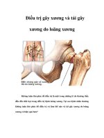
Điều trị gãy xương doc
Bạn đang xem bản rút gọn của tài liệu. Xem và tải ngay bản đầy đủ của tài liệu tại đây (116.13 KB, 7 trang )
Among all wrist injuries, the inci-
dence of fractures of the scaphoid is
second only to that of fractures of
the distal radius.
1
Scaphoid frac-
tures constitute 60% to 70% of all
carpal bone fractures. It has been
estimated that there are 17,250 to
34,500 nonunions per year despite
proper treatment.
2
Nonunions have been attrib-
uted to delay in beginning
treatment, inadequate immo-
bilization, displacement of the
fragments, instability due to lig-
amentous injury, and inade-
quate blood supply of the
proximal fragment.
3
Biome-
chanical studies have demon-
strated that the scaphoid plays a
key role as the stabilizing link
between the proximal and distal
carpal rows.
4
Patients with
scaphoid nonunions are likely
to develop traumatic arthritis
with increasing pain, decreased
wrist mobility, and weakness.
5
However, the data concerning
the natural history of scaphoid
nonunions are largely anecdotal
and difficult to interpret.
6
Classification
Scaphoid fractures can be clas-
sified according to the time after
injury as (1) acute fractures (less
than 3 weeks old), (2) delayed
unions (4 to 6 months old), and (3)
nonunions (more than 6 months
old). However, many clinicians
diagnose these fractures as
nonunions regardless of the time
period if sclerosis, cyst formation,
or bone resorption is present.
Herbert devised an alphanumeric
classification scheme that com-
bines fracture anatomy, stability,
and history; this system was
designed to facilitate prognostic
evaluation. Russe
7
classified frac-
tures into three types according to
the fracture line relative to the
long axis of the scaphoid: hori-
zontal oblique, transverse, and
vertical oblique. Scaphoid frac-
tures have also been classified as
distal, waist, and proximal. Frac-
tures of the middle third of the
scaphoid are the most common
type and have shown a high per-
centage of delayed unions and
nonunions. Proximal-pole frac-
tures have a slower rate of healing
than more distal fractures.
3,7
Diagnostic Imaging
Early Diagnosis
Because of the incidence of
nonunions after occult scaphoid
fractures, new methods have
recently been investigated to fur-
ther image the posttraumatic
scaphoid. Traditionally, scaphoid
fracture is assumed in the post-
traumatic situation if there is ten-
derness in the anatomic snuffbox
despite normal radiographs. Radi-
ographs are then repeated at 10 to
14 days to determine whether a
fracture is indeed present. A stan-
dard radiographic examination of
the suspected scaphoid fracture
includes neutral, ulnar deviation,
posteroanterior, and lateral views,
as well as oblique views obtained
with the wrist in pronation.
In an effort to decrease the delay
in diagnosis, King and Turnbull
8
rec-
ommended that a technetium bone
Vol 2, No 4, July/Aug 1994 185
Scaphoid Nonunion
Peter T. Simonian, MD, and Thomas E. Trumble, MD
Dr. Simonian is Resident, Department of
Orthopaedics, University of Washington, Seat-
tle. Dr. Trumble is Chief of Hand Surgery and
Microsurgery, Department of Orthopaedics,
University of Washington.
Reprint requests: Dr. Trumble, Department of
Orthopaedic Surgery, University of Washington,
Seattle, WA 98195.
Copyright 1994 by the American Academy of
Orthopaedic Surgeons.
Abstract
The natural history and treatment of scaphoid fractures and subsequent
nonunions have occupied a substantial portion of the orthopaedic literature. The
authors examine the role of modern diagnostic tools in making an earlier diagno-
sis of scaphoid nonunion, in more accurately determining the displacement and
angulation of the fragments, and in identifying the presence of avascular necro-
sis. They also consider the various available treatment modalities, including
immobilization, electrical stimulation, both conventional and vascularized bone
grafting, and internal fixation. Finally, a brief review of salvage procedures and
the authors’ preferred treatment are presented.
J Am Acad Orthop Surg 1994;2:185-191
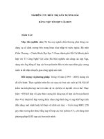
scan be obtained 24 hours after injury
(Fig. 1). Because this technique is
very sensitive (reported 100% sensi-
tivity),
8
it can obviate unnecessary
casting and allow early medical
clearance for return to work. How-
ever, the procedure is relatively time-
consuming and costly, and it exposes
the patient to radiation. The low
specificity (75%) of bone scans is
improved with clinical correlation.
A vibratory instrument has
recently been used as a means of
screening for scaphoid fractures. It
is reported to be reliable, inexpen-
sive, noninvasive, and easy to use
and involves no ionizing radiation.
9
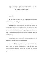
Displacement and Angulation
Precise imaging of the fracture
fragments is difficult because of the
complex shape of the scaphoid. Col-
lapse of the fracture fragments is a
concern and can be seen on plain
radiographs (Fig. 2,A) and in more
detail with computed tomography
(CT) (Fig. 2,B). The scaphoid is visu-
alized most completely when six to
eight CT sections are obtained along
the longitudinal axis of the scaphoid.
Computed tomographic scans have
been used to create three-dimen-
sional images and models. The vol-
ume of bone loss as determined from
computer models can vary from 6%
to 15% and does not show a linear
relationship with the duration of the
nonunion. The missing-bone space is
consistent in configuration, exhibit-
ing a prismatic shape with a quadri-
lateral base, and is oriented palmarly.
The proximal scaphoid fracture com-
ponent is extended, radially devi-
ated, and supinated in relation to the
distal fracture component.
10
Avascular Necrosis
Avascular necrosis of the proxi-
mal pole of the scaphoid is an impor-
tant predictive factor in the success
of surgery to correct scaphoid
nonunion.
11
This is of particular
importance in higher-risk elderly
patients and in patients with long-
standing nonunions. The correla-
tion between gross examination of
the osseous blood supply at surgery
and success following bone grafting
is controversial.
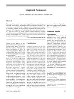
Magnetic resonance (MR) imaging
studies can be used to detect avascu-
lar necrosis in carpal bones and to aid
in patient selection
11
(Fig. 3). It is
essential to use a label for bone for-
mation in order to provide dynamic
evidence of bone viability that can be
correlated with the MR imaging
appearance. Trumble
11
obtained his-
tologic confirmation of the MR
findings consistent with avascular
necrosis by administering a tetracy-
cline label preoperatively and then
using a vital staining technique. This
helped predict whether the scaphoid
was unlikely to unite following bone
grafting and internal fixation. In vivo
labeling of bone samples is a reliable
method for assessing the presence or
absence of bone turnover.
Nonoperative Treatment
No Intervention
According to some reports, estab-
lished nonunions, particularly if sta-
ble and without carpal collapse, may
not require any operative treatment,
186 Journal of the American Academy of Orthopaedic Surgeons
Scaphoid Nonunion
Fig. 1 Bone scan demonstrates scaphoid
fracture not evident on plain radiographs in
a patient with tenderness in the anatomic
snuffbox.
Fig. 2 A, Anteroposterior radiograph reveals collapse and resultant angulation of the
scaphoid. B, Lateral intrascaphoid angle on a sagittal CT scan of another patient indicates a
significant (45-degree) displacement at the fracture secondary to collapse.
AB
for they can remain essentially
symptom-free. Clearly, patients
older than 40 years of age, patients
with nonunions of more than 2 years’
duration, and patients with evidence
of avascular necrosis (without a
decrease in carpal height or an
increase in the scapholunate angle)
may not require any treatment.
Casting
Cast immobilization has been
shown to promote union of stable
nondisplaced nonunions (i.e., those
with no evidence of sclerosis, bone
resorption, or carpal collapse and
no prior history of casting). Casting
can also be used in combination
with other forms of treatment,
including electrical stimulation.
Immobilization for prolonged peri-
ods of time (longer than 6 months)
can have a significant impact on a
patient’s wrist motion, as well as
quality of life and productivity.
Electrical Stimulation
Electrical stimulation has been
used as an alternative or adjunct to
surgical treatment, but its use and
effectiveness have been highly con-
troversial. In some studies it has
not been shown to be more useful
than other nonoperative methods.
12
Furthermore, the efficacy of this
type of treatment is difficult to eval-
uate objectively with double-blind
studies because of the many vari-
ables associated with scaphoid
fractures.
Although the current support-
ing evidence is not conclusive,
pulsed electromagnetic fields have
also been recommended as a treat-
ment modality,
12
for example, for
nondisplaced nonunions without
carpal instability of less than 5
years’ duration.
13
Pulsed electro-
magnetic field treatment is not
inexpensive, however; the cost
compares with that of surgical
treatment and hospitalization.
Frykman et al
13
treated 44 non-
unions of at least 6 months’ dura-
tion with a combination of
electromagnetic field treatment
and plaster immobilization and
found that 35 (80%) healed after a
mean of 4.3 months. According to
this and other reports, this treat-
ment is not as effective as bone-
graft techniques, nor can it correct
scaphoid collapse; however, the
results seem satisfactory enough to
justify its consideration as an alter-
native treatment.
More recently, Adams et al
14
reported that a successful outcome
with pulsed electromagnetic field
treatment and casting is less likely
than they had previously believed.
13
They proposed that pulsed electro-
magnetic field treatment should be
second choice to bone-grafting pro-
cedures until more controlled stud-
ies have been done.
14
Operative Treatment
Indications and Options
There is now considerable evi-
dence to suggest that the incidence
of posttraumatic osteoarthritis
increases in patients with scaphoid
fractures treated with immobiliza-
tion, because of the increased inci-
dence of nonunion; however, the
exact incidence is unknown.
5,15
The
severity of osteoarthritis and the
rapidity of its progression are
increased for displaced fractures
and for fractures with coexistent
carpal instability. Several reports
indicate that few nonunions remain
stable or nondisplaced and free of
arthritis after 10 years.
5,15
Accord-
ingly, even asymptomatic patients
with stable nondisplaced nonunions
should be advised of the possibility
of late degenerative changes. For
these reasons, we believe that frag-
ments that are grossly displaced or
unstable because of ligamentous or
osseous disruption should be
treated with open reduction and
internal fixation as soon as possi-
ble.
1,7
Because of the evidence link-
ing scaphoid nonunions with
osteoarthritis,
5
surgery is recom-
mended for most young, healthy
patients even if they are free of
symptoms and have normal wrist
mobility.
Operative techniques used to
manage scaphoid nonunion at its
various stages of presentation
include bone grafting, vascularized
bone grafting, internal fixation, and
salvage procedures.
Surgical Approach
Studies of the arterial anatomy of
the carpal scaphoid have provided
relevant information on the various
operative approaches that have been
designed to preserve the critical
intraosseous blood supply. They have
generally confirmed that the palmar
approach is least injurious to the vas-
cular supply of the proximal pole.
3,16
Vol 2, No 4, July/Aug 1994 187
Peter T. Simonian, MD, and Thomas E. Trumble, MD
Fig. 3 Magnetic resonance
image demonstrates avascu-
lar necrosis of the proximal
scaphoid fragment.
Gelberman and Menon
3
demon-
strated that 70% to 80% of the
intraosseous vascularity and the
entire vascular supply of the proxi-
mal pole are from branches of the
radial artery entering through the
dorsal ridge. In the region of the dis-
tal tuberosity, 20% to 30% of the
bone receives its blood supply from
volar radial artery branches. There
is excellent collateral circulation to
the scaphoid by way of the dorsal
and volar branches of the anterior
interosseous artery.
More recently, Botte et al
16
reported the effects of the dorsal and
the palmar operative approaches on
the internal vascularity of the
scaphoid. They found the palmar
approach to be safer with respect to
preserving the dorsal nutrient
branches. The dorsal operative
approach placed the vessels of the
dorsal ridge at higher risk, particu-
larly when the vascular leash was
not visualized directly and pro-
tected.
Another important considera-
tion is the location of the nonunion.
Waist fractures should be ap-
proached through the volar inci-
sion to protect the vascular supply.
However, proximal-pole fractures
are best approached through a dor-
sal incision. This allows the small
proximal fragment to be stabilized
to the larger distal fragment with a
screw or Kirschner wire. Because
the blood supply has usually been
completely divided in this fracture,
the dorsal approach will not likely
add additional injury to the bone
vascularity.
Bone Grafting
Traditionally, bone grafting has
been the most popular surgical
treatment and remains the proce-
dure of choice for scaphoid
nonunion. It was recommended in
1928 by Adams and Leonard, who
inserted a graft into the major cavity
of the proximal fragment and laid
the distal portion of the graft in a
trough in the distal fragment. The
technique was later refined by Mur-
ray, who used a cortical peg from
the tibia and passed the graft
through the intramedullary por-
tion of both fragments in a proxi-
mal direction.
The concept of an inlay bone graft
was introduced in 1937 by Matti. He
described resection of sclerotic bone
from the nonunion side approached
dorsally. He then filled the defect
with cancellous graft. In 1960
Russe
7
described a similar tech-
nique of inlay graft using a volar
approach in which a corticocancel-
lous graft was set in a cavity made in
the proximal and distal fragments to
serve as osteogenic material and sta-
bilize the fracture. He believed that
a palmar surgical approach was less
likely to cause further damage to the
bone circulation. Russe reported a
90% union rate, which has become
the benchmark for the surgical man-
agement of scaphoid nonunions.
The high predictability of bone
grafting in achieving bone union
(80% to 90%) is well established.
1
The disadvantage of this technique
is the prolonged period of postop-
erative immobilization, precluding
an early return to work and poten-
tially causing a loss of wrist
motion. Green
17
has pointed out
that the Matti-Russe technique has
a lower success rate when the prox-
imal pole is avascular, as docu-
mented intraoperatively by the
absence of punctate bleeding sites
in the bone. When dorsal interca-
lated segment instability is pres-
ent, an anterior wedge graft after the
method of Fisk and Fernandez
18
is
the preferred option, as it allows
restoration of scaphoid height.
If bone grafting has not been suc-
cessful in treating a scaphoid
nonunion, the procedure should be
repeated if the criteria for the origi-
nal surgery still exist (e.g., there are
no secondary arthritic changes).
Although the rate of healing after a
second or third bone graft is lower
than after a primary graft, it remains
a viable option.
19
Vascularized Bone Grafting
Vascularized bone grafting has
been attempted to decrease the pro-
longed period of immobilization
required after surgery, improve the
rate of union, and provide an alter-
native if previous bone grafting has
not been successful for a scaphoid
nonunion. Zaidemberg et al
20
recently utilized a vascularized
bone-graft source from the distal
dorsoradial radius and had a 100%
union rate in 11 cases, with an aver-
age time to union of 6.2 weeks. They
consider this dorsal approach to be
technically easier than implanting a
vascularized bone graft from the
volar approach. Furthermore, it
does not require sacrifice of the
radial or ulnar artery.
Internal Fixation
Internal fixation can decrease the
duration of immobilization
required to achieve union, thus
allowing early range of motion, and
can also correct collapsed deformi-
ties of the scaphoid. Many of the
same devices recommended for use
in the treatment of acute scaphoid
fractures have been used in the
treatment of scaphoid nonunions.
In 1954 McLaughlin reported the
use of a cobalt-chrome alloy screw,
but its insertion was cumbersome
and the union rate was unaccept-
able. Other devices that have been
utilized include Kirschner wires,
21
pneumatically inserted staples, the
Ender plate,
22
the ASIF screw,
23,24
and the Herbert screw.
25,26
A can-
nulated ASIF screw and a cannu-
lated Herbert-Whipple screw have
recently been introduced (Fig. 4).
The advantages of Kirschner
wires include the ease of insertion
and removal and the lack of a need
for extended incisions or radial sty-
188 Journal of the American Academy of Orthopaedic Surgeons
Scaphoid Nonunion
loidectomy. They can also be used in
the presence of vascular changes in
the proximal fragment. Kirschner
wires have been utilized in conjunc-
tion with screws as derotational
devices to provide torsional stability
(Fig. 4, A). However, they do not
provide compression of the fracture
site.
Huene and Huene
22
demon-
strated union in 19 of 20 cases of
scaphoid nonunion treated with
the Ender compression blade plate.
They reported that this implant is
helpful in the presence of compli-
cating factors such as vascular
necrosis, cystic degeneration, and
bone-size disparity. The disadvan-
tages of this implant include the
necessity of late removal and the
possibility of articular impinge-
ment. Also decreasing the popular-
ity of this implant is the inability to
achieve compression of the fracture
fragments; a similar problem exists
with use of pneumatic staples.
Leyshon et al
23
described a satis-
factory experience treating delayed
unions and nonunions of the
scaphoid with the ASIF lag screw
and use of an extended lateral and
volar bayonet-shaped incision. A
radial styloidectomy was not required,
and they could directly visualize the
reduction.
Using the ASIF screw, Sukul et al
24
achieved a greater than 90% union
rate at an average of 26.9 weeks in 42
patients with established nonunions.
A dorsolateral incision and a cortic-
ocancellous bone graft were used in
these cases. The advantages of this
implant include excellent compres-
sion without disruption of ligaments,
which is often needed with other
devices to achieve the same degree of
compression. The disadvantages of
the ASIF screw include the con-
straints due to the size of the fracture
fragments and the possibility of intra-
articular screw head placement. To
help alleviate the possibility of intra-
articular damage, Sukul developed a
“dynamic compression screw for the
scaphoid bone” that is totally con-
tained within the bone, similar to the
Herbert screw.
The Herbert screw was specifi-
cally designed for internal fixation of
the scaphoid.
25
This provided the
theoretical advantages of other
forms of internal fixation. Its unique
double-threaded design, relatively
narrow diameter, and differential
pitch allow complete subchondral
containment, thereby decreasing the
likelihood of hardware impinge-
ment. This usually eliminates the
need for later removal and mini-
mizes the host response to the
implant. Most important, the Her-
bert screw allows early range of
motion prior to the achievement of
union. The Herbert screw can also
be used from the dorsal side for
small proximal-pole fractures.
Potential disadvantages of the Her-
bert screw include the technical dif-
ficulty of its insertion,
26
the need to
violate the scaphotrapezial ligament
to allow screw insertion,
26
and its
inferior ability to provide bone com-
pression (when used without the
jig) compared with the ASIF
screw.
27
We have recently compared the
cannulated ASIF screw with the Her-
bert screw in scaphoid nonunions.
The time to union averaged 3.8
months for the cannulated ASIF
screw and 7.2 months for the Herbert
screw. We believe that this differ-
ence in time to union between the
implants is related to the increased
accuracy of screw placement in the
proximal fragment with the cannu-
lated ASIF screw (Fig. 5).
Biomechanical studies have ana-
lyzed the strength of these internal
fixation devices. The following
implants designed for internal
fixation of the scaphoid are listed in
descending order of strength: the
noncannulated ASIF screw, the can-
nulated ASIF screw, the Herbert-
Whipple screw, two 0.045-mm
Kirschner wires, and the Herbert
screw.
28
The ultimate goals of internal
fixation are to provide immediate
stability to correct deformity, to pro-
mote union, and to allow early
return to function.
Salvage Procedures
There are a variety of operative
salvage procedures, including exci-
sion of the proximal fragment or
both fragments, proximal-row
Vol 2, No 4, July/Aug 1994 189
Peter T. Simonian, MD, and Thomas E. Trumble, MD
Fig. 4 Devices used for internal fixation. A, Cannulated ASIF screw with a derotational
Kirschner wire in position. B, Cannulated Herbert-Whipple screw.
A
B
carpectomy, intercarpal fusion with
scaphoid excision, arthrodesis of the
wrist, replacement of the scaphoid
with a metal or silicone prosthesis,
radial styloidectomy, and interposi-
tion of soft tissue into the nonunion
site or fascial arthroplasty. Some
procedures, such as drilling of the
bone, are of historic interest only and
have little relevance to contempo-
rary hand surgery.
Excising the proximal fragment is
a useful procedure if the fragment is
small (usually not exceeding one
fourth of the length of the bone). A
small fracture fragment can also be
removed by using arthroscopic tech-
niques. However, some of the mod-
ern implants are designed to
incorporate small fragments of bone.
Excising fragments larger than one
third of the length of the scaphoid
should be avoided because the
surgery is likely to produce inter-
carpal instability. Excising the prox-
imal carpal row as a salvage
procedure should be considered
with partial and total wrist arthrode-
sis if secondary arthritic changes
have developed.
Replacing the scaphoid with a
prosthesis is another option. Sili-
cone carpal implants have been
associated with numerous compli-
cations, including dislocation,
breakage, and synovitis, and we
do not recommend their use.
Interposing a soft-tissue flap
between the nonunited fragments
was recommended by Bentzon in
1940 and is still used, mainly in the
Scandinavian countries. It might
be considered if postoperative
immobilization is contraindicated.
Partial and total arthrodesis are
also salvage options, depending on
the degree and location of arthritis.
For the common combination of
radioscaphoid and midcarpal arthri-
tis often seen in chronic nonunions,
a combination of midcarpal
arthrodesis and scaphoid excision
can be considered. This has been
called the scapholunate advanced
collapse (SLAC) procedure.
Authors’ Preferred
Treatment
We define scaphoid nonunion as
being present when radiographic
signs consistent with inability of the
fracture to heal (sclerosis, cyst for-
mation, collapse, and bone resorp-
tion) are present or union has not
occurred over a period of 6 months
despite treatment.
If the patient is less than 40 years
old or the fracture is of less than 2
years’ duration, we recommend
bone grafting with internal fixation.
When the nonunion is proximal, a
dorsal approach is used. When the
nonunion is at the waist, a volar
approach is used.
If the patient is more than 40
years old or if the fracture is of more
than 2 years’ duration, treatment
depends on the symptoms. Asymp-
tomatic patients are observed. If
symptomatic and avascular necro-
sis is evident, either excision arthro-
plasty with intercarpal fusion or
wrist fusion is performed, depend-
ing on the extent and location of
osteoarthritis. When there is no
radiolunate osteoarthritis, SLAC
fusion is done. Addition of radio-
scaphoid and capitate-lunate joint
osteoarthritis prompts considera-
tion of a complete wrist fusion.
If there is uncertainty about the
presence of avascular necrosis, MR
imaging is performed. When avas-
cular necrosis is not evident on MR
imaging, bone grafting with internal
fixation is undertaken. If the
nonunion is proximal, a dorsal
approach is used; if the nonunion
is at the waist, a volar approach is
used. When MR imaging is positive
for avascular necrosis, SLAC fusion
is recommended for active patients;
a similar scaphoid excision is done in
elderly patients.
We favor use of the Herbert-
Whipple screw and a derotational
Kirschner wire as the means of inter-
nal fixation.
190 Journal of the American Academy of Orthopaedic Surgeons
Scaphoid Nonunion
Fig. 5 Radiograph demonstrates the
increased accuracy of screw placement in the
proximal fragment of the scaphoid with a
cannulated screw.
References
1. Cooney WP III, Dobyns JH, Linscheid
RL: Nonunion of the scaphoid: Analy-
sis of the results from bone grafting. J
Hand Surg 1980;5:343-354.
2. Osterman AL, Mikulics M: Scaph-
oid nonunion. Hand Clin 1988;14:
437-455.
3. Gelberman RH, Menon J: The vascular-
ity of the scaphoid bone. J Hand Surg
1980;5:508-513.
4. Smith DK, Cooney WP III, An KN, et al:
The effects of simulated unstable
Vol 2, No 4, July/Aug 1994 191
Peter T. Simonian, MD, and Thomas E. Trumble, MD
scaphoid fractures on carpal motion. J
Hand Surg [Am] 1989;14:283-291.
5. Ruby LK, Stinson K, Belsky MR: The
natural history of scaphoid non-union:
A review of fifty-five cases. J Bone Joint
Surg Am 1985;67:428-432.
6. Kerluke L, McCabe SJ: Nonunion of the
scaphoid: A critical analysis of recent
natural history studies. J Hand Surg
[Am] 1993;18:1-3.
7. Russe O: Fracture of the carpal navicu-
lar: Diagnosis, non-operative treatment,
and operative treatment. J Bone Joint
Surg Am 1960;42:759-768.
8. King JB, Turnbull TJ: An early method
of confirming scaphoid fracture. J Bone
Joint Surg Br 1981;63:287-288.
9. Finkenberg JG, Hoffer E, Kelly C, et al:
Diagnosis of occult scaphoid fractures
by intrasound vibration. J Hand Surg
[Am] 1993;18:4-7.
10. Belsole RJ, Hilbelink DR, Llewellyn JA,
et al: Computed analyses of the patho-
mechanics of scaphoid waist nonunions.
J Hand Surg [Am] 1991;16:899-906.
11. Trumble TE: Avascular necrosis after
scaphoid fracture: A correlation of mag-
netic resonance imaging and histology.
J Hand Surg [Am] 1990;15:557-564.
12. Bora FW Jr, Osterman AL, Woodbury
DF, et al: Treatment of nonunion of the
scaphoid by direct current. Orthop Clin
North Am 1984;15:107-112.
13. Frykman GK, Taleisnik J, Peters G, et al:
Treatment of nonunited scaphoid frac-
tures by pulsed electromagnetic field
and cast. J Hand Surg [Am] 1986;11:
344-349.
14. Adams BD, Frykman GK, Taleisnik J:
Treatment of scaphoid nonunion with
casting and pulsed electromagnetic
fields: A study continuation. J Hand
Surg [Am] 1992;17:910-914.
15. Mack GR, Bosse MJ, Gelberman RH, et
al: The natural history of scaphoid non-
union. J Bone Joint Surg Am 1984;66:504-
509.
16. Botte MJ, Mortensen WW, Gelberman
RH, et al: Internal vascularity of the
scaphoid in cadavers after insertion of
the Herbert screw. J Hand Surg [Am]
1988;13:216-222.
17. Green DP: The effect of avascular
necrosis on Russe bone grafting for
scaphoid nonunion. J Hand Surg [Am]
1985;10:597-605.
18. Fernandez DL: A technique for anterior
wedge-shaped grafts for scaphoid
nonunions with carpal instability. J
Hand Surg [Am] 1984;9:733-737.
19. Carrozzella JC, Stern PJ, Murdock PA:
The fate of failed bone graft surgery for
scaphoid nonunions. J Hand Surg [Am]
1989;14:800-806.
20. Zaidemberg C, Siebert JW, Angrigiani
C: A new vascularized bone graft for
scaphoid nonunion. J Hand Surg [Am]
1991;16:474-478.
21. Stark HH, Rickard TA, Zemel NP, et al:
Treatment of ununited fractures of the
scaphoid by iliac bone grafts and
Kirschner-wire fixation. J Bone Joint
Surg Am 1988;70:982-991.
22. Huene DR, Huene DS: Treatment of
nonunions of the scaphoid with the
Ender compression blade plate system.
J Hand Surg [Am] 1991;16:913-922.
23. Leyshon A, Ireland J, Trickey EL: The
treatment of delayed union and non-
union of the carpal scaphoid by screw
fixation. J Bone Joint Surg Br 1984;66:
124-127.
24. Sukul DMKSK, Johannes EJ, Marti RK:
Corticocancellous grafting and an
AO/ASIF lag screw for nonunion of the
scaphoid: A retrospective analysis. J
Bone Joint Surg Br 1990;72:835-838.
25. Herbert TJ, Fisher WE: Management of the
fractured scaphoid using a new bone
screw. J Bone Joint Surg Br 1984;66:114-123.
26. Adams BD, Blair WF, Reagan DS, et al:
Technical factors related to Herbert
screw fixation. J Hand Surg [Am] 1988;
13:893-899.
27. Shaw JA: A biomechanical comparison
of scaphoid screws. J Hand Surg [Am]
1987;12:347-353.
28. Butler T, McCormack T, Jayaraman, et
al: Comparisons of scaphoid internal
fixation techniques with cyclic bending.
Presented at the 11th Annual Residents’
and Fellows’ Conference on Hand
Surgery of the American Society for
Surgery of the Hand, Kansas City, Kan,
Sep 28, 1993.