Khối u sụn: Đánh giá và điều trị pptx
Bạn đang xem bản rút gọn của tài liệu. Xem và tải ngay bản đầy đủ của tài liệu tại đây (249.92 KB, 13 trang )
Journal of the American Academy of Orthopaedic Surgeons
292
Cartilaginous neoplasms are rela-
tively common tumors that can
involve almost any bone.
1
These
tumors vary in presentation and can
range from a latent enchondroma to
a high-grade or dedifferentiated
chondrosarcoma. The major dilemma
facing the surgeon is clinically and
radiologically differentiating an en-
chondroma from a low-grade chon-
drosarcoma. Occasionally, even the
histologic diagnosis can be difficult.
The diagnosis and treatment options
for these tumors are dependent on a
combination of clinical, radiologic,
and histologic findings.
Most musculoskeletal surgeons,
radiologists, and pathologists can
readily distinguish an enchondro-
ma from a high-grade chondrosar-
coma. Enchondromas are benign
intramedullary tumors that are
usually asymptomatic and do not
metastasize.
1
They are most com-
monly located in the short tubular
bones in the hands but are also
found in long bones. Radiographs
usually demonstrate a small (<5
cm) cartilaginous lesion with in-
tramedullary calcifications without
cortical involvement or soft-tissue
extension.
1-4
Histologically, enchon-
dromas exhibit discrete islands of
hyaline cartilage surrounded by
lamellar bone. Multinucleated cells
are rare. An asymptomatic enchon-
droma usually does not require
treatment beyond observation.
Occasionally, symptomatic enchon-
dromas are treated by intralesional
excision. The incidence of local
recurrence is extremely low.
4
High-grade chondrosarcomas
are malignant neoplasms that com-
monly recur and metastasize.
5-9
This tumor is usually painful and
often demonstrates a range of radio-
graphic findings, including cortical
destruction, significant endosteal
scalloping, cortical thickening, and
soft-tissue extension. High-grade
chondrosarcomas are characterized
by marked atypia, mitotic figures,
and some spindle elements. A
wide excision is necessary to obtain
local control of these tumors.
Enchondromas and high-grade
chondrosarcomas have distinct clini-
copathologic and radiologic appear-
ances, which can be used to easily
distinguish one entity from the
other. However, enchondromas and
intramedullary low-grade chon-
drosarcomas of long bones can
resemble each other clinically, radio-
logically, and histologically. Intra-
medullary low-grade chondrosarco-
mas are usually painful. They are
most commonly located in the me-
taphyses of the humerus, femur, or
tibia and are usually larger (>5 cm)
than an enchondroma. Endosteal
scalloping and lysis are common.
2,10
Cortical thickening, expansion, or
disruption and soft-tissue masses
are uncommon findings.
10,11
Be-
cause low-grade chondrosarcomas
can have cytologic features similar
to those of enchondromas, histologic
evaluation is important.
12,13
Dr. Marco is Assistant Professor of Surgery,
M.D. Anderson Cancer Center, Houston. Dr.
Gitelis is Professor of Orthopaedic Surgery,
Rush Medical College, Chicago. Dr. Brebach is
Instructor in Orthopaedic Surgery, Rush
Medical College. Dr. Healey is Professor of
Orthopaedic Surgery, Weill/Cornell University
and Memorial Sloan-Kettering Cancer Center,
New York.
Reprint Requests: Dr. Gitelis, Suite 440, 1725
W. Harrison Street, Chicago, IL 60612.
Copyright 2000 by the American Academy of
Orthopaedic Surgeons.
Abstract
The proper treatment of cartilaginous tumors is dependent on the clinicopatho-
logic and radiologic findings. Enchondroma is a benign tumor that is usually
asymptomatic and thus should be treated nonoperatively. Symptomatic
enchondromas are often treated by intralesional excision. Intramedullary low-
grade chondrosarcoma is a malignant tumor that is usually painful. The treat-
ment of low-grade chondrosarcoma may range from intralesional excision with
or without adjuvant therapy to wide excision. Although intralesional excisions
have a higher bone and joint preservation rate than wide excisions, they may be
associated with a higher local recurrence rate. Intermediate- and high-grade
chondrosarcomas are treated with wide excisions. The treatment of these carti-
laginous lesions should involve a multidisciplinary team including a muscu-
loskeletal surgeon, a radiologist, and a pathologist.
J Am Acad Orthop Surg 2000;8:292-304
Cartilage Tumors: Evaluation and Treatment
Rex A. W. Marco, MD, Steven Gitelis, MD, Gregory T. Brebach, MD, and John H. Healey, MD
Rex A. W. Marco, MD, et al
Vol 8, No 5, September/October 2000
293
Low-grade chondrosarcomas
rarely metastasize, but frequently
recur if inadequate surgery is per-
formed.
7-9
Most authors therefore
recommend a wide excision to erad-
icate a low-grade chondrosarcoma,
although some have advocated
intralesional therapy. Wide exci-
sions are associated with low local
recurrence rates, whereas intrale-
sional excisions are associated with
high local recurrence rates. Intra-
lesional excisions combined with
adjuvant therapy, however, are
associated with low mortality and
local recurrence rates in carefully
selected patients with low-grade
chondrosarcomas.
14
Intralesional
excisions preserve the adjacent bone
and joint surfaces, which probably
improves the functional outcome.
The primary dilemma is determin-
ing which intramedullary low-
grade chondrosarcomas can be
treated by intralesional excision
rather than wide excision. A thor-
ough evaluation of the clinical pre-
sentation, radiographic findings,
and histologic appearance is neces-
sary to determine the most appro-
priate treatment (Table 1).
Clinical Presentation
Enchondroma involving a metacar-
pal or phalanx of the hand may pre-
sent as pathologic fracture in a
young adult. Enchondromas in-
volving long bones are usually
asymptomatic and are commonly
an incidental finding identified on a
radiograph obtained to evaluate the
chest or an adjacent joint. Regional
pain about an enchondroma is more
frequently related to a nearby joint
or a local soft-tissue disorder than to
the tumor itself and may be the
cause for incidental discovery of an
asymptomatic enchondroma.
A common scenario is a patient
with shoulder pain in whom there
is a completely intramedullary car-
tilaginous lesion in the proximal
humerus, which could represent an
enchondroma or a low-grade chon-
drosarcoma. A thorough history
and physical examination are nec-
essary to evaluate the shoulder for
other causes of the pain. Subacro-
mial or acromioclavicular injection
of a local anesthetic agent can help
identify the origin of the pain. If
Table 1
Characteristics of Cartilage Tumors and Treatment Recommendations
Adaptive or
Aggressive Radio-
Tumor Type Pain logic Changes* Bone Scan Histology Treatment
Enchondroma −− −Enchondroma Observation
Atypical enchondroma
†
(chondrosarcoma in situ) + − +/− Enchondroma Observation or intra-
lesional excision
Chondrosarcoma in situ +/−− +/− Grade I Observation, intralesional
chondrosarcoma excision, or (occasion-
ally) wide excision
Low-grade
chondrosarcoma + + + Grade I Wide excision
chondrosarcoma
Intermediate-grade
chondrosarcoma + + + Grade II Wide excision
chondrosarcoma
High-grade
chondrosarcoma + + + Grade III Wide excision
chondrosarcoma
Dedifferentiated
chondrosarcoma + + + Dedifferentiated Wide excision
chondrosarcoma
*
Adaptive radiologic changes include cortical thickening and expansion. Aggressive changes include cortical disruption and the pres-
ence of a soft-tissue mass.
†
Synonymous with grade 0.5 chondrosarcoma, low-grade I chondrosarcoma, or borderline chondrosarcoma.
Cartilage Tumors
Journal of the American Academy of Orthopaedic Surgeons
294
the pain resolves, it was likely sec-
ondary to an inflammatory syn-
drome in the shoulder, rather than
being due to the proximal humerus
lesion. The shoulder disorder
should be treated appropriately,
and the lesion, which is likely an
enchondroma, should be periodical-
ly monitored for the development of
clinical or radiographic signs or
symptoms of tumor progression. If
the pain persists despite appropri-
ate treatment of the presumed
shoulder disorder, the symptoms
may be from the lesion, which can
be either an enchondroma or a low-
grade chondrosarcoma, necessitat-
ing further evaluation to differenti-
ate between them.
Most patients with chondrosar-
coma have pain.
9,10,12,14
In a study
of 58 patients with intramedullary
low-grade chondrosarcoma, Marco
et al
14
found that 60% (35) had rest
or night pain, 21% had vague
regional pain, and 19% had lesions
that were detected incidentally.
Nearly 80% of patients with inter-
mediate- or high-grade chondrosar-
coma have pain.
6
Pathologic frac-
tures occur in 3% to 8% of patients
with chondrosarcoma.
6,9,14
Radiologic Findings
Enchondromas (Fig. 1) and low-
grade intramedullary chondrosar-
comas (Fig. 2) of long bones can
have similar radiologic appear-
ances. Both types of tumors dem-
onstrate stippled calcifications, and
both may display endosteal scal-
loping on plain radiographs.
1,15
They are commonly located in the
metaphysis of the humerus, femur,
or tibia. Calcification is manifested
by punctate mineralization or pop-
cornlike calcification. The margins
of the tumor should be examined
for osteolysis and endosteal scal-
loping. The extent and degree of
endosteal scalloping correlate with
the likelihood of the lesion being
a chondrosarcoma.
10
In one study,
Murphey et al
10
found that 71 (75%)
of 95 patients with chondrosarco-
ma had endosteal scalloping of
more than two thirds of the cortical
thickness, compared with 8 (9%) of
92 patients with enchondroma.
Chondrosarcoma can demon-
strate adaptive and aggressive radio-
logic signs. Cortical expansion and
thickening are adaptive changes, and
cortical disruption and soft-tissue
masses are aggressive changes asso-
ciated with chondrosarcoma.
1,10
Rosenthal et al
15
summarized the
plain-radiographic and computed
tomographic (CT) findings in low-
and high-grade chondrosarcoma.
Low-grade features include (1) dense
calcifications forming rings or spic-
ules, (2) widespread or uniformly
distributed calcifications, and (3) ec-
centric lobular growth of a soft-tissue
mass. High-grade features include
(1) faint amorphous calcification,
(2) large noncalcified areas, and
(3) concentric growth of a soft-tissue
mass. Lysis within a previously calci-
fied area may be a sign of tumor pro-
gression. The primary exception to
these radiologic findings is enchon-
droma in a short tubular bone of the
hand, which frequently demonstrates
marked endosteal scalloping, large
areas of lysis, and cortical expansion.
A technetium-99m diphospho-
nate whole-body bone scan can
provide some useful information
about an intramedullary cartilagi-
nous lesion. A whole-body bone
scan with a high degree of radionu-
clide uptake within the lesion com-
pared with an internal standard,
such as the anterior superior iliac
spine or acromioclavicular joint, is
more consistent with chondrosarco-
ma than enchondroma.
10
Murphey
et al
10
graded radionuclide uptake
from grade 1 to grade 3, with grade
1 indicating uptake less than that in
the anterior iliac crest; grade 2, up-
take similar to that in the anterior
iliac crest; and grade 3, uptake
greater than that in the anterior iliac
Figure 1 A, Anteroposterior radiograph of the left proximal humerus and shoulder of an
82-year-old man without any pain. Note the calcified lesion without evidence of cortical
erosion. B, T1-weighted (repetition time, 350 msec; echo time, 12 msec [350/12]) MR
image of the left humerus shows tumor lobules present, with multiple satellites. The
tumor did not destroy bone and was consistent with an enchondroma. Follow-up plain
radiographs showed no evidence of progression.
A B
Rex A. W. Marco, MD, et al
Vol 8, No 5, September/October 2000
295
crest. In their study of 51 patients
with chondrosarcoma, 42 (82%) had
grade 3 uptake, compared with 14
of 67 patients (21%) with enchon-
droma. However, most enchondro-
mas demonstrate some activity on
bone scan; therefore, that finding
alone is not particularly worrisome.
The bone scan can also help identify
polyostotic disease.
Axial imaging with CT or mag-
netic resonance (MR) imaging can
be helpful in evaluating the depth
of endosteal scalloping and the size
of the lesion and its soft-tissue com-
ponent. Computed tomography is
the study of choice to evaluate the
osseous architecture for endosteal
scalloping and bone disruption.
Magnetic resonance imaging is par-
ticularly useful in determining the
nonmineralized intramedullary
extent of the tumor and soft-tissue
extension. The axial and coronal
images accurately demonstrate
marrow replacement by tumor,
providing measurements that can
guide the surgeon when either an
intralesional or a wide excision is
performed. The relationship of a
soft-tissue mass to important para-
osseous structures, such as the joint
capsule and the neurovascular bun-
dle, is accurately demonstrated on
MR images. The percentage of med-
ullary fill of the lesion visualized on
MR imaging is also useful informa-
tion. Medullary fill greater than 90%
is predictive of chondrosarcoma.
11
Noncontiguous foci of cartilage, or
satellites (Fig. 1, B), are predictive of
enchondroma if the medullary fill is
less than 90%. Finally, a chest radio-
graph and usually a CT scan of the
chest are obtained for staging.
Biopsy
The biopsy of a chondrosarcoma
can be performed with closed or
open techniques. Closed biopsy
techniques with fine (20- to 23-
gauge) or core needles are com-
monly utilized to confirm the diag-
nosis of a cartilaginous tumor that
is clinically and radiographically a
chondrosarcoma. A fine-needle
biopsy directed by fluoroscopy or
CT can be utilized if there is a soft-
tissue component. Imaging may
not be required if the soft-tissue
mass is palpable. This procedure
primarily yields material for cyto-
logic and, to a lesser extent, histo-
logic examination. If the tumor is
located within bone, a core needle
penetrates the bone more readily
than a fine needle. A core-needle
biopsy provides a cylinder of tissue,
which can be examined both cyto-
logically and histologically. Biopsy
specimens should be taken from
the areas of most concern, such as
areas of bone destruction and those
demonstrating a high degree of
endosteal scalloping and lysis.
Experienced musculoskeletal
pathologists can usually diagnose a
high-grade chondrosarcoma if ma-
lignant cartilaginous cells are noted.
A major drawback of needle-biopsy
techniques, however, is sampling
error due to tumor heterogene-
ity.
16,17
A high-grade cartilaginous
tumor often contains low-grade or
benign hyaline cartilage material.
Figure 2 A, Anteroposterior radiograph of the left proximal humerus of a 43-year-old man with progressively increasing shoulder pain,
which was present at rest. Note the calcification with minimal endosteal scalloping. B, T2-weighted (3,500/16) MR image of the lesion in
the proximal humerus. Biopsy revealed a low-grade chondrosarcoma. C, The patient was treated with intralesional excision, cauteriza-
tion with phenol, and insertion of methylmethacrylate. The pain resolved completely.
A B C
Cartilage Tumors
Journal of the American Academy of Orthopaedic Surgeons
296
The final pathologic study could con-
ceivably reveal a chondrosarcoma
despite a needle-biopsy diagnosis of
enchondroma. Differentiating an
enchondroma from a low-grade
chondrosarcoma is often difficult, if
not impossible, with the small
amount of material obtainable by
needle biopsy.
An open biopsy usually pro-
vides adequate tissue for diagnosis
but is associated with surgical-site
contamination and other complica-
tions associated with open proce-
dures and general anesthesia. Con-
firmation of the viability of the
tumor and the adequacy of the tis-
sue sample should be obtained by
frozen-section diagnosis at the time
of the procedure.
Symptomatic intramedullary car-
tilaginous tumors that display nei-
ther adaptive radiologic changes
(cortical thickening or expansion)
nor aggressive radiologic changes
(cortical disruption or soft-tissue
mass) are likely to be enchondromas
or low-grade chondrosarcomas. If
the clinical presentation warrants
further evaluation, a biopsy is rec-
ommended before definitive treat-
ment. If an intermediate- or a high-
grade cartilage tumor is identified
on the basis of frozen-section analy-
sis, the procedure should be termi-
nated, and treatment deferred until
a final pathology report is made. If
the frozen section is consistent with
an enchondroma or a low-grade
chondrosarcoma, some surgeons
would proceed with intralesional
excision with or without adjuvant
therapy.
Performing a simultaneous in-
tralesional excision can obviate a
second operative procedure, pro-
vide curative treatment, and mini-
mize bleeding with subsequent
seeding of tumor cells within the
incision.
15
However, the patient
must be counseled preoperatively
that the tumor grade (and thus the
optimal treatment) may change
with the final diagnosis on perma-
nent sections. Definitive treatment
should be based on the highest grade
of tumor present. If the diagnosis is
an enchondroma or a low-grade
chondrosarcoma, close observation
is appropriate. If intermediate- or
high-grade chondrosarcoma is
identified within any portion of the
tumor, a secondary wide excision
may be required. To minimize local
contamination of the tissues by
chondrosarcoma cells, it is impor-
tant to protect the surrounding tis-
sues during the curettage and
achieve meticulous hemostasis after
intralesional treatment. If the biop-
sy and intralesional excision are
performed properly, the definitive
oncologic procedure and outcome
should not be adversely affected if
more aggressive surgical interven-
tion is required.
Although simultaneously per-
forming a biopsy and an intralesion-
al excision for an intramedullary
cartilaginous tumor has advan-
tages, most surgeons prefer to wait
for the final pathologic diagnosis
before further treatment. An intra-
lesional or wide excision with re-
moval of the entire biopsy track
and previously exposed tissue is
then performed. However, the
pathologist may identify higher-
grade tumor in the specimen re-
moved at the definitive excision
than was originally found at biopsy.
Delaying the definitive treatment
while waiting for a final biopsy
diagnosis does not completely
avoid the possibility that a change
in the preoperative diagnosis may
occur once the entire specimen is
examined.
Some authors have advocated not
obtaining biopsy specimens of carti-
laginous tumors that are clinically
and radiographically chondrosarco-
mas, although this is not a widely
held opinion.
18
These chondrosarco-
mas are painful and may have an
associated soft-tissue mass. A high
degree of endosteal scalloping and
adaptive and aggressive radiologic
findings are seen. Although these
tumors can be low-grade chon-
drosarcomas, they are more often
intermediate- or high-grade chon-
drosarcomas. Chondrosarcomas
demonstrating these clinical and
radiographic signs should be treated
with wide excision. Some tumor
surgeons would proceed with a
wide excision without performing a
biopsy, thereby avoiding the in-
evitable contamination of the biopsy
site with tumor cells. The specimen
is then sent for final gross and histo-
logic diagnosis. However, although
this procedure is theoretically better,
only a very experienced tumor sur-
geon should make these decisions.
Clinicopathologic
Grading
Chondrosarcomas are graded on
the basis of the cytologic and histo-
logic appearance
8,12,13,15,19
(Fig. 3),
combined with the clinical and radio-
logic presentation. Most authors
grade chondrosarcomas from grade
I to grade III.
8,12,13,15,19
The diagno-
sis of grade II (intermediate-grade)
and grade III (high-grade) chon-
drosarcoma can usually be made on
the basis of either cytologic or histo-
logic features.
12,13
Grade I (low-
grade) chondrosarcoma, however,
has cytologic features similar to
those of enchondroma. Therefore,
histologic criteria must be combined
with clinical and radiologic findings
to differentiate enchondroma (Fig. 4)
from low-grade chondrosarcoma.
12,13
Histologically, both enchondromas
and low-grade chondrosarcomas are
composed of hyaline cartilage cells.
A low-grade chondrosarcoma should
be suspected if there are (1) many
cells with plump nuclei, (2) more
than an occasional binucleate cell,
and (3) giant cartilage cells with
large nuclei or with clumps of chro-
matin.
19
Further differentiation
between an enchondroma and a
low-grade chondrosarcoma is then
Rex A. W. Marco, MD, et al
Vol 8, No 5, September/October 2000
297
possible by examining the tissue
pattern of the cartilage cells and the
lamellar bone, as described by Mirra
et al.
12
The enchondroma pattern
consists of nodules of hyaline carti-
lage that are encased by lamellar
bone. These nodules are separated
from each other by normal marrow.
The low-grade chondrosarcoma
pattern consists of cartilage cells
that permeate marrow spaces and
completely replace the marrow fat.
The cartilage cells directly abut and
surround the lamellar bone in the
chondrosarcoma pattern. Other his-
tologic findings of chondrosarcoma
include (1) malignant bands of
fibrosis, (2) chondrosarcomatous
invasion of marrow fat, (3) malig-
nant invasion of the haversian sys-
tem, and (4) a soft-tissue mass.
Occasionally, a painful cartilagi-
nous lesion in a long bone has the
radiologic appearance of a low-
grade chondrosarcoma (e.g., lytic
areas or high-grade endosteal scal-
loping without adaptive or aggres-
sive radiographic changes) and the
histologic appearance of an enchon-
droma. This lesion is referred to as a
grade 0.5 chondrosarcoma by some
authors; others may describe it as a
borderline chondrosarcoma, low
grade 1 chondrosarcoma, grade 0
chondrosarcoma, painful enchon-
droma, or atypical enchondroma.
We prefer the term “chondrosarcoma
in situ,” which implies that the
lesion is benign and should not
metastasize unless there is malig-
nant transformation. We also be-
lieve that tumors with both the
radiologic and the histologic appear-
ance of a low-grade chondrosarco-
ma should be considered chon-
drosarcomas in situ because these
lesions do not metastasize if treated
properly.
4,14,20,21
Cartilaginous lesions in the hand
and pelvis behave differently than
intramedullary cartilaginous le-
sions of the long bones with similar
histologic appearances.
1
Enchon-
dromas of the short tubular bones
in the hand frequently have multi-
nucleated cells, as well as increased
cellularity that resembles the appear-
ance of grade 1 chondrosarcoma. Al-
though these tumors occasionally
recur after intralesional treatment,
they do not metastasize. However,
most patients with a histologically
similar lesion in the pelvis will have
a local recurrence after intralesional
excision.
21-23
Staging
Chondrosarcomas are staged ac-
cording to the system described by
Enneking.
24
Nonmetastatic low-
grade chondrosarcomas are consid-
ered stage I neoplasms. Nonmeta-
static intermediate- and high-grade
chondrosarcomas are stage II. Met-
astatic chondrosarcomas are stage
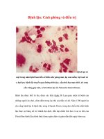
Figure 3 A, Low-grade chondrosarcoma (hematoxylin-eosin, original magnification ×100). This tumor is well-differentiated. Hyper-
cellularity is noted, but the cartilage matrix may be easily identified. There are numerous binucleate cells within lacunae and few atypi-
cal cells. B, Higher-magnification view of the same tumor (hematoxylin-eosin, original magnification ×250). Mild pleomorphism and
hyperchromatism are apparent, and binucleate cells are seen. The tumor had a well-differentiated cartilage matrix. C, Intermediate-
grade chondrosarcoma (hematoxylin-eosin, original magnification ×250). The tumor displays distinct pleomorphism, with some very
large hyperchromatic cells.
A B C
Figure 4 Enchondroma (hematoxylin-
eosin, original magnification ×100). Note
the hypocellularity of the lesion and the
uniformity in size and staining features of
the cells. The hyaline cartilage matrix is
readily apparent.
Cartilage Tumors
Journal of the American Academy of Orthopaedic Surgeons
298
III. Tumors are then subclassified as
either stage A or stage B on the basis
of whether they are located within
the bone or extend outside the bone.
For example, a low-grade intramed-
ullary chondrosarcoma without
metastases is stage IA, whereas a
high-grade nonmetastatic chon-
drosarcoma with an associated soft-
tissue mass is stage IIB.
Enchondromas may be staged by
using the Enneking staging system
for benign tumors.
24
In that system,
a stage 1 tumor is latent (i.e., a
tumor that does not progress or that
heals spontaneously). A stage 2
tumor is active (i.e., it progresses
but respects natural barriers, such as
the bone cortex). A stage 3 tumor is
aggressive (i.e., it progresses and
will ultimately destroy natural bar-
riers). Enchondromas are usually
stage 1 but are occasionally stage 2.
Types of Surgical Excisions
Enneking
24
defined surgical mar-
gins for bone tumors. An intrale-
sional excision is a procedure that
enters the tumor during removal.
Intralesional excisions may be
planned or inadvertent (i.e., those
that occur during attempted wide
excision). A planned intralesional
excision grossly debulks the tumor
through a large cortical window,
which conceivably leaves micro-
scopic and macroscopic tumor in
the tumor bed. Intralesional mar-
gins can be extended by use of an
adjuvant, such as phenol or liquid
nitrogen. A marginal excision passes
through the reactive zone around
the tumor, which probably contains
microscopic satellite lesions of the
tumor. These microscopic deposits
remain in the excision bed. A wide
margin includes a cuff of normal tis-
sue completely encircling the tumor.
Wide excisions remove the reactive
zone with its microscopic satellites.
The margin definitions are the same
for limb salvage and amputation.
Treatment of
Enchondromas
Enchondroma is a benign latent
lesion or, at worst, an active lesion
that does not metastasize and rarely
undergoes malignant degeneration.
Enchondromas can be treated non-
operatively unless they are sympto-
matic or enlarging or unless there is
an impending or existing fracture.
Most patients with an enchondroma
are asymptomatic and are best fol-
lowed up by sequential clinical
assessments and radiographic evalu-
ations (i.e., a set of orthogonal plain
radiographs) in 3 months. If there is
no clinical or radiographic change at
that time, another set of radiographs
is obtained 6 months later. In the
absence of progressive changes (e.g.,
increased endosteal scalloping or
osteolysis), obtaining repeat clinical
and radiographic examinations once
a year is reasonable. Patients are told
to return for examination if symp-
toms develop. Bone scanning, CT,
and MR imaging are usually not nec-
essary for the evaluation of well-
calcified lesions. Extensive noncalci-
fied or lytic areas should be followed
with serial MR imaging studies.
A few patients with enchondro-
mas present with vague regional
pain about the involved bone. The
pain is usually related to joint or
soft-tissue pathologic changes.
Nonoperative measures, such as
physical therapy and differential
injections, can be used. If the pain
persists or worsens despite nonop-
erative treatment or if there is radio-
graphic evidence of tumor progres-
sion, the pain may be originating
from the lesion.
The most worrisome symptoms
are rest pain and night pain (often
termed “nonmechanical pain”), which
are considered an ominous sign sug-
gesting the presence of a malignant
neoplasm. Patients with these symp-
toms or lesional progression should
undergo further evaluation with
axial imaging and a biopsy.
Enchondromas involving the
short tubular bones of the hand usu-
ally present as pathologic fractures.
If a fracture is present, the digit is
immobilized until union occurs. If
the lesion is large and another path-
ologic fracture is expected, an in-
tralesional excision and reconstruc-
tion with autogenous or allograft
bone can be performed. Local re-
currence is unusual. Some surgeons
prefer to treat the fracture and the
tumor at the time of presentation.
Occasionally, internal fixation is
required to help stabilize the frac-
ture. Adjuvant therapy may help
decrease local recurrence rates but is
not routinely utilized.
Treatment of
Chondrosarcomas in Situ
The treatment of low-grade chon-
drosarcomas without adaptive or
aggressive radiologic changes is con-
troversial. Most authors recommend
a wide excision for treatment of low-
grade chondrosarcoma. In three
studies,
6,7,22
wide excisions were as-
sociated with lower local recurrence
rates compared with intralesional
excisions. However, the authors of
those studies combined low-grade
and high-grade chondrosarcomas, as
well as axial and appendicular chon-
drosarcomas, in their analyses of the
surgical margin.
There is a subset of patients with
low-grade chondrosarcomas that
can be treated with intralesional
excision with adjuvant therapy
without compromise of the oncologic
outcome.
4,14,20,21
Adjacent bone and
joint preservation and improved
function are the major advantages
of an intralesional excision com-
pared with a wide excision, which
usually requires bone and joint sac-
rifice. These patients have intra-
medullary low-grade chondrosarco-
ma (stage IA) of the appendicular
skeleton, which can demonstrate a
high degree of endosteal scalloping
Rex A. W. Marco, MD, et al
Vol 8, No 5, September/October 2000
299
but not adaptive or aggressive radio-
logic signs (Fig. 2). These tumors
are usually painful. They are histo-
logically low-grade chondrosarco-
mas and do not metastasize when
treated properly. Thus, they are
more appropriately described as
chondrosarcomas in situ.
In a large retrospective review of
the data on 58 patients with intra-
medullary low-grade chondrosar-
coma of a long bone treated with
intralesional excision with or with-
out adjuvant therapy, Marco et al
14
demonstrated low local recurrence
rates. There were no local recur-
rences or metastases in the 57 pa-
tients who met criteria for the diag-
nosis of chondrosarcoma in situ
after a minimum follow-up interval
of 5 years. The only local recurrence
developed in a patient with cortical
disruption, thickening, and expan-
sion, as well as a soft-tissue mass.
By definition, this patient did not
have a chondrosarcoma in situ. The
joint was preserved in 92% of the
patients when it was in jeopardy.
Bauer et al
20
reported on 22 pa-
tients with intramedullary low-
grade chondrosarcoma (chondrosar-
coma in situ) of a long bone treated
by an intralesional excision. One pa-
tient had a local recurrence, and
there were no metastases.
Schreuder et al
4
treated 9 patients
with intramedullary low-grade
chondrosarcoma (chondrosarcoma
in situ) with intralesional excision
plus adjuvant liquid nitrogen. They
had no local recurrences at a mean
follow-up interval of 26 months.
Marcove et al
21
reported on in-
tralesional excision plus cryosur-
gery for low- and medium-grade
chondrosarcoma. There were no
local recurrences in the four pa-
tients who met criteria for the diag-
nosis of chondrosarcoma in situ of a
long bone. Recurrences were seen
in three of nine patients with grade
II chondrosarcoma of a long bone
or a grade I or grade II tumor of the
axial skeleton.
The combined local recurrence
rate in these studies was 1% (1 of 92
patients) for patients with tumors
that met the criteria for diagnosis of
chondrosarcoma in situ. None of
these patients had metastases or
died of disease.
It should be noted that chondro-
sarcoma in situ can demonstrate
malignant behavior. Lee et al
5
noted
that 2 of 16 patients with atypical
enchondroma had metastases, and 1
patient died of the disease. Chon-
drosarcoma in situ is thus an appro-
priate designation for a sympto-
matic intramedullary cartilaginous
tumor without adaptive or aggres-
sive radiologic changes but with his-
tologic findings consistent with an
enchondroma or a low-grade chon-
drosarcoma. The term implies that
the tumor is a premalignant lesion
that will not metastasize if properly
treated. Appropriate intervention
and follow-up are justified, yet the
patient is not given the diagnosis of
a malignant condition.
Technique for Intralesional
Excision
Intralesional excisions may be
used in carefully selected individu-
als. The exposure is limited initially
until the biopsy has been performed.
Sponges are used to protect the
exposed muscle and soft tissues
from contamination with tumor
cells. A high-speed burr is used to
open the humerus. Alternatively, a
trephine can be used to procure a
sample that preserves the interface
between the tumor and the cortical
endosteum. Care should be taken to
minimize spillage. Biopsy speci-
mens are obtained from the most
worrisome areas with a curette. A
frozen section is also obtained. The
surgeon should discuss the case
with the pathologist before the bi-
opsy to factor in the clinical and
radiologic features. If the frozen-
section findings are consistent with
an intermediate- or high-grade chon-
drosarcoma, the defect is filled with
bone wax or methylmethacrylate to
prevent tumor spillage, and the
wound is closed after meticulous he-
mostasis has been established. After
the final pathologic diagnosis, the
definitive procedure is performed. If
the frozen section is consistent with
an enchondroma or a low-grade
chondrosarcoma, the surgeon can
stop and wait for the final pathologic
diagnosis or proceed with an intrale-
sional excision.
The intralesional excision re-
quires a slightly more extensile
exposure than the biopsy. Sponge
protection is augmented to cover all
exposed muscle and soft tissue,
which helps prevent implantation
of sarcoma cells. Avoiding unnec-
essary dissection and exposure is
critical so that a salvage procedure
can be performed if the final diag-
nosis warrants a wide excision. A
burr is used to unroof the tumor
cavity. Another technique is to con-
nect multiple drill holes with an
osteotome. A Kerrison rongeur is
effective in enlarging the hole until
there is complete visualization of
the entire cavity. The lesion is
excised with progressively smaller
instruments until all gross tumor
has been removed. Internal burring
is then performed throughout the
cavity, thereby extending the mar-
gins by another millimeter. A fiber-
optic light is used for direct visual-
ization of the entire tumor cavity.
Adjuvant Therapy
Most authors believe that adju-
vant therapy is required to kill re-
maining microscopic foci of tu-
mor.
3,4,14,21
Some prefer to cauterize
the cavity with both electrocautery
and phenol. A phenol and glycerol
solution is dabbed on the bone with
a cotton-tipped applicator. Phenol
percentages as high as 80% are
used. The phenol is removed by
lavaging the cavity with absolute
alcohol. Further lavage with a high-
pressure pulsatile system is then
performed.
Cartilage Tumors
Journal of the American Academy of Orthopaedic Surgeons
300
An alternative to phenol cauteri-
zation is cryosurgery.
21
Cryosur-
gery effectively extends the margin
of resection beyond that achieved
by mechanical curettage and burr-
ing. This method kills tumor cells
by mechanically disrupting the cell
membrane with intracellular ice
crystals and poisoning them by cre-
ating intracellular electrolyte imbal-
ances. Cryosurgery also causes cap-
illary scarring, which necroses both
tumor cells and host bone. It is
most effective when the lesion is
frozen rapidly and thawed slowly.
One treatment consists of three
cycles in succession. The depth of
freeze is governed by the size of the
defect, the volume of liquid nitro-
gen delivered, the effectiveness of
local heat-exchange mechanisms
(e.g., blood flow) in dissipating the
cold, and the duration of the freeze.
Some surgeons monitor the depth of
the freeze with multiple tempera-
ture probes around the lesion.
Freezing can usually be assessed on
the basis of the amount of frost or
the size of the ice ball created.
For selected stage IA chondro-
sarcomas (chondrosarcomas in
situ), successful local control is
obtained after freezing the bone
until the periosteum starts to
frost.
14,21
The general technique is
as follows: Hemostasis is obtained
by using a tourniquet when possi-
ble; alternatively, electrocautery,
argon-beam laser, or a thin layer of
bone wax may be used. The bone
cavity should be kept horizontal to
avoid spillage of the liquid nitro-
gen. The soft tissues are retracted
widely so that the skin is not inad-
vertently frozen. Liquid nitrogen is
instilled rapidly by pouring it in the
cavity or by using a spray gun. The
liquid is then allowed to evaporate.
The bone window must not be oc-
cluded, because nitrogen emboliza-
tion can occur when trapped nitro-
gen expands during its conversion
from liquid to gas. Ice or frozen
blood bubbles are broken up to re-
lease captured nitrogen. The bone
is thawed slowly, and the process is
then repeated twice. In selected
cases, two cycles may be sufficient.
25
The remaining shell of bone con-
tains some necrotic bone, which is
left in place as autogenous graft.
The cortical defect weakens the
bone. The use of adjuvant cryother-
apy may cause increased fracture
rates during the revascularization
phase of bone healing compared
with untreated intralesional defects.
Protection of the bone during the
remodeling and revascularization
phase is recommended to decrease
the risk of pathologic fractures.
Defect reconstruction and activity
modification help protect the bone.
Partial weight bearing with crutches
is utilized to protect lower-extremity
bone defects. Avoidance of twisting
of both upper and lower extremities
is also recommended. Most sport-
ing activities are prohibited for 2
years to allow remodeling and re-
vascularization. Although most pa-
tients feel that they can resume nor-
mal activity, they must be reminded
that the bone will be weak for as
long as 2 years after the procedure.
Reconstruction After
Intralesional Excision
Although an intralesional excision
usually preserves the adjacent joint
and most of the bone cylinder, recon-
struction is required to prevent frac-
tures through the weakened bone.
Methylmethacrylate reconstruction
provides immediate stability, avoids
the morbidity of autogenous bone
graft, facilitates the postoperative
radiologic evaluation for signs of re-
currence, and may kill residual mi-
croscopic tumor cells with thermo-
therapy. The cement is molded into
the cavity, creating a smooth cortical
margin. If the osseous defect is large,
internal fixation with threaded pins
embedded into the cement can be
added. Alternative reconstructions
include autogenous or allogeneic
bone graft or bone-graft substitutes
(Fig. 5). Plate-and-screw fixation
may be used to reinforce this recon-
struction. Although long intramed-
ullary devices may decrease the risk
of fracture, this type of fixation may
spread tumor cells within the bone
and adjacent soft tissue. The wound
is closed in the usual manner over
closed suction.
Gentle, early range-of-motion ex-
ercises of the joint are encouraged.
The fracture rate ranges from 10% to
20% after intralesional excision.
14,25
Patients should therefore modify
their activity until the bone strength
is restored, which may require up to
2 years of bone remodeling.
Final Diagnosis and Follow-up
The final diagnosis and tumor
grade are determined after the
pathologist has evaluated the entire
specimen. Proper treatment is dic-
tated by the highest grade of tumor
present in the excised tissue. If a
diagnosis of chondrosarcoma in situ
is rendered, careful follow-up with
clinical and radiologic examinations
is recommended to monitor for
local recurrence or distant metas-
tases. If an intermediate- or high-
grade tumor is seen, wide excision is
recommended. If the intralesional
excision was done properly, so as to
minimize tumor contamination, a
wide excision with limb preserva-
tion can then be performed.
Treatment of
Chondrosarcomas With
Adaptive or Aggressive
Radiologic Changes
Several studies have demonstrated
that adequate surgical margins
lower the risk of local recurrence in
patients with chondrosarcoma.
5,7-9,23
Gitelis et al
7
reported a 6% local
recurrence rate if adequate margins
were achieved, compared with a
69% local recurrence rate in patients
with inadequate surgical margins.
Although an intralesional excision
Rex A. W. Marco, MD, et al
Vol 8, No 5, September/October 2000
301
with adjuvant therapy provides ade-
quate margins in patients with chon-
drosarcoma in situ, this method
does not provide adequate margins in
most patients with higher grades of
chondrosarcoma. A wide excsion is
thus recommended for intermediate-
and high-grade chondrosarcomas of
long bones.
Marcove et al
21
reported a 33%
local recurrence rate in nine patients
with intermediate-grade chondro-
sarcoma in a long bone treated with
intralesional excision plus cryosur-
gery. Metastases developed in one
of these patients, and only one re-
mained disease-free after a subse-
quent wide excision. Wide margins
are probably required to obtain ade-
quate local control even in the case
of low-grade chondrosarcomas in
long bones with adaptive or aggres-
sive radiologic findings (Fig. 6).
Marco et al
14
reported that one
patient with a low-grade chon-
drosarcoma with cortical expan-
sion, thickening, and disruption, as
well as a soft-tissue mass, had a lo-
cal recurrence after an intralesional
excision combined with cryosur-
gery. The local recurrence was a
dedifferentiated chondrosarcoma.
Wide excisions of chondrosarco-
mas involving the axial skeleton are
associated with lower local recur-
rence rates (13% to 25%)
26,27
com-
pared with intralesional procedures
(67% to 100%).
21-23
Tsuchiya et al
22
treated two patients with border-
line chondrosarcoma (chondrosar-
coma in situ) of the pubis. One
patient underwent an intralesional
A B C D
Figure 5 A, Lateral radiograph of the right proximal tibia of a 43-year-old woman with leg pain shows a calcified lesion in the tibial di-
aphysis, as well as mild endosteal erosion associated with the tumor. B, T2-weighted (1,900/80) MR image demonstrates mild endosteal
erosion and the full extent of the tumor. C, Postoperative radiograph after biopsy and excision of a low-grade chondrosarcoma (grade I,
stage IA). The bone was cauterized with phenol and filled with a bone-graft substitute (calcium sulfate). D, Radiograph obtained 2 years
postoperatively shows bone repair with dense ossification. The patient’s pain had resolved.
Figure 6 Anteroposterior (A) and lateral (B) radiographs of the right proximal femur of a
41-year-old man with a painful right hip show adaptive changes of cortical thickening and
expansion. The grade I chondrosarcoma was treated by wide resection.
A B
Cartilage Tumors
Journal of the American Academy of Orthopaedic Surgeons
302
excision without adjuvant therapy,
and one patient underwent a mar-
ginal excision. Recurrent disease
developed in both patients, and one
patient died of the disease.
Ozaki et al
26
had a 67% local re-
currence rate in nine patients with
low-grade chondrosarcomas of the
pelvis and sacrum that had been
contaminated intraoperatively dur-
ing an attempted wide excision.
Soft-tissue extension was noted in
eight of these patients.
Marcove et al
21
treated two pa-
tients with intermediate-grade
chondrosarcoma of the sacrum and
pelvis with intralesional excisions
combined with cryosurgery. Both
patients had local recurrences.
Wide excisions are also recom-
mended for chondrosarcomas in-
volving the ribs, the proximal fibu-
la, or the distal clavicle, because
resection of these bones can be
accomplished without significant
morbidity.
Wide excisions create intercalary
or articular defects. This type of
procedure requires major recon-
struction (Fig. 7). The options for
intercalary reconstruction include
allograft, autograft, vascularized
autograft, and implant. The options
for joint reconstruction include
arthrodesis with autograft or allo-
graft, arthroplasty with a modular
oncology prosthesis, allograft pros-
thetic composite, and osteoarticular
allograft.
26,28,29
Major intercalary or joint recon-
struction after a wide excision is
associated with very significant
morbidity and functional limita-
tions. The reported complications
include infection, allograft non-
union, allograft fracture, allograft
dissolution, implant fracture, and
implant loosening.
14
Most oncolog-
ic surgeons permanently restrict
the function of their patients after
major joint reconstruction. Patients
are usually limited to low-impact
stress to improve the durability of
the replaced joint.
Summary
The diagnosis and treatment of carti-
laginous tumors is dependent on the
clinical presentation, the location of
the lesion, the radiologic findings,
and the histologic grade of the tumor.
Redefining the current diagnostic ter-
minology should help determine the
proper treatment for these tumors.
The term enchondroma should be
utilized to describe an asymptomatic
intramedullary cartilaginous lesion.
In the hand, an enchondroma can
exhibit cortical expansion, lysis, and
endosteal scalloping. Enchondroma
A B
C D
Figure 7 A, Anteroposterior radiograph of the left proximal humerus of a 74-year-old
man with a painful shoulder. Note the calcified lesion involving the humeral metaphysis.
There is marked endosteal scalloping and some bone destruction. B, CT scan of the
humerus demonstrates a large area of lysis and cortical thinning. C, Technetium bone scan
reveals intense uptake in the proximal humerus. D, Postoperative radiograph of the proxi-
mal humerus after wide excision of a low-grade (grade I) chondrosarcoma. There was cor-
tical breakthrough by tumor (stage IB). The proximal humerus was replaced by an
osteoarticular allograft.
Rex A. W. Marco, MD, et al
Vol 8, No 5, September/October 2000
303
ation pattern. A wide excision is rec-
ommended for these lesions.
Intermediate, high-grade, and
dedifferentiated chondrosarcomas
are more aggressive tumors. They
are associated with higher local re-
currence and mortality rates. These
tumors are usually painful and dem-
onstrate adaptive and aggressive
radiologic changes. A soft-tissue
mass is often seen. The cytologic and
histologic features are readily distin-
guished from those of enchondroma
and low-grade chondrosarcoma. A
wide excision is recommended to
minimize the risk of local recurrence.
Limb salvage with reconstruction is
possible in most cases.
Cartilaginous lesions in the pelvis
and sacrum are worrisome. These
tumors frequently recur after intrale-
sional procedures even if the histo-
logic appearance is benign or sug-
gestive of a low-grade neoplasm.
Therefore, wide excisions are recom-
mended for nearly all cartilaginous
tumors of the pelvis and sacrum.
The diagnosis and treatment of
cartilaginous tumors can be com-
plex. Ideally, treatment involves a
multidisciplinary team composed of
a musculoskeletal surgeon, a radio-
logist, and a pathologist.
in a long bone can exhibit some
endosteal scalloping but should not
demonstrate lysis, cortical expansion,
thickening, disruption, or associated
soft-tissue masses. Histologically, en-
chondromas are composed of islands
of hyaline cartilage encased by lamel-
lar bone. Most are treated appropri-
ately by periodic observation.
Chondrosarcoma in situ is a
symptomatic, intramedullary carti-
laginous tumor of a long bone with-
out radiologic evidence of either
adaptive changes (cortical expan-
sion or thickening) or aggressive
changes (cortical disruption or soft-
tissue mass) and with the histologic
features of an enchondroma or a
low-grade chondrosarcoma (stage
IA). Occasionally, a chondrosarcoma
in situ is asymptomatic but has a
high degree of endosteal scalloping
or medullary fill.
Chondrosarcoma in situ is a pre-
malignant lesion that warrants close
observation with clinical and radio-
graphic evaluation. Patients with
persistent pain or progressive radio-
logic findings can be treated with in-
tralesional excision combined with
adjuvant therapy. This procedure
preserves the joint and provides im-
proved functional outcome poten-
tial compared with wide excision.
Chondrosarcoma in situ does not
metastasize or recur if treated prop-
erly. This designation is preferable
to grade 0.5 chondrosarcoma, low
grade 1 chondrosarcoma, or border-
line chondrosarcoma because those
terms imply that the patient has a
malignant condition rather than a
premalignant one. We also prefer
the term chondrosarcoma in situ to
the term atypical enchondroma
because the latter implies benignity,
which can downplay the necessity
for treatment or long-term follow-
up. Careful follow-up for 10 years is
recommended to monitor for local
recurrence.
A cartilaginous tumor of a long
bone that is histologically a low-
grade chondrosarcoma and exhibits
a soft-tissue mass or cortical expan-
sion, thickening, or disruption is de-
signated a low-grade chondrosar-
coma. Such tumors are commonly
located in the long bones and pel-
vis. They rarely occur in the hand.
Cytologically, low-grade chondro-
sarcomas resemble enchondromas.
Histologically, however, they ex-
hibit the criteria described by Mirra
et al.
12
The most common finding
is the chondrosarcomatous perme-
References
1. Unni KK: Dahlin's Bone Tumors: General
Aspects and Data on 11,087 Cases, 5th ed.
Philadelphia: Lippincott-Raven, 1996.
2. Geirnaerdt MJ, Hermans J, Bloem JL,
et al: Usefulness of radiography in
differentiating enchondroma from
central grade 1 chondrosarcoma. AJR
Am J Roentgenol 1997;169:1097-1104.
3. Quint U, Pingsmann A: Surgical treat-
ment of enchondroma in long tubular
bones: Preservation of function versus
extensive excision in the humerus. Arch
Orthop Trauma Surg 1995;114:352-356.
4. Schreuder HWB, Pruszczynski M,
Veth RPH, Lemmens JAM: Treatment
of benign and low-grade malignant
intramedullary chondroid tumours
with curettage and cryosurgery. Eur J
Surg Oncol 1998;24:120-126.
5. Lee FY, Mankin HJ, Fondren G, et al:
Chondrosarcoma of bone: An assess-
ment of outcome. J Bone Joint Surg Am
1999;81:326-338.
6. Pritchard DJ, Lunke RJ, Taylor WF,
Dahlin DC, Medley BE: Chondrosar-
coma: A clinicopathologic and statisti-
cal analysis. Cancer 1980;45:149-157.
7. Gitelis S, Bertoni F, Picci P, Campa-
nacci M: Chondrosarcoma of bone:
The experience at the Istituto Ortope-
dico Rizzoli. J Bone Joint Surg Am
1981;63:1248-1257.
8. Evans HL, Ayala AG, Romsdahl MM:
Prognostic factors in chondrosarcoma
of bone: A clinicopathologic analysis
with emphasis on histologic grading.
Cancer 1977;40:818-831.
9. Bjornsson J, McLeod RA, Unni KK,
Ilstrup DM, Pritchard DJ: Primary
chondrosarcoma of long bones and
limb girdles. Cancer 1998;83:2105-2119.
10. Murphey MD, Andrews CL, Flemming
DJ, Temple HT, Smith WS, Smirnio-
topoulos JG: From the archives of the
AFIP: Primary tumors of the spine—
Radiologic pathologic correlation.
Radiographics 1996;16:1131-1158.
11. Colyer RA, Sallay P, Buckwalter K, Van
Bastelaer F: MRI assessment of chon-
droid matrix tumours, in Limb Salvage:
Current Trends—Proceedings of the 7th
International Symposium. Singapore:
International Symposium of Limb
Salvage, 1993, pp 89-93.
12. Mirra JM, Gold R, Downs J, Eckardt JJ:
A new histologic approach to the dif-
ferentiation of enchondroma and
Cartilage Tumors
Journal of the American Academy of Orthopaedic Surgeons
304
chondrosarcoma of the bones: A clini-
copathologic analysis of 51 cases. Clin
Orthop 1985;201:214-237.
13. Sanerkin NG: The diagnosis and grad-
ing of chondrosarcoma of bone: A
combined cytologic and histologic
approach. Cancer 1980;45:582-594.
14. Marco RAW, Lane J, Huvos A, Kawai
A, Healey JH: Intralesional excision of
intramedullary low grade chondrosar-
coma of the extremity. Presented at the
67th Annual Meeting of the American
Academy of Orthopaedic Surgeons,
Orlando, Fla, March 15-19, 2000.
15. Rosenthal DI, Schiller AL, Mankin HJ:
Chondrosarcoma: Correlation of radio-
logical and histological grade. Radiology
1984;150:21-26.
16. Tunç M, Ekinci C: Chondrosarcoma
diagnosed by fine needle aspiration
cytology. Acta Cytol 1996;40:283-288.
17. Ayala AG, Ro JY, Fanning CV, Flores
JP, Yasko AW: Core needle biopsy and
fine-needle aspiration in the diagnosis
of bone and soft-tissue lesions. Hematol
Oncol Clin North Am 1995;9:633-651.
18. Barnes R, Catto M: Chondrosarcoma of
bone. J Bone Joint Surg Br 1966;48:729-764.
19. Lichtenstein L, Jaffe HL: Chondrosarco-
ma of bone. Am J Pathol 1943;19:553-574.
20. Bauer HCF, Brosjö O, Kreicbergs A,
Lindholm J: Low risk of recurrence of
enchondroma and low-grade chon-
drosarcoma in extremities: 80 patients
followed for 2-25 years. Acta Orthop
Scand 1995;66:283-288.
21. Marcove RC, Stovell PB, Huvos AG,
Bullough PG: The use of cryosurgery
in the treatment of low and medium
grade chondrosarcoma: A preliminary
report. Clin Orthop 1977;122:147-156.
22. Tsuchiya H, Ueda Y, Morishita H, et
al: Borderline chondrosarcoma of long
and flat bones. J Cancer Res Clin Oncol
1993;119:363-368.
23. Ozaki T, Lindner N, Hillmann A, Rödl
R, Blasius S, Winkelmann W: Influ-
ence of intralesional surgery on treat-
ment outcome of chondrosarcoma.
Cancer 1996;77:1292-1297.
24. Enneking WF: A system of staging
musculoskeletal neoplasms. Clin
Orthop 1986;204:9-24.
25. Aboulafia AJ, Rosenbaum DH, Sicard-
Rosenbaum L, Jelinek JS, Malawer
MM: Treatment of large subchondral
tumors of the knee with cryosurgery
and composite reconstruction. Clin
Orthop 1994;307:189-199.
26. Ozaki T, Hillmann A, Lindner N,
Blasius S, Winkelmann W: Chondro-
sarcoma of the pelvis. Clin Orthop 1997;
337:226-239.
27. Kawai A, Healey JH, Boland PJ, Lin PP,
Huvos AG, Meyers PA: Prognostic fac-
tors for patients with sarcomas of the
pelvic bones. Cancer 1998;82:851-859.
28. Eriksson AI, Schiller A, Mankin HJ:
The management of chondrosarcoma
of bone. Clin Orthop 1980;153:44-66.
29. van Loon CJM, Veth RPH, Pruszczynski
M, Wobbes T, Lemmens JAM, van
Horn J: Chondrosarcoma of bone: On-
cologic and functional results. J Surg
Oncol 1994;57:214-221.