Điều trị tổn thương sụn khớp doc
Bạn đang xem bản rút gọn của tài liệu. Xem và tải ngay bản đầy đủ của tài liệu tại đây (223.45 KB, 10 trang )
Journal of the American Academy of Orthopaedic Surgeons
180
Treatment of full-thickness articu-
lar surface lesions in the knee re-
mains a challenge for the practicing
orthopaedic surgeon. These lesions
may be small and asymptomatic at
the time of discovery, yet may
increase in size and become painful
at a later date if left untreated. De-
cisions about whether and how to
treat an individual lesion are prob-
lematic.
Untreated articular surface le-
sions have little or no potential to
spontaneously heal with normal
hyaline surface cartilage. Curl et al
1
found a 63% incidence of chondral
lesions (averaging 2.7 lesions per
knee) when they reviewed more
than 31,000 arthroscopic surgical
procedures. Grade IV (modified
Outerbridge classification system)
articular cartilage lesions were noted
in 20% of patients, with 5% of these
occurring in patients less than age 40
years. Seventy-five percent of the
patients in this group who were
less than 40 years old had solitary
lesions, and only 35% of them had
no accompanying meniscal or liga-
mentous lesion. With such a high
incidence of articular surface lesions,
the orthopaedic surgeon should
expect to see a high percentage of
symptomatic patients in his or her
office. However, Messner and Mal-
etius
2
reported that 22 of 28 patients
with isolated chondral lesions had
good or excellent clinical results
without treatment 14 years after
diagnosis. Although this might
imply that most chondral lesions are
asymptomatic, the majority of their
patients had abnormal radiographic
findings, suggesting that some
asymptomatic lesions do go on to
permanently damage the knee.
Maletius and Messner
3
also re-
ported on a 12- to 15-year follow-up
of 42 matched patients with chon-
dral damage who were treated with
or without partial meniscectomy.
Radiographic follow-up revealed
more significant changes (P<0.03)
in patients with both meniscectomy
and chondral damage; however,
those with chondral damage alone
still had some radiographic evi-
dence of joint-space narrowing.
While this limited evidence sug-
Dr. Browne is Clinical Associate Professor of
Orthopaedic Surgery and Director of the
Orthopaedic Sports Medicine Fellowship
Program, University of Missouri, Kansas City.
Dr. Branch is Director, University Orthopaedic
Clinic, Decatur, Ga.
Reprint requests: Dr. Browne, Orthopaedic and
Sports Medicine Clinic, Suite 400, 6675
Holmes Road, Kansas City, MO 64131.
One or more of the authors or the departments
with which they are affiliated have received
something of value from a commercial or other
party related directly or indirectly to the sub-
ject of this article.
Copyright 2000 by the American Academy of
Orthopaedic Surgeons.
Abstract
Articular cartilage injuries in the knee are common; fortunately, full-thickness
articular cartilage defects constitute only a small portion of this group. These
lesions may be incidentally encountered during ligament or meniscal surgery,
having been silent or asymptomatic for an unknown period of time. However,
when they are large and symptomatic, the surgeon may choose from a wide
array of techniques available for treatment. The relatively small number of nat-
ural history studies regarding full-thickness articular surface lesions compli-
cates the decision-making process. Accurate evaluation and classification of the
anatomic defect aids in the development of a clinical algorithm for treatment.
Surgical techniques are either reparative or restorative in nature. Reparative
techniques fall short of complete reestablishment of the articular cartilage; how-
ever, the resultant repairs may remain quite functional for varying periods of
time. Restorative techniques attempt to reestablish the native articular surface.
To date, no peer-reviewed, prospective, randomized, controlled studies of opera-
tive versus nonoperative treatment for full-thickness articular cartilage lesions
have been published. Even though the long-term results of surgical treatment
for full-thickness articular surface lesions remain unknown, the early results are
encouraging.
J Am Acad Orthop Surg 2000;8:180-189
Surgical Alternatives for Treatment of
Articular Cartilage Lesions
Jon E. Browne, MD, and Thomas P. Branch, MD
Jon E. Browne, MD, and Thomas P. Branch, MD
Vol 8, No 3, May/June 2000
181
gests that chondral damage in the
knee predicts early development of
osteoarthritis, there is a decided
absence of matched controlled nat-
ural history studies.
It is important that arthroscopic
surgeons be familiar with the cur-
rent techniques available for the
treatment of full-thickness articular
surface lesions and the guidelines
for treatment of both symptomatic
and asymptomatic lesions. The
techniques and guidelines dis-
cussed in this review are limited to
those applicable to chondral defects
that are traumatic in origin and are
not related to osteoarthrotic and in-
flammatory arthritic conditions.
Anatomy
Knowledge of the microanatomy of
the articular surface cartilage pro-
vides a framework on which the
surgeon can base selection of the
appropriate surgical procedure.
The goal is to reestablish the articu-
lar surface to normal biomechanical
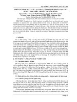
and histologic integrity. The basic
structural components of articular
cartilage include chondrocytes, col-
lagen, extracellular matrix proteo-
glycans, noncollagenous proteins,
and water. The distribution of each
component varies within four dis-
tinct histologic zones: superficial,
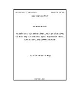
middle, deep, and calcified (Fig. 1).
The basic building block of the
articular surface is the chondrocyte,
which originates from undifferenti-
ated mesenchymal marrow stem
cells. These cells in turn propagate
through the calcified cartilage zone
to become chondroblasts. When the
chondroblasts become isolated in
lacunae, they become chondro-
cytes, which receive their nutritional
support from the synovial fluid. In
skeletally mature articular carti-
lage, chondrocytes no longer di-
vide but still remain alive via the
glycolytic anaerobic metabolism
pathway. Skeletally immature
articular cartilage chondrocytes
undergo cell division and an in-
crease in cell matrix volume. As
chondrocytes age, they exhibit a
decrease in cellular activity, espe-
cially production of both collagen
and proteoglycan. Although chon-
drocytes constitute only 5% of the
wet weight of articular cartilage,
they are the major source for new
synthesis and maintenance of its
components. This includes the
production of collagen, proteogly-
cans, and noncollagenous proteo-
glycans as well as enzymes. They
maintain the balance of synthesis
and degradation of the protein
macromolecular complex.
Water constitutes approximately
75% of the weight of articular carti-
lage. Because of its role as a cation,
water is one of the most important
components of cartilage. Collagen,
predominantly type II, underlies
the form and tensile strength of ar-
ticular cartilage. It makes up ap-
proximately 10% of the weight of
cartilage. Proteoglycans, with their
structural subunits, glycosamino-
glycans, provide the compressive
strength of articular cartilage. They
account for the remaining 10% of
cartilage weight. Proteoglycans
trap and hold water within articu-
lar cartilage. Like other systems
within the body, articular cartilage
Figure 1 Basic structural anatomy of articular cartilage.
Zones
Superficial
Middle
Deep
Flat,
parallel
Flatter,
more rounded
Random,
oblique
Spherical,
in columns
Tidemark
Smaller-
volume cells
Cortical bone
Cancellous
bone
Mesenchymal
stem cells
Calcified
Chondrocyte AppearanceCollagen Orientation
Articular Cartilage Lesions
Journal of the American Academy of Orthopaedic Surgeons
182
contains special subunits, which
interact with cytokines and growth
factors. Interleukin-1, insulinlike
growth factor-1, and transforming
growth factor-β1 combine with
articular cartilage in an anabolic, a
catabolic, or a mixed fashion.
The microarchitecture of articular
cartilage is unique. The outermost
layer, or superficial zone, which con-
tains a relatively small amount of
proteoglycan, is thin, noncellular,
and porous. In this layer, called the
lamina splendens, the fibers are
arranged parallel to the joint surface.
Farther down in the articular carti-
lage, the collagen fibers are oriented
perpendicular to the joint surface.
In the middle zone, the collagen
fibrils have a larger diameter com-
pared with those in the superficial
zone, with a higher concentration of
proteoglycans and lower amounts
of water and collagen. In the third
layer, or deep zone, the largest-
diameter collagen fibrils, the high-
est concentration of proteoglycans,
and the lowest concentration of
water are noted. The collagen fi-
brils eventually pass through the
tidemark boundary and extend into
the remaining area, the calcified
zone that separates the noncalcified
zone from the underlying subchon-
dral bone.
The biomechanics of articular car-
tilage utilize this microanatomy to
reduce the forces of friction across
the joint to extremely low values.
This system incorporates three ma-
jor avenues to lessen the friction in
the joint. First, the parallel fibers of
the lamina splendens provide a flat
surface for the joint to roll or slide
across during motion. Second, the
porous nature of the lamina splen-
dens in combination with the water-
attracting characteristics of the pro-
teoglycans allow fluid flow through
the surface of articular cartilage dur-
ing compression. This fluid flow pro-
duces hydrostatic pressure, which
helps decrease the forces of friction
across the joint. Third, the lamina
splendens surface becomes coated
with phospholipids, which have a
hydrophobic head attracted to the
collagen surface and a hydrophilic
tail pointed toward the opposite
articular surface. This creates an
electrostatic pressure similar to that
of magnets opposing one another.
Recreating this complex microstruc-
ture makes surgical reconstruction
of articular cartilage very difficult, as
all three parts of this biomechanical
system must work together for opti-
mal function.
4,5
Articular cartilage lacks vascu-
lar, neural, and lymphatic access
networks, which creates a limited
environment for spontaneous re-
pair. Injuries that do not penetrate
into the subchondral bone show lit-
tle sign of spontaneous repair,
whereas those that extend into the
depth of subchondral bone initiate
a vascular proliferative response
that produces only fibrocartilage.
Current surgical procedures either
incorporate penetration into the
subchondral bone as part of their
technique or utilize it as a bound-
ary or base for surface restoration.
Clinical Presentation of
Articular Cartilage Lesions
The most common clinical presen-
tation of a full-thickness articular
cartilage lesion is a loose body. It
may be associated with an acute
injury, with a concomitant large
knee effusion, or, more likely, it
may have an insidious onset with
no effusion. Other patients may
have a progressive onset of joint-
line and/or patellofemoral pain
with occasional mechanical symp-
toms of locking or catching. Com-
mon scenarios for the presentation
of full-thickness articular surface
injuries include patellar dislocation
with lateral femoral condylar and
medial-patella facet lesions, Òdash-
boardÓ injuries in which the patella
is driven into the trochlea, and liga-
ment injuries, most often to the
anterior cruciate ligament.
The physical examination usually
does not elicit a distinct consistent
finding other than localized pain
with or without an effusion. The
presence of a loose body should be
considered predictive of the occur-
rence of an articular surface injury
until proven otherwise. A routine
complete examination should be
performed to rule out other factors,
such as malalignment and other
meniscal, ligamentous, and extensor
mechanism problems. Various sub-
jective and objective criteria related
to articular surface injury and repair
may be used to categorize the status
of the knee in both the history and
the physical examination.
Plain radiographs, including
standing posteroanterior flexion
views, may visualize compartment
joint-space narrowing or an osteo-
chondritis dissecansÐtype defect,
with or without a loose body. With
full-thickness articular cartilage
lesions, plain radiography might
not reveal any changes; in that set-
ting, magnetic resonance (MR)
imaging may be more helpful.
Herzog
6
reviewed current MR
imaging techniques for assessing
chondral injury and concluded that
proton-density imaging of thin (3-
to 4-mm) sections and T2-weighted
imaging with fat-saturation sequences
optimize resolution of the articular
chondral surface. High-resolution
gradient-echo imaging has also been
proposed to allow more careful
evaluation of the articular surface
of the patella. Defects are best ana-
lyzed with three orthogonal planes.
In this way, the image obtained will
be perpendicular to the area of con-
cern. With the known high inci-
dence of subchondral bone contu-
sions associated with ligamentous
injuries, the identification of edema
in the subchondral bone should
serve as a flag to carefully review
and analyze the overlying articular
surface.
Jon E. Browne, MD, and Thomas P. Branch, MD
Vol 8, No 3, May/June 2000
183
Although MR imaging remains
the benchmark for musculoskeletal
soft-tissue imaging, its usefulness
in consistently analyzing changes
in the articular surface has been
questioned. Arthroscopy is a more
accurate technique for diagnosing
articular surface lesions. Ochi et al
7
prospectively and retrospectively
analyzed preoperative MR imaging
studies of 65 patients who under-
went surgical procedures and were
found to have 72 articular surface
defects. The overall prospective
sensitivity of MR imaging for these
defects was 40%, with a retrospec-
tive sensitivity of 70%.
The role of the bone scan remains
controversial. Isolated articular sur-
face defects that do not penetrate
the subchondral bone might not be
identified by bone scanning. Dye
and Chew
8
stressed that the change
in joint homeostasis occurring with
any significant joint injury will be
reflected in a persistent increase in
scintigraphic activity. The return to
the normal state is concomitant with
the return of a normal scintigraphic
appearance. Bone scanning has not
been used to document joint homeo-
stasis during the treatment of artic-
ular surface lesions; however, it
may ultimately provide the best tool
for evaluating whether surgical
intervention has restored the joint to
its normal state.
Documentation of
Arthroscopic Findings
Classifying the condition of the
joint and the nature of a chondral
lesion necessitates a documentation
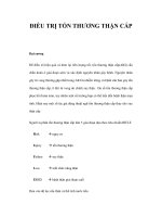
system. The grading system de-
vised by Outerbridge
9
is the sim-
plest working tool for describing
chondral lesions (Fig. 2). Other sys-
tems may be more elaborate and
specific, but the clinical usefulness
of the Outerbridge system in daily
practice makes it still a practical
working approach. This must be
combined with an accurate notation
of the location, size (i.e., surface
area), and shape (i.e., circular, rec-
tangular, or elliptical) of the articu-
lar surface lesion and a description
of the walls (i.e., whether they are
contained, partially contained, or
open). The depth of the lesionÑ
designated as mild (partial thick-
ness), moderate (characterized by
extension to subchondral bone), or
severe (extending into subchondral
bone)Ñmay be the major determi-
nant in the final selection of the sur-
gical technique to be utilized.
The appropriate treatment for
the asymptomatic patient with an
incidental finding of a full-thickness
articular cartilage lesion is problem-
atic. If such a lesion is left untreated,
will it then go on to be symptomatic
within a short period of time? Con-
versely, if it is treated, will it be-
come symptomatic as a result?
Without treatment, might it have
remained asymptomatic? The ab-
sence of a documented natural his-
tory makes these decisions difficult.
Until the natural history of the sur-
gically treated symptomatic lesion
is confirmed, surgical treatment
cannot be recommended; however,
continual reevaluation and follow-
up monitoring are warranted.
Nonoperative Treatment
The goal of nonoperative treatment
is to reduce symptoms related to
the articular cartilage lesion, not to
restore anatomy. Physical therapy
for muscle strengthening, gait
training, and application of appro-
priate bracing or use of an orthotic
device may eliminate some of the
symptoms. Use of intra-articular
viscosupplementation products
and oral chondroprotective agents
for the treatment of osteoarthritis
may also provide symptomatic
relief, but to date there has been
no evidence of structural improve-
ment.
Operative Choices
The various techniques available
for surgical intervention result in a
tissue response that is either repar-
ative or restorative (Table 1). The
ultimate response to surgical inter-
vention may be correlated with the
numbers and kinds of cells used and
how closely the surgical reconstruc-
tion seeks to emulate the micro-
anatomy of the articular cartilage.
The chondrocytes for all of these
procedures are facilitated from
mesenchymal stem cells induced
Figure 2 System for grading the status of
the articular cartilage, as described by
Outerbridge.
9
In grade I, the articular sur-
face is swollen and soft and may be blis-
tered. Grade II is characterized by the
presence of fissures and clefts measuring
less than 1 cm in diameter. Grade III is
characterized by the presence of deep fis-
sures extending to the subchondral bone,
measuring more than 1 cm in diameter.
Loose flaps and joint debris may also be
noted. In grade IV, subchondral bone is
exposed.
Grade I
Clefts
Blister
Subchondral bone
Deep fissures
Subchondral bone exposed
Grade II
Grade III
Grade IV
Articular Cartilage Lesions
Journal of the American Academy of Orthopaedic Surgeons
184
from periosteum or perichondrium,
harvested as autologous chondro-
cytes, or transplanted as allogeneic
chondrocytes.
The goal of restorative surgical
techniques is complete reconstruc-
tion of the microarchitecture of
articular cartilage, with restoration
of all biomechanical and physiolog-
ic functions and resultant complete
relief of symptoms. In contrast, a
reparative surgical technique re-
constructs the defect in a manner
that does not necessarily restore the
articular cartilage architecture but
still may relieve symptoms. Conse-
quently, only some of the biome-
chanical functions of the articular
cartilage are restored, which com-
promises the longevity of the artic-
ular surface due to a higher coeffi-
cient of friction.
There are also some operative
techniques that have no impact on
the articular cartilage defect itself.
For example, arthroscopic lavage
and/or debridement (chondroplas-
ty) may lessen symptoms, but the
effects diminish with time.
10
Pa-
tients with angular deformity and
articular surface lesions (generally
due to osteoarthritis) may show
signs of clinical improvement and
increased joint-space widening
after osteotomy; however, biopsy
specimens obtained from the ar-
thritic compartment consistently
show proliferation of a fibrocarti-
laginous response with little hya-
linelike cartilage restoration.
11
Sim-
ilarly, varus or valgus bracing may
offer symptomatic relief to the
patient with a malaligned knee
without changing the damaged
articular surface structure.
Truly restorative procedures for
the treatment of full-thickness ar-
ticular surface lesions are limited to
single-plug osteochondral auto-
graft transfer (i.e., with the use of
plugs measuring 5 to 12 mm in
diameter) and osteochondral allo-
graft reconstruction. The other avail-
able procedures attempt to achieve
full restoration of only the articular
surface and therefore should be
considered merely reparative.
Abrasion arthroplasty and micro-
fracture rely on facilitation of local
mesenchymal stem cells for articu-
lar cartilage reconstruction; unfor-
tunately, the repair tissue is pre-
dominantly fibrocartilaginous in
nature. Surgery utilizing periosteal
or perichondrial tissue can achieve
a biologic response that is closer to
full restoration, with induction of
chondroneogenic cells; neverthe-
less, the result falls short of full
restoration because microfracture is
still a key component in the tech-
nique. Mosaicplasty is a technique
that involves the use of multiple
donor osteochondral dowel plugs.
This procedure would approach
being restorative if it were not for
the fibrocartilage that invariably
grows between the plugs. Autol-
ogous chondrocyte implantation
appears to offer the best potential
for restoration, involving as it does
reimplantation of the patientÕs own
cultured chondrocytes; however,
core biopsy specimens include
residual periosteum from the artic-
ular surface and therefore repre-
sent some fibrocartilage mixture.
Table 1
Goals and Source of Chondrocytes for Surgical Treatment of Articular Cartilage Lesions
Goals Source of Chondrocytes
Facilitated Intra- Extra-
Procedure Reparative Restorative MSC
*
articular articular Cultured Allogeneic
Chondroplasty
(debridement)
ÐÐ ÐÐÐÐÐ
Laser chondroplasty
ÐÐ ÐÐÐÐÐ
Abrasion arthroplasty +
Ð
+
ÐÐÐ Ð
Microfracture +
Ð
+
ÐÐÐ Ð
Periosteum/
perichondrium +
ÐÐÐ
+
ÐÐ
Autologous chondro-
cyte implantation
++
ÐÐ
++
Ð
Osteochondral auto-
graft transfer
Ð
+
Ð
+
ÐÐ Ð
Mosaicplasty + + + +
ÐÐ Ð
Allograft
Ð
+
ÐÐÐÐ
+
*
MSC = mesenchymal marrow stem cells.
This procedure has both reparative and restorative qualities, but it is predominantly restorative in nature.
Jon E. Browne, MD, and Thomas P. Branch, MD
Vol 8, No 3, May/June 2000
185
Surgical Procedures
Arthroscopic Debridement
Arthroscopic debridement (chon-
droplasty) to remove loose flaps or
edges that mechanically impinge on
the joint will temporarily improve
symptoms. On the basis of a 1-year
follow-up on 15 patients, Levy et al
12
noted 100% good or excellent results
from simple arthroscopic debride-
ment. In their study, they limited
surgical intervention to debridement
of the lesion to a stable rim and
removal of the calcified cartilage
base. Remarkably, 33% of the lesions
found in this homogeneous popula-
tion of soccer players were less than
10 mm in diameter and were consid-
ered to be the source of their symp-
toms. Repeat biopsy specimens
obtained from 4 patients revealed
fibrocartilage in the lesions, suggest-
ing a reparative response. Longer
follow-up is necessary to decide
whether this form of treatment car-
ries the longevity of modern articular
cartilage repair techniques.
Abrasion Arthroplasty
Popularized in the early 1980s by
Johnson, abrasion arthroplasty is
indicated in the treatment of an
exposed sclerotic degenerative ar-
thritic joint lesion. It involves careful
intracortical superficial abrasion to
create a vascular response not medi-
ated by the subchondral bone mar-
row elements, but rather by cells
within the joint itself. At the follow-
up evaluation of 10 patients 1 year
after treatment, Johnson
13
found that
1 patient had 100% hyaline type II
collagen formation; biopsy specimens
from the remaining patients showed
predominantly fibrocartilaginous re-
sponses, with varying amounts of
hyaline articular cartilage. Repara-
tive tissue appears to be the domi-
nant result of this technique.
Microfracture Techniques
Microfracture techniques, such
as drilling of sclerotic subchondral
exposed bone,
14
stimulate the for-
mation of a smooth fibrocartilagi-
nous surface. Steadman et al
15
ex-
panded their use for the treatment
of full-thickness traumatic chondral
injuries. In a series of more than
200 treated patients, the authors
found that 75% had an improve-
ment in pain at a minimum follow-
up interval of 7 years. This tech-
nique involves the use of surgical
awls (rather than drilling, which
generates heat) to create several
subchondral puncture holes 3 to 4
mm apart. Important technical
adjuncts are careful debridement of
the calcified cartilage layer and the
use of postoperative continuous
passive motion (CPM) with protected
weight bearing for 6 to 8 weeks.
Long-term follow-up histologic
analysis is needed to allow evalua-
tion of the repair tissue.
Laser Chondroplasty
Laser chondroplasty allows pre-
cise molding and contouring of
soft-tissue joint structures. How-
ever, there is concern about poten-
tial cellular necrosis of chondro-
cytes near the directed laser beam.
Therefore, care should be taken
when using this technique.
16
Periosteal and Perichondrial
Grafting
Periosteal and perichondrial
grafts have been demonstrated to
effect chondroneogenesis in vitro
from their cambium layer.
17
Hom-
minga et al
18
implanted 30 costal
perichondrial grafts in 25 knees
and noted very good early func-
tional rating scores. At 5 to 7 years
postoperatively, 20 of 30 grafts had
developed enchondral ossification.
Lorentzon et al
19
reported on 26
tibia-based periosteal grafts im-
planted into patellar defects that
had been concurrently treated with
microfracturing and debridement
accompanied by an aggressive
postoperative regimented CPM
program. At an average follow-up
interval of 42 months, 16 excellent
and 9 good results were noted; only
1 patient had a poor result. Biopsy
specimens obtained randomly from
5 patients revealed a hyalinelike car-
tilage appearance. To date, clinical ex-
periences with isolated periosteal
transplants in humans remain limited.
Autologous Chondrocyte
Implantation
Autologous chondrocyte implan-
tation was first reported in 1994 by
Brittberg et al.
20
They initially har-
vested autologous chondrocytes
from 23 patients and then expanded
and manipulated these cells in cul-
ture, prior to reimplantation under
a periosteal flap. The mean follow-
up of these procedures was 39
months. Second-look arthroscopy
and biopsy was performed on 15 of
16 treated femoral lesions in 16 pa-
tients. Hyalinelike tissue repair
was found in 11 lesions. Fourteen
patients rated their results as either
good or excellent. The patellar
defects fared worse, with only 2 of
7 patients rating their knees as
excellent or good, and 1 having
hyalinelike tissue on second-look
arthroscopy and biopsy. Unfortu-
nately, the results noted in in vivo
animal models are conflicting.
21
Improvement was noted in rabbit
models with periosteum plus chon-
drocyte implantation versus perios-
teum implantation only; however,
these results were not replicated in
a canine model.
22
The United States and European
experience in 50 patients (not in-
cluding the Swedish experience)
with at least 2 years of postopera-
tive follow-up has been reported
(Cartilage Repair Registry Report,
vol 4, Genzyme Tissue Repair,
Cambridge, Mass, February 1998).
Clinicians noted a good to excellent
result in 86% of their patients, and
79% of the patients also rated their
results as good to excellent. A total
of 891 transplants are included in
this report. There was a 12.6% com-
Articular Cartilage Lesions
Journal of the American Academy of Orthopaedic Surgeons
186
plication rate (112 patients), and 88
patients (9.9%) required a second
operative procedure. Treatment fail-
ures were noted in 18 patients (2%).
The cumulative index rate of failure
at 2 years was estimated at 5.8%.
Autologous chondrocyte im-
plantation (Fig. 3) is indicated for the
younger (aged 20 to 50 years) active
patient with an isolated traumatic
femoral chondral defect greater than
2 to 4 cm
2
. Care should be taken to
ensure that the lesion is not so deep
(i.e., 3 to 6 mm into the subchondral
boundary) that an initial repair of the
subchondral base might be neces-
sary. Accompanying ligamentous
and meniscal lesions, joint malalign-
ment, and patellofemoral instability
must be corrected concurrently.
Absence of a meniscus may preclude
such treatment even with a meniscal
allograft due to the persistence of
residual high joint-reaction forces.
23
Bipolar lesions of the articular sur-
face also militate against its use.
Osteochondral Autograft
Osteochondral autograft was
first reported by Outerbridge et al
24
for treatment of osteochondritis
dissecans defects in the femur.
They used the lateral patellar facet
as an autograft. As much as one
third of the surface width may be
removed. A follow-up study of 10
patients an average of 6.5 years after
the procedure revealed satisfactory
functional results with decreased
symptoms. Postoperatively, the pa-
tients had mild donor-site patello-
femoral pain.
The mosaicplasty procedure
popularized by Hangody and co-
workers provides treatment op-
tions for much larger and deeper
femoral condylar or patellar de-
fects. In their series of 227 cases,
25
the follow-up interval for 57 pa-
tients was more than 3 years. As
evaluated with use of a modifica-
tion of the Hospital for Special
Surgery scoring system, 91% of
these 57 patients achieved a good
or excellent result. Twelve patients
underwent second-look arthroscop-
ic biopsy, which revealed that the
transplanted cartilage remained
hyaline in character and that donor-
graft bonding sites were fibrocarti-
laginous.
The use of autografts is appeal-
ing; however, there is a limited
amount of donor-graft tissue avail-
able for transfer and a potential
risk of donor-site morbidity. Mo-
saicplasty currently is dependent
on surgical skill to recreate the nor-
mal radius of curvature in the
femoral condyle. This is particular-
ly true when multiple small grafts
(2.7 to 4.5 mm in diameter) must be
press-fitted together to repair a
large defect. The two-dimensional
surface area can be covered with
this technique, but it is difficult to
reproduce the three-dimensional
surface of the femoral condyle.
Collapse of the osteochondral dow-
els by migration or degradation
leads to flattening in the area of the
mosaicplasty. This procedure may
also result in additional damage to
the subchondral bone structure of
the femur, resulting in a change in
the osseous contour of the femoral
condyle in those cases in which the
original lesion affected only chon-
dral tissue.
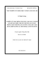
Osteochondral Allografts
Osteochondral allografts (Fig. 4)
may be used for larger (>10 cm
2
)
full-thickness lesions after the fail-
ure of one or two previous surgical
procedures. Fresh allografts (i.e.,
obtained within 24 to 72 hours)
provide the greatest likelihood of
chondrocyte survivability, but also
carry a higher risk of immunogenic
and transmissible disease. Incuba-
tion periods for infection screening
may be too long to allow implanta-
tion of a fresh graft within the 72-
hour time limit. Use of a ÒshellÓ
graft (one with <1 cm of subchon-
dral bone base) reduces immuno-
genicity of the graft by decreasing
exposure of white cells found in can-
cellous bone.
A factor contributing to the fail-
ure of osteochondral allografts is
the host-directed tissue remodeling
of the graft by Òcreeping substitu-
tion.Ó The speed of this substitu-
tion is reduced by ÒcorkÓ fixation
of the allograft (in which a graft
shaped like a tapered cone is press-
fitted into the site) into the host
knee but is increased when trans-
graft stabilization (e.g., with screws
or absorbable pins) is needed.
The technical constraints of sur-
gical implantation of fresh osteo-
chondral allografts are extremely
Figure 3 Arthroscopic images obtained 1 year after autologous chrondrocyte implantation
show an intact graft (arrows) in the lateral femoral condyle of a 35-year-old man who had
had a 6-cm
2
lesion. An acute lateral meniscus tear was noted during examination (A) and
was subsequently resected (B).
A B
Jon E. Browne, MD, and Thomas P. Branch, MD
Vol 8, No 3, May/June 2000
187
demanding. Fresh tissue from a
young donor (<30 years old) must
be available; the recipient patient
must be on call; and the surgeon
must be able to transplant the graft
at all hours of the day and night.
Gross
26
considers the best indica-
tion for the procedure to be a post-
traumatic or osteochondritis disse-
cans defect. Any associated angular
deformity must be corrected by oste-
otomy, usually performed at the
same time. A total of 126 proce-
dures on 123 knees were reviewed at
an average follow-up interval of 7.5
years. The success rate (defined on
the basis of achieving good or excel-
lent results) at 5 years was 95%; at 10
years, 71%; and at 20 years, 66%.
Failures were most common in bipo-
lar grafts and in workerÕs compensa-
tion cases. This procedure is not rec-
ommended for the patient with
osteoarthritis.
Typically, patients present with a
large monopolar traumatic lesion,
which, if left untreated, will result in
permanent damage to the opposite
side of the joint (becoming a bipolar
lesion). Failed primary reconstruc-
tions of intra-articular femoral
condylar fractures or of complex lat-
eral tibial plateau fractures are
appropriate situations. Even when
these patients are maintained in
non-weight-bearing status on
crutches, rapid deterioration of the
other side of the joint occurs. In
these situations, fresh allografts may
be the answer. Although fresh-
frozen allografts have a decreased
risk of immunogenic response and
viral transmission, there is concern
that the viability of chondrocytes in
the donor articular surface will be
reduced, potentially decreasing the
longevity of the osteochondral allo-
graft.
Rehabilitation Guidelines
All of these procedures except
debridement require protected
weight bearing for varying time
periods (a minimum of 6 weeks).
Continuous passive motion may be
helpful for improving surface con-
tour during the postoperative peri-
od. Limitation of the arc of motion
may be necessary, but its value in
articular surface nutrition and
function has been well documented
by Salter.
27
Allograft techniques
usually require longer periods of
protected weight bearing (3 to 4
months). Return to functional
work and sports activities is possi-
ble with all the procedures, but
allograft transplantation necessi-
tates consideration of permanent
moderation of activities.
Authors’ Preference
Our preferred approach for treating
full-thickness articular surface inju-
ries assumes that six basic criteria
have been satisfied: (1) age range
from skeletal maturity to 50 years;
(2) stable knee ligaments, with either
preoperative or concurrent recon-
struction of any defects; (3) stable
neutral tracking extensor mecha-
nism; (4) intact menisci (meniscal
allograft may be necessary); (5) sin-
gle or multiple full-thickness fe-
moral condyle or patellar articular
surface defects without bipolar
defect (i.e., femoral tibial and/or
patellofemoral joint-surface changes
A B
C D
Figure 4 A, Preoperative photograph of the knee of a 50-year-old patient with a failed
osteochondral autograft transfer. B, Implanted osteochondral allograft. C, Osteochondral
allograft and meniscus. D, Photograph obtained during second-look arthroscopy shows
restored contour of the articular surface.
Articular Cartilage Lesions
Journal of the American Academy of Orthopaedic Surgeons
188
greater than grade 2); and (6) a
defect that is not osteoarthrotic or
associated with inflammatory joint
disease. The algorithm shown in
Figure 5 outlines an approach to
simplification of decision making in
the surgical treatment of these
defects.
Traumatic joint injuries that have
resulted in loss of surface contour
involving more than half of the joint
compartment are best handled with
fresh allograft even as a primary
surgical procedure. To date, little
information regarding resurfacing
of tibial defects (other than with al-
lograft) has been published. There-
fore, at this time, smaller lesions
should be treated with debride-
ment, with or without microfractur-
ing. Lesions with loss of surface
contour congruity should be man-
aged with fresh allograft. As for the
tibial articular surface, technical
details of the osteochondral auto-
graft transfer and the mosaicplasty
procedure limit their use to the ante-
rior third of each compartment.
Summary
Articular cartilage is a nearly fric-
tionless system that can provide
maintenance-free service for de-
cades of activity. Unfortunately, its
intrinsic reparative processes can-
not cope with full-thickness injury.
It is difficult to predict which full-
thickness chondral lesion will
progress to become symptomatic,
but current reparative or restora-
tive surgical procedures provide an
opportunity to return the surface to
its normal or nearly normal func-
tional status. Obviously, many
associated factors, such as accom-
panying joint abnormalities, body
weight, job description, and activi-
ty level may influence the necessity
of treating these defects.
Postoperative evaluation of all of
these techniques requires constant
documentation of patient progress.
Occasionally, the need for second-
look arthroscopy will arise. Assess-
ment of the defect should include
inspection, instrumented indenta-
tion probing to measure cartilage
stiffness (compared with the oppo-
site surrounding normal tissue
walls), and biopsy. Surgical biopsy
establishes a more dynamic picture
with histologic evaluation, particu-
larly when it extends to the zonal
base of the calcified and noncalci-
fied subchondral bone as well as
the junction between normal tissue
and the treated defect. Collagen
typing, weight-bearing plain films,
MR imaging, and possibly bone
scanning may also be useful.
New developments might influ-
ence the ease and reproducibility of
articular-surface restoration proce-
dures. Growth factors, adhesives,
artificial bioabsorbable scaffolding
matrices, and gene therapy manip-
ulation are being investigated as
possible adjuncts to the current
standard surgical techniques. Also
being explored is the use of mar-
row aspiration to obtain pluripo-
tential mesenchymal marrow stem
cells, which can then be injected
into the defect and covered by bio-
absorbable artificial matrices or
scaffolding.
Early results need to be carefully
assessed over many years with
continual monitoring and updating
before clinical recommendations
about the durability of results can
be made.
Acknowledgment: The authors would like
to thank Spencer P. Browne for his techni-
cal assistance in manuscript preparation.
Lesions
<1.5 cm
2
Lesions
>1.5 cm
2
but
<4 cm
2
Restorative
• Single-plug OAT
Reparative
• Microfracture
• Chondroplasty
Restorative
• ACI
• Allograft
• Mosaicplasty
Reparative
• Microfracture
• Mosaicplasty
• Microfracture plus
periosteal flap
Restorative
• ACI
• Allograft
Reparative
• Possibly microfracture
• Possibly mosaicplasty
Restorative
• Allograft
• Possibly ACI
Reparative
• None
Lesions
>4 cm
2
but
<8 cm
2
Lesions
>8 cm
2
Symptomatic full-thickness articular surface
lesion (assuming that all preoperative
requirements have been satisfied)
Salvage procedures for all of these techniques should be limited to allograft reconstruction
for lesions >1.5 cm
2
. Lesions <1.5 cm
2
may merit attempts at other surgical techniques
without risking transformation to a bipolar lesion.
Figure 5 Algorithm for the treatment of articular cartilage lesions. ACI = autologous
chondrocyte implantation; OAT = osteochondral autograft transfer.
Jon E. Browne, MD, and Thomas P. Branch, MD
Vol 8, No 3, May/June 2000
189
References
1. Curl WW, Krome J, Gordon ES,
Rushing J, Smith BP, Poehling GG:
Cartilage injuries: A review of 31,516
knee arthroscopies. Arthroscopy 1997;
13:456-460.
2. Messner K, Maletius W: The long-
term prognosis for severe damage to
weight-bearing cartilage in the knee: A
14-year clinical and radiographic fol-
low-up in 28 young athletes. Acta
Orthop Scand 1996;67:165-168.
3. Maletius W, Messner K: The effect of
partial meniscectomy on the long-term
prognosis of knees with localized,
severe chondral damage: A twelve- to
fifteen-year follow-up. Am J Sports
Med 1996;24:258-262.
4. Buckwalter JA, Mankin HJ: Articular
cartilage: Part I. Tissue design and
chondrocyte-matrix interactions. J
Bone Joint Surg Am 1997;79:600-611.
5. Buckwalter JA, Mankin HJ: Articular
cartilage: Part II. Degeneration and
osteoarthrosis, repair, regeneration,
and transplantation. J Bone Joint Surg
Am 1997;79:612-632.
6. Herzog RJ: Radiologic imaging in reha-
bilitation, in Kibler WB, Herring SA,
Press JM, Lee PA (eds): Functional
Rehabilitation of Sports and Musculo-
skeletal Injuries. Gaithersburg, Md:
Aspen Publishers, 1998, pp 20-56.
7. Ochi M, Sumen Y, Kanda T, Ikuta Y,
Itoh K: The diagnostic value and limi-
tation of magnetic resonance imaging
on chondral lesions in the knee joint.
Arthroscopy 1994;10:176-183.
8. Dye SF, Chew MH: The use of scintig-
raphy to detect increased osseous
metabolic activity about the knee. J
Bone Joint Surg Am 1993:75:1388-1406.
9. Outerbridge RE: The etiology of chon-
dromalacia patellae. J Bone Joint Surg
Br 1961;43:752-757.
10. Jackson RW: Arthroscopic surgery
and a new classification system. Am J
Knee Surg 1998;11:51-54.
11. Odenbring S, Egund N, Lindstrand A,
Lohmander LS, WillŽn H: Cartilage
regeneration after proximal tibial
osteotomy for medial gonarthrosis: An
arthroscopic, roentgenographic, and
histologic study. Clin Orthop 1992;277:
210-216.
12. Levy AS, Lohnes J, Sculley S, LeCroy
M, Garrett W: Chondral delamination
of the knee in soccer players. Am J
Sports Med 1996;24:634-639.
13. Johnson LL: Arthroscopic abrasion
arthroplasty, in McGinty JB, Caspari RB,
Jackson RW, Poehling GG (eds): Oper-
ative Arthroscopy, 2nd ed. Philadelphia:
Lippincott-Raven, 1996, pp 427-446.
14. Pridie KH: A method of resurfacing
osteoarthritic knee joints [abstract]. J
Bone Joint Surg Br 1959;41:618-619.
15. Steadman JR, Rodkey WG, Singleton
SB, Briggs KK: Microfracture technique
for full-thickness chondral defects:
Technique and clinical results. Opera-
tive Techniques Orthop 1997;7:300-304.
16. Vangsness CT Jr, Ghaderi B: A litera-
ture review of lasers and articular car-
tilage. Orthopedics 1993;16:593-598.
17. OÕDriscoll SW, Keeley FW, Salter RB:
The chondrogenic potential of free
autogenous periosteal grafts for bio-
logical resurfacing of major full-thick-
ness defects in joint surfaces under the
influence of continuous passive
motion: An experimental investigation
in the rabbit. J Bone Joint Surg Am
1986;68:1017-1035.
18. Homminga GN, Bulstra SK, Bouw-
meester PSM, van der Linden AJ:
Perichondral grafting for cartilage
lesions of the knee. J Bone Joint Surg Br
1990;72:1003-1007.
19. Lorentzon R, Alfredson H, Hildings-
son C: Treatment of deep cartilage
defects of the patella with periosteal
transplantation. Knee Surg Sports
Traumatol Arthrosc 1998;6:202-208.
20. Brittberg M, Lindahl A, Nilsson A,
Ohlsson C, Isaksson O, Peterson L:
Treatment of deep cartilage defects in
the knee with autologous chondrocyte
transplantation. N Engl J Med 1994;
331:889-895.
21. Grande DA, Pitman MI, Peterson L,
Menche D, Klein M: The repair of
experimentally produced defects in
rabbit articular cartilage by autologous
chondrocyte transplantation. J Orthop
Res 1989;7:208-218.
22. Breinan HA, Minas T, Hsu HP, Nehrer S,
Sledge CB, Spector M: Effect of cultured
autologous chondrocytes on repair of
chondral defects in a canine model. J
Bone Joint Surg Am 1997;79:1439-1451.
23. Paletta GA Jr, Manning T, Snell E,
Parker R, Bergfeld J: The effect of allo-
graft meniscal replacement on intraar-
ticular contact area and pressures in the
human knee: A biomechanical study.
Am J Sports Med 1997;25:692-698.
24. Outerbridge HK, Outerbridge AR,
Outerbridge RE: The use of a lateral
patellar autologous graft for the repair
of a large osteochondral defect in the
knee. J Bone Joint Surg Am 1995;77:65-72.
25. Hangody L, Kish G, K‡rp‡ti Z, Udvar-
helyi I, Szigeti I, BŽly M: Mosaicplasty
for the treatment of articular cartilage
defects: Application in clinical prac-
tice. Orthopedics 1998;21:751-756.
26. Gross AE: Fresh osteochondral allo-
grafts for post-traumatic knee defects:
Surgical technique. Operative Tech-
niques Orthop 1997;7:334-339.
27. Salter RB: The biologic concept of con-
tinuous passive motion of synovial
joints: The first 18 years of basic
research and its clinical application.
Clin Orthop 1989;242:12-25.