Báo cáo y học: "Use of the float-moor-crush approach for subtotal mid-segment collapse of a protruding aorto-ostial vein graft stent: a case report" pps
Bạn đang xem bản rút gọn của tài liệu. Xem và tải ngay bản đầy đủ của tài liệu tại đây (1.28 MB, 4 trang )
Case report
Open Access
Use of the float-moor-crush approach for subtotal mid-segment
collapse of a protruding aorto-ostial vein graft stent: a case report
Lieuwe H Piers*, Gillian AJ Jessurun and Rutger L Anthonio
Address: Department of Cardiology, Thoraxcenter, University Medical Center Groningen, Hanzeplein, 9700 RB, Groningen, The Netherlands
Email: LHP* - ; GAJJ - ; RLA -
* Corresponding author
Received: 5 June 2008 Accepted: 27 May 2009 Published: 8 September 2009
Journal of Medical Case Reports 2009, 3:8497 doi: 10.4076/1752-1947-3-8497
This article is available from: />© 2009 Piers et al.; licensee Cases Network Ltd.
This is an Open Access article distributed under the terms of the Creative Commons Attribution License (
/>which permits unrestricted use, distribution, and reproduction in any medium, provided the original work is properly cited.
Abstract
Introduction: Aorto-ostial stenting remains one of the most demanding and risky types of
angioplasty to perform. We report a case outlining a creative solution for the reengagement of a
protruding aorto-ostial stent.
Case presentation: A 69-year-old Caucasian man was admitted to our hospital’s coronary care
unit with progressive unstable angina five years following coronary artery bypass grafting and three
years after percutaneous coronary intervention of the graft. Several attempts to engage the
protruding part of the aorto-ostial stent in the graft failed. A catheter was eventually floated towards
the protruding part using a wire to moor the catheter to the stent through the side-strut. The
proximal part of the protruding stent was subsequently crushed with a new stent. Stent patency was
observed 12 months after the procedure was performed.
Conclusion: Although careful cannulation of a aorto-ostial stent during repeat coronary angiography
coupled with the placement of a guidewire and stent through the true stent lumen during repeat
intervention remains the ideal approach for aorto-ostial in-stent restenosis, this case report confirms
the feasibility of the side-strut stenting technique in reaching a long-term positive outcome.
Introduction
Aorto-ostial stenting remains one of the most demanding
types of angioplasty to perform. Anticipation of risks such
as pot ential stent loss, imprecise or malposed stent
delivery and stent recoil or collapse should guide the
technical approach of the procedure [1]. Aorto-ostial
stenting after bypass surgery adds an additional risk to
the overall technical outcome as the anastomotic area may
be vulnerable especially during the early post-surgical
period. Restenosis or occlusion of the aorto-ostial stent
may render appropriate access to the stent difficult.
Aorto-ostial stenting carries a significant risk and aortic
manipulation should be minimized. We present a case
that demonstrates a creative solution for reengaging a
protruding aorto-ostial stent.
Case presentation
A 69-year-old Caucasian man was admitted to the
coronary care unit with unstable ang ina five years
following coronary artery bypass grafting (CABG) of his
left internal mammary artery to his left anterior descend-
ing artery and a saphenous vein graft from the aorta to the
Page 1 of 4
(page number not for citation purposes)
diagonal, obtuse marginal branch and right descending
posterior artery. Three years prior to presentation, the
patient also underwent a percutaneous coronary interven-
tion (PCI) of the stenotic ostium of the saphenous vein
graft supplying the obtuse marginal coronary artery, with a
Lekton motion 4.0 × 15 mm stent (Biotronik, AG, Bülach,
Switzerland) at 16 atmosphere (Figure 1). One year after
PCI, the patient still suffered from stable angina (NYHA
III), and a second attempt to engage the stent in the vein
graft ostium was performed. Unfortunately, the engage-
ment was unsuccessful and the procedure was aborted.
Another conservative approach was proposed and a PCI of
the native left main and left circumflex artery was
recommended in the event of progressive anginal com-
plaints. Anti-anginal drug treatment was optimized with
long acting nitrates.
Three months later the patient was readmitted with
unstable angina pectoris (NYHA IV). A third attempt to
reengage the protruding stent in the vein graft ostium was
discussed and planned. The protruding part of the aorta
was long and pointed towards the aortic valve at an angle
of about 45 degrees from the aortic wall. This made
reengagement unsuccessful despite multiple attempts with
the use of many 6F guiding catheters. Thereafter, an attempt
to dilate the angulated and calcified left main coronary
artery was aborted when rupture of the balloon compli-
cated the procedure. Instead of trying to engage the stent by
its true lumen, a maneuver that had repeatedly failed, the
treating doctor chose to float a 3.5-mm coronary catheter
(Medtronic) towards the protruding part of the stent until it
stabilized. Subsequently, a rather supportive Pilot 150 wire
(Guidant, Santa Clara, CA, USA) was used to moor the
catheter to the stent through a side strut (Figure 2) and
further advanced the wire to the peripheral portion of the
graft. After a new channel was created by predilatation of
the strut, a 3.5 × 15 mm Endeavour stent (Medtronic,
Minneapolis, MN, USA) was delivered and post-dilated
Figure 1. Final result after aorto-ostial stenting of a vein graft
showing significant protrusion of the stent (marked as = = =)
into the aortic lumen.
Figure 2. Visualization of an aorto-ostial stenting procedure
using the float-moor-crush technique. The treating doctor
chose to float a 3.5-mm coronary catheter towards the
ostium stent. Subselective engagement of the vein graft shows
the collapsed ostium stent (marked as = = =) at the mid-
segment, visible as an hourglass aspect, and the mooring stage
of the procedure (A). After predilatation of the strut, a stent
(====) was delivered and postdilated crushing the protruding
stent at the aorto-ostial side; the crushing phase of the
technique (B). The crushed twin stent is visible as it points at
an 80-degree angle of the new channel (C).
Page 2 of 4
(page number not for citation purposes)
Journal of Medical Case Reports 2009, 3:8497 />with a 4.0 × 15 mm balloon through the newly placed
stent at 18 atmospheres, allowing the proximal part of the
protruding stent to become crushed at the ostial side
(Figure 3). Care was taken not to leave a large part of the
stent protruding into the aorta (Figure 3). Clinical follow-
up at 12 months showed stent patency after selective vein
graft cannulation (Figure 3).
Discussion
PCI of aorto-ostial coronary lesions is confronted by
unique technical challenges not offered by other lesion
subtypes [1]. These include poor guiding catheter support,
difficult stent placement and incomplete stent expansion.
These challenges are further enh anced during repeat
interventions by poor visualization of the intra-aortic
component of stent struts, non-coaxial guiding catheter
engagement [2] and difficulty in placing the guidewire in
the true stent lumen [3]. Furthermore, causal attempts at
engaging the protruded stent and coronary ostium may
deform the stent struts, making further coaxial guiding
catheter engagement impossible.
Stenting for de novo aorto-ostial coronary lesions is
recommended to prevent the strong elastic recoil inherent
to these lesions and to reduce the risk of restenosis. Precise
stent placement, however, is hampered by a lack of
guiding catheter support and poor visualization during
nonselective angiography. Despite these limitations, it is
imperative that a stent should be placed across the
coronary ostium with only 1mm to 2mm of the proximal
stent segment protruding into the aorta to allow complete
lesion coverage and minimize the risk of stent
deformation during subsequent procedures. Similarly,
when encountering a previously placed aorto-ostial stent,
cautious catheter manipulation is essential to establish
coaxial guiding catheter alignment without deforming the
protruded stent struts. Placement of a guidewire in the true
lumen of the stent is often not difficult after a coaxial
guiding catheter has been engaged, but intricate guidewire
techniques may be required if the protruded stent segment
in the aorta is longer than a few millimeters.
As evident from this case, failure to achieve coaxial guiding
catheter alignment despite cautious attempts with multi-
ple guiding catheters, as well as the inability to advance the
guidewire through the true stent lumen, may be the only
signs of occult stent strut deformity. We managed these
difficulties by placing the guiding catheter on top of the
protruded stent and advancing a Pilot 150 (Guidant)
guidewir e th ough the struts of the intra-aortic stent
seg ment. Serial balloon dilatations with sequentially
larger, low-profile balloons were performed to widen the
stent cell opening and dilate the collapsed stent. This
facilitated the passage and placement of a second
Endeavour stent across the lesion through the widened
stent opening. PCI through stent struts has been reported
for treatment of aorta-ostial in-stent restenosis [4,5]. Redo
surgery, however, should be considered. The creative
percutaneous approach employed in our patient was
judged as safe and feasible. The decision for redo surgery
would have probably been facilitated in the presence of
more target lesions. Moreover, the presence of local
adhesions and scar tissue growth during redo surgery
remains a substantial limiting factor for clinical success.
Current literature supports percutaneous intervention as a
good clinical alternative to the surgical indications used in
the past [6].
This case shows a side-strut stenting technique for
complete aorto-ostial collapsed stent at the mid-segment.
Side-strut stenting represents a modification of the
culotte technique [7 ], and displaces the intra- aortic
segment of the previously placed lateral stent, thus
creating a new entry site into the coronary arter y.
Although careful cannulation of the aorto-ostial stent
duringrepeatcoronaryangiography and placement of the
guidewire and stent through the true stent lumen during
repeat intervention remains the ideal approach for aort o-
ostial in-stent restenosis, this report confirms the
feasibility of the side-strut stenting technique in reaching
a long-term positive outcome.
Conclusions
The float-moor-crush approach may be described as a
strategy combining both the side strut and culotte
techniques, and should always be considered as a bail-
out intervention in challenging aorto-ostial lesions.
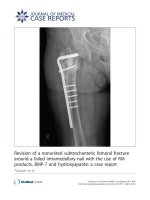
Figure 3. Control angiogram of a vein graft 12 months after
an aorto-ostial stenting procedure using the float-moor-crush
technique shows deep intubation into a patent stent
(marked as ====) and a good backflow.
Page 3 of 4
(page number not for citation purposes)
Journal of Medical Case Reports 2009, 3:8497 />Abbreviations
CABG, coronary artery bypass grafting; PCI, percutaneous
coronary intervention; NYHA, New York Heart Association.
Competing interests
The authors declare that they have no competing interests.
Consent
Written informed consent was obtained from the patient
for publication of this case report and any accompanying
images. A copy of the written consent is available for
review by the Editor-in-Chief of this journal.
Authors’ contributions
GJ and RA analyzed the pro cedure describe in this
manuscript. LP and RA wrote the manuscript. All authors
read and approved the final manuscript.
References
1. Satler LF: Aorto-ostial disease and aorto-ostial in-stent rest-
enosis: poorly recognized but very complex lesion subsets.
Catheter Cardiovasc Interv 2002, 56:220-221.
2. Chetcuti SJ, Moscucci M: Double-wire technique for access into
a protruding aorto-ostial stent for treatment of in-stent
restenosis. Catheter Cardiovasc Interv 2004, 62:214-217.
3. Jain D, Kurowski V, Katus HA, Richardt G: A unique pitfall in
percutaneous coronary angioplasty of in-stent restenosis:
guidewire passage out of the stent. Catheter Cardiovasc Interv
2001, 53:229-233.
4. Abhyankar A, Gai L, Bailey BP: Angioplasty through a stent side
door. Int J Cardiol 1996, 55:107-110.
5. Burstein JM, Hong T, Cheema AN: Side-strut stenting technique
for the treatment of aorto-ostial in-stent restenosis and
deformed stent struts. J Invasive Cardiol 2006, 18:e234-e237.
6. Serruys PW, Morice MC, Kappetein AP, Colombo A, Holmes DR,
Mack MJ, Ståhle E, Feldman TE, van den Brand M, Bass EJ, Van Dyck N,
Leadley K, Dawkins KD, Mohr FW: SYNTAX Investigators:
percutaneous coronary intervention versus coronary-artery
bypass grafting for severe coronary artery disease. N Engl J
Med 2009, 360:961-972.
7. Chevalier B, Glatt B, Royer T, Guyon P: Placement of coronary
stents in bifurcation lesions by the “culotte” technique. Am J
Cardiol 1998, 82:943-949.
Page 4 of 4
(page number not for citation purposes)
Journal of Medical Case Reports 2009, 3:8497 />Do you have a case to share?
Submit your ca se report today
• Rapid peer review
• Fast publication
• PubMed indexing
• Inclusion in Cases Database
Any patient, any case, can teach us
something
www.casesnetwork.com