Báo cáo y học: " Invasive thyroglossal duct cyst papillary carcinoma: a case report" docx
Bạn đang xem bản rút gọn của tài liệu. Xem và tải ngay bản đầy đủ của tài liệu tại đây (428.39 KB, 4 trang )
BioMed Central
Page 1 of 4
(page number not for citation purposes)
Journal of Medical Case Reports
Open Access
Case report
Invasive thyroglossal duct cyst papillary carcinoma: a case report
Leila Aghaghazvini
1
, Habib Mazaher
1
, Hashem Sharifian
1
,
Shirin Aghaghazvini
1
and Majid Assadi*
2
Address:
1
Department of Radiology, Amiralam Hospital, Tehran University of Medical Science, Tehran, Iran and
2
Department of Nuclear Medicine
and Oncology, The Persian Gulf Biomedical Research Institute, Busheher University of Medical Sciences, Bushehr, Iran
Email: Leila Aghaghazvini - ; Habib Mazaher - ;
Hashem Sharifian - ; Shirin Aghaghazvini - ; Majid Assadi* -
* Corresponding author
Abstract
Introduction: A thyroglossal duct cyst is the most common congenital anomaly of the thyroid
gland and midline masses in childhood (70% abnormality in childhood, 7% in adult). Carcinomas
arising from a thyroglossal duct cyst are rare (only 1% of thyroglossal duct cyst cases) and
characterized by relatively non-aggressive behavior and rare lymphatic spread. They are also
diagnosed mostly during the third and fourth decades of life. About 85% to 92% of all thyroglossal
duct cyst carcinomas are papillary carcinomas.
Case presentation: We present the case of a 44-year-old Iranian woman with Cacausian
ethnicity with a painless anterior neck mass that appeared gradually over three months. She had a
history of frequent painful swelling of the anterior part of her neck, which subsided with antibiotic
therapy. Thyroid functional tests were normal and a thyroid scinitigraphy showed a cold nodule in
the left lobe of her thyroid. A computed tomography scan revealed a large, heterogeneous
enhancing soft tissue mass with cystic components in the midline of the anterior neck space. This
extended from the base of the tongue,(completely separated from its muscles, to the inferior
aspect of the thyroid gland and showed the destruction of the hyoid bone and the thyroid cartilage.
The diagnosis of a thyroglossal duct cyst with malignant transformation was maintained. A fine
needle aspiration revealed papillary carcinoma.
Conclusion: This patient's case is presented because of its rare, aggressive, and invasive nature
and rare and unusual manifestation, as well as its rapid increase in size, the destruction of the hyoid
bone, chondrolysis of the thyroid cartilage, lymph adenopathy and the existence of a cold nodule
in the thyroid gland.
Introduction
Thyroglossal duct cysts (TDCs) are the most common
anomaly in thyroid development. In general, duct cysts
are benign, but 1% of cases can be malignant [1]. A review
of the literature showed that 250 cases of malignant thy-
roglossal cysts have been reported [2]. The percentages of
different types of neoplasia in reported cases of TDC are:
papillary carcinoma 81.7%; mixed papillary-follicular car-
cinoma 6.9%; squamous cell carcinoma 5.2%; follicular
and adenocarcinoma, 1.7% each; and malignant struma,
Published: 1 December 2009
Journal of Medical Case Reports 2009, 3:9308 doi:10.1186/1752-1947-3-9308
Received: 7 September 2008
Accepted: 1 December 2009
This article is available from: />© 2009 Aghaghazvini et al; licensee BioMed Central Ltd.
This is an Open Access article distributed under the terms of the Creative Commons Attribution License ( />),
which permits unrestricted use, distribution, and reproduction in any medium, provided the original work is properly cited.
Journal of Medical Case Reports 2009, 3:9308 />Page 2 of 4
(page number not for citation purposes)
epidermoid carcinoma and anaplastic carcinoma, 0.9%
each [1]. Carcinomas arising from a TDC are rare and are
usually characterized by non-aggressive behavior and rare
lymphatic spread [3]. Most cases of TDC carcinoma are
diagnosed during the third and fourth decades of life, and
rarely in children under 14 years of age [4]. We present the
case of a TDC papillary carcinoma because of its rarity,
unusual manifestation, aggressive nature with invasion to
adjacent structures, and interesting history and clinical
findings.
Case presentation
A 44-year-old Iranian woman with Cacausian ethnicity
presented with an anterior midline neck mass that gradu-
ally appeared without tenderness over three months. The
patient had a history of frequent painful swelling of the
anterior part of her neck, which subsided with antibiotics
therapy. A physical examination of the patient revealed a
100 × 55 mm mass that was painless, smooth and hard.
The mass was located on the anterior part of the patient's
neck and extended from the suprahyoid portion to the
thyroid gland. The thyroid gland could not be separated
from the mass. Thyroid functional tests (serum thyroxine,
triiodothyronine and thyroid stimulating hormone) were
within normal limits. A thyroid scan with technetium
pertechnetate detected a cold nodule corresponding to the
mass in the left lobe of the thyroid gland.
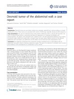
A computed tomography scan (CT) revealed a relatively
large (100 × 55 × 48 mm), heterogeneous enhancing soft
tissue mass with a cystic component in the midline of the
anterior neck space, that extended from the base of the
tongue to the inferior aspect of the thyroid gland and the
bilateral aspect of the submandibular gland (Figure 1, Fig-
ure 2 and Figure 3). The mass was completely separated
from the tongue muscles. The destruction of the hyoid
bone and chondrolysis of the thyroid cartilage were also
seen. A hypodense lesion in the left thyroid lobe and
some adenopathy in the submandibular space were
detected. A fine needle aspiration (FNA) revealed a papil-
lary carcinoma. The tumor mass, together with the thyroid
gland, the hyoid bone and the bilateral cervical lymph
node were therefore removed. Although the thyroid gland
was not involved, some micrometastases in the cervical
lymph nodes were seen. The pathological report revealed
a papillary carcinoma arising with a 100 × 53 mm TDC.
The pathologic report described in detail the occurrence of
complex, branching and randomly oriented papillae with
a central fibrovascular core and a single or stratified lining
of cuboidal cells with a zoom in of 10 (×10). In addition,
nuclear features showed optically clear (ground glass)
nuclei and nuclear grooves (×40), indicating a malignant
papillary carcinoma.
A large heterogenous enhancing mass with cystic component in the midline portion of the anterior aspect of the neckFigure 1
A large heterogenous enhancing mass with cystic
component in the midline portion of the anterior
aspect of the neck. Invasion of the hyoid bone is noted.
A large heterogenous enhancing mass with cystic component in the midline portion of the anterior aspect of the neckFigure 2
A large heterogenous enhancing mass with cystic
component in the midline portion of the anterior
aspect of the neck. Invasion of the hyoid bone is noted.
Journal of Medical Case Reports 2009, 3:9308 />Page 3 of 4
(page number not for citation purposes)
Postoperative radioactive iodine treatment and thyroid
hormone supplements were recommended. The patient
was followed up with a clinical examination, thyroid scin-
tigraphy and ultrasonography of the operation site. The
tumor had not recurred one year after the operation.
Discussion
Thyroglossal duct cysts usually develop in the midline of
the neck. The duct extends upward from the cyst with a
few branches and secretory glands. These ducts or
branches merge into a single duct at the level of the hyoid
bone.
TDCs result from the dilatation of a remnant at the base
of the tongue (foramen cecum), where the primitive thy-
roid originally descended, to its permanent location at the
lower part of the neck. Failure of this tract to close predis-
poses the formation of a thyroglossal cyst [3-7]. Thy-
roglossal duct cysts most often present with a palpable
asymptomatic midline neck mass at the level of or below
the hyoid bone. Suprahyoid thyroglossal duct cysts are
located in the midline of the neck. The more common inf-
rahyoid thyroglossal duct cysts often have both midline
and off-midline components, with the latter embedded in
the strap muscles. The presence of a solid mass along it
should raise the suspicion of ectopic thyroid tissue, in
which occult malignancy is more likely [8].
The neck mass moves with swallowing. Some patients will
have neck or throat pain, or dysphagia and the spectrum
of clinical symptoms may be varied. Diagnosis is usually
made clinically. Antibiotics are indicated if infection is
suspected. Definitive surgical management requires exci-
sion not only of the cyst but also of the path's tract and
branches [9,10].
Carcinomas arising from a TDC are rare (only 1% of
reported TDC cases) and are characterized by relatively
non-aggressive behavior and rare lymphatic spread [2-4].
There are different theories regarding the origin of these
malignancies. Some authors believed that these carcino-
mas were metastases of thyroid carcinomas. However, fol-
lowing demonstrations of normal thyroid tissue
occurrence in the wall of thyroglossal duct cysts, it is now
almost universally accepted that a carcinoma may arise
from thyroglossal remnants [11].
Among the vairous types of neoplasia in TDC, a papillary
thyroglossal duct cyst carcinoma has the best favorable
prognosis, with occurrence of metastatic lesions in fewer
than 2% of cases, while a squamous cell carcinoma has
the worst prognosis [12].
Regional lymph node metastasis of TDC carcinoma occur
in only 7.7% of reported cases, and local invasion rarely
occurs [1-4]. A rapid increase in size, the occurrence of
pain, and the presence of enlarged lymph nodes may sug-
gest malignancy [3,4]. CT scans have demonstrated
unilocular or multilocular low density masses, 2 cm to 4
cm in diameter, presented anywhere from the base of
tongue to the superior margin of the thyroid gland. A well-
circumscribed, low density mass, and occasional periph-
eral rim enhancements or internal septations were seen on
contrast enhanced CTs. The presence of nodules of
enhancing tissue within the cyst raised the possibility of
concurrent malignancy, but this may have only repre-
sented the ectopic thyroid tissue. Magnetic resonance
imaging (MRI) signal characteristics vary depending on
the protein contents of fluid within cysts [8].
In managing patients with carcinomas of TDC before a
surgical procedure, it is important to identify whether the
normally functioning thyroid tissue is in its usual location
or not. Thyroid scans and thyroid function studies should
be ordered preoperatively [3,8]. A CT or MRI scan is usu-
ally performed in cases of suspected thyroglossal duct
cysts in adults to confirm the diagnosis and to exclude
other nodal masses. Definitive surgical management
requires excision not only of the cyst but also of the path's
tracts and branches. The intimate association between the
tract and the hyoid bone requires the simultaneous
removal of the central portion of the hyoid bone to ensure
the complete removal of the tract (Sistrunk procedure).
Recurrence is unlikely after such an operation, except in
cases with skin involvement and intraoperative cyst rup-
ture. There is still controversy about the removal of the
thyroid gland in the case of a papillary carcinoma of TDC
[9-12]. A thyroidectomy is recommended in cases where:
A large heterogenous enhancing mass with cystic component in the midline portion of the anterior aspect of the neckFigure 3
A large heterogenous enhancing mass with cystic
component in the midline portion of the anterior
aspect of the neck. Invasion of the hyoid bone is noted.
Publish with BioMed Central and every
scientist can read your work free of charge
"BioMed Central will be the most significant development for
disseminating the results of biomedical research in our lifetime."
Sir Paul Nurse, Cancer Research UK
Your research papers will be:
available free of charge to the entire biomedical community
peer reviewed and published immediately upon acceptance
cited in PubMed and archived on PubMed Central
yours — you keep the copyright
Submit your manuscript here:
/>BioMedcentral
Journal of Medical Case Reports 2009, 3:9308 />Page 4 of 4
(page number not for citation purposes)
(1) the thyroid gland is found to be nodular with a cold
nodule in the thyroid scan; (2) enlarged lymph nodes are
present; or (3) a history of neck irradiation exists [3].
Conclusion
This case has been presented because of the rare, aggres-
sive and invasive nature, and rare and unusual manifesta-
tion, of the TDC described, as well as the rapid increase in
its size, the associated destruction of the hyoid bone,
chondrolysis of the thyroid cartilage, lymphadenopathy
and the existence of a cold nodule of the thyroid gland.
The patient has been disease-free one year after the opera-
tion.
Abbreviations
CT: computed tomography; FNA: fine needle aspiration;
MRI: magnetic resonance imaging; TDC: thyroglossal duct
cyst;
Consent
Written informed consent was obtained from the patient
for publication of this case report and any accompanying
images. A copy of the written consent is available for
review by the Editor-in-Chief of this journal.
Competing interests
The authors declare that they have no competing interests.
Authors' contributions
LG helped in the design and coordination of the study
and also contributed to writing the draft of the manuscript
and interpreting the radiological figures. HM, HS and SA
supervised the process of acquiring data, and also inter-
preted the radiological images. MA helped draft the man-
uscript and revised it for intellectual content. All authors
read and approved the final manuscript.
Acknowledgements
We are indebted to Mrs Elham Seidali for her helpful suggestions. Thanks
are also extended to our colleagues at the Institute for their technical help
and assistance in data acquisition.
References
1. Weiss SD, Orlich CC: Primary papillary carcinoma of thy-
roglossal duct cyst report of a case and review literature. Br
J Surg 1992, 79:1248-1249.
2. Geok Chin T, Mohd Sidik Sh, Manickam S, Seng Phang K, Clarence-Ko
CH, Jeevanan J: Papillary carcinoma of the thyroglossal duct
cyst in a 15-year-old girl. Int J Otorhinolaryngology 2007, 2:732-735.
3. Kazemi M, Assadi m, Kazemi AA, Ghazvini LA: Primary papillary
carcinoma of thyroglossal duct cyst. Hell J Nucl Med 2006,
9:39-40.
4. Martin-Peres E, Larranaga E, Marron C: Primary papillary carci-
noma arising in thyroglossal duct cyst. Eur J Surg 1997,
163:143-145.
5. Dedivitis RA, Guimaraes AV: Papillary thyroid carcinoma in thy-
roglossal duct cyst. Int Surg 2000, 85:198-201.
6. Qzturk O, Demirci L, Egeli E, Cukur S, Belenli O: Papillary carci-
noma of thyroglossal duct cyst in childhood. Eur Arch Otorhi-
nolaryngol 2003, 260:541-543.
7. Sebastian BJ, Sreekumar S, Vaidyanathan S: Thyroglossal duct cyst
carcinoma - report of a case and review of literature. Thyroid
Research and Practice 2004, 1:27-29.
8. Haaga J: CT and MRI imaging of whole body Ohio: Mosby; 2003.
9. Dedivitis RA, Camargo DL, Peixoto GL, Weissman L, Guimaraes AV:
Thyroglossal duct: a review of 55 cases. J AM Coll Surg 2002,
194:274-277.
10. Hilgar AW, Thomson SD, Smallman LA, Watkinson JC: Papillary
carcinoma arising in a thyroglossal duct cyst: a case report
and review of literature. J Laryngol Otol 1995, 109:112.
11. Sorrenti G, Cavazzuti PP, Zanetti G: Papillary carcinoma arising
in thyroglossal duct cyst: a case report and review of the lit-
erature. Acta Otorhinolaryngol Ital 1995, 15:460-464.
12. Martins AS, Melo GM, Tincani AJ, Lage HT, Matos PS: Papillary car-
cinoma in a thyroglossal duct: case report.
Sao Paulo Med J
1999, 117:248-250.