Phục hồi chức năng đóng chuỗi chi trên doc
Bạn đang xem bản rút gọn của tài liệu. Xem và tải ngay bản đầy đủ của tài liệu tại đây (1.23 MB, 10 trang )
Journal of the American Academy of Orthopaedic Surgeons
412
Orthopaedic surgeons are intimately
involved in the rehabilitation pro-
cess, in that they establish the ana-
tomic diagnosis of the injury, deter-
mine the timing of entrance into and
exit from the rehabilitation program,
and select the modalities and exer-
cises that are used. Even though they
usually do not physically demon-
strate or personally supervise the
exercises, they must understand the
basic concepts underlying the vari-
ous types of exercises and the timing
of exercise progressions, in order to
effectively communicate with the
physical therapists and coordinate
the best program for the individual
patient’s needs.
Most current rehabilitation pro-
grams emphasize functional resto-
ration of the injured part, which
requires not only repair and healing
of injured tissues but also restoration
of correct positioning and move-
ment of joints as well as activation of
muscles in the proper sequence so as
to achieve normal function. Closed-
chain protocols have been advocated
in rehabilitation because they have
characteristics that encourage more
complete functional restoration. These
exercises have been used extensively
for anterior cruciate ligament (ACL)
injuries as a major component of
“accelerated” knee rehabilitation, as
well as for rehabilitation of patients
with patellofemoral and shoulder
injuries.
Despite increasing usage, there is
still controversy about what closed-
chain exercises are and how and
when to use them. Neither is there
a common understanding of what
defines a closed-chain exercise, how
closed-chain exercises promote
functional restoration, and what the
best closed-chain exercises are for
different stages of the rehabilitation
process. To be able to appropriately
prescribe and utilize these tech-
niques, the physician must first un-
derstand the underlying physiology
and biomechanics of closed-chain
rehabilitation.
Definitions
The human body produces motions
and performs complex skills through
sequential activation of muscles and
movement of body segments, or
links.
1,2
This link activation, which
may be activity- or sport-specific, is
termed a “kinetic chain.” There are
two broad-based classes of kinetic
chains—”open,” in which the termi-
nal link of the chain is not loaded
and is freely movable (mobile end,
no load [MNL]), and “closed,” in
which the terminal link is con-
strained or immovable due to a
fixed position or large load (fixed
end, external load [FEL]). The mo-
tion of the foot of a kicking leg is an
example of an MNL kinetic chain.
A pure FEL kinetic chain is exempli-
fied by a fixed foot during a squat
exercise. Force generation, force
distribution, joint motion, muscle ac-
tivation, and resultant tissue stress
can be quite different in the two
classes.
Dr. Kibler is Medical Director, Lexington
Sports Medicine Center, Lexington, Ky. Mr.
Livingston is Clinical Specialist, Lexington
Sports Medicine Center.
Reprint requests: Dr. Kibler, Lexington Sports
Medicine Center, 1221 South Broadway,
Lexington, KY 40504.
Copyright 2001 by the American Academy of
Orthopaedic Surgeons.
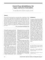
Abstract
Closed-chain exercise protocols are used extensively in rehabilitation of knee
injuries and are increasingly used in rehabilitation of shoulder injuries. They
are felt to be preferable to other exercise programs in that they simulate normal
physiologic and biomechanical functions, create little shear stress across
injured or healing joints, and reproduce proprioceptive stimuli. Because of
these advantages, they may be used early in rehabilitation and have been inte-
gral parts of “accelerated” rehabilitation programs. The authors review the
important components of a closed-chain rehabilitation program and provide
examples of specific exercises that are used for rehabilitation of knee and shoul-
der injuries.
J Am Acad Orthop Surg 2001;9:412-421
Closed-Chain Rehabilitation for
Upper and Lower Extremities
W. Ben Kibler, MD, and Beven Livingston, MS, PT
W. Ben Kibler, MD, and Beven Livingston, MS, PT
Vol 9, No 6, November/December 2001
413
Steindler
2
was the first to de-
scribe the differences in muscle
activation and joint motion that
occur when the distal end of the
arm or leg in a kinetic chain meets
considerable resistance compared
with when it is freely movable. His
definition of a closed-chain condi-
tion required that the foot or hand
meet enough resistance to prohibit
or restrain its free motion, and that
the resultant extremity muscle acti-
vation was sequential from distal
to proximal in the extremity.
Physical therapy protocols have
been developed to take advantage
of the force-generation and loading
characteristics of closed-chain exer-
cises. There has been wide variety
in their application, but in general,
closed-chain protocols include a
progression of exercises that are
based on application of a load to the
distal end of an extremity that does
not move freely due to either posi-
tioning (e.g., on a wall or on the
ground [Fig. 1]) or the load charac-
teristics (e.g., axially applied heavy
load). Subsequent joint motion takes
place in multiple planes while the
limb is supporting weight. These
conditions differentiate these exer-
cises from “open chain” exercises,
such as knee extensions, trunk ex-
tensions against gravity, and isolated
rotator cuff exercises with tubing or
weights.
Dillman et al
3
described condi-
tions that define closed-chain exer-
cises. They realized that the effect
of the exercise on joint positions
and muscle activations is the critical
point in defining a closed-chain
exercise. They felt that closed-chain
exercises have to include relatively
small joint movements, low joint
accelerations, large resistance
forces, joint compression, decreased
joint shear, stimulation of joint pro-
prioception, and enhanced dynamic
stabilization through muscle coac-
tivation. They also recognized that
the amount of load at the terminal
end of the extremity is as important
as the motion of the extremity.
Even if the distal end of the extrem-
ity is somewhat mobile, a large
enough load can still create physio-
logic conditions that replicate
closed-chain characteristics. This
enlarged the concept of a closed-
chain exercise from being only an
FEL activity to include mobile
end–external load (MEL) activities
as well, making it more applicable
to the upper extremity. For exam-
ple, a military press (an MEL exer-
cise) can have closed-chain effects
similar to those of a pushup (an
FEL exercise). This concept has
been validated by Blackard et al,
4
who found that there was no differ-
ence in muscle activation between
equivalently loaded upper-extremity
exercises with either fixed or mobile
ends. They concluded that external
loading characteristics were more
important than arm motion in de-
scribing and simulating human
activity.
Lephart and Henry
5
modified the
MEL condition to include two differ-
ent external load conditions—axial
load (e.g., military press) and rotary
load (e.g., arm rotations with dumb-
bells). This helped to define types of
exercises but did not change the con-
cept that closed-chain joint loading
and muscle activation characteristics
can be obtained with a movable dis-
tal end. However, it enlarged the
scope of the types of exercises that
can be employed in a closed-chain
rehabilitation program.
Livingston
6
developed an opera-
tional definition to guide implemen-
tation of closed-chain exercises and
to determine whether exercises have
the characteristics described by
Dillman et al.
3
As Livingston de-
fines closed-chain rehabilitation
exercises, the activities require a
sequential combination of joint
motions; the distal end of the kinetic
chain meets considerable resistance
(MEL or FEL conditions); and move-
ment of the individual joints of the
kinetic chain sequence and transla-
tion of their instant centers of rota-
tion occur in a predictable manner
determined by the distribution of
forces throughout the chain. This
definition implies control by the
physician or therapist of (1) extrem-
ity position, (2) distal segment mo-
tion and position, (3) application of
forces and loads, and (4) movement
of the entire extremity.
Physiology and
Biomechanics of Closed-
Chain Rehabilitation
Most lower-extremity occupational
and athletic activities involve kinetic
chain activity. The large majority of
these activities start with the feet on
the ground, which gives a base of
stability, allows generation of a
ground-reaction force, and initiates
a sequence of segment activity to
provide optimal position and mo-
tion for the distal aspect of the ter-
minal segment in the chain.
1,6
Force
production is governed by the
“summation of speed” principle, in
Figure 1 Positioning for early-stage lower-
extremity exercises. With one-leg support,
notice hip-trunk extension posture. The
arms may be used to help balance the
trunk initially.
Closed-Chain Rehabilitation
Journal of the American Academy of Orthopaedic Surgeons
414
which the total energy or force in a
kinetic chain is summated from the
contributions of individual seg-
ments.
1
Kinetic-chain segment motions
and positions are created by mus-
cle activation patterns. Length-
dependent patterns operate locally
around a joint, using co-contraction
force couples to control joint per-
turbations. Force-dependent pat-
terns harmonize segment motions
by operating around two or more
joints and using agonist-antagonist
force couples to generate or trans-
fer force.
7
These two types of mus-
cle activations result in coordinated
segment motions that allow kinetic-
chain activity to produce the de-
sired forces needed for occupa-
tional or athletic purposes. The
resultant synergistic patterns create
postural stability throughout the
entire extremity while allowing
voluntary muscle activity at the
distal segment.
1,2,8
These synergis-
tic patterns include increased acti-
vation of biarticular muscles (i.e.,
hamstrings, quadriceps) by mon-
articular muscles (i.e., gluteus
medius, soleus)
9
and coordination
of arm and scapular movements to
produce glenohumeral stability
through the range of motion, al-
lowing maximum arm motion.
10
They are highly dependent on
joint- and angle-specific proprio-
ceptive feedback.
11
In the leg, the hamstrings act as
part of a length-dependent force cou-
ple to control anterior tibial transla-
tion.
8
They also work as part of a
force-dependent pattern to coordi-
nate hip and knee motion,
1
stabilize
the hip, and transfer loads up and
down the leg.
9
In the shoulder, the
rotator cuff acts as part of a length-
dependent force couple to increase
glenohumeral concavity and com-
pression,
10
but also works as part of
a force-dependent pattern to link
trunk extension, scapular rotation,
and arm internal rotation.
8,10
Closed-chain exercise protocols
have characteristics that simulate
these biomechanical and physiolog-
ic requirements. Mechanically, they
initiate joint movements from the
ground or a base of support, em-
phasize sequential control of seg-
ment position or motion, place the
segments in functionally correct
positions, and control the transfer of
generated loads. Physiologically, they
utilize both length-dependent and
activity-specific force-dependent
activation patterns, emphasize
position-specific proprioceptive
feedback to initiate and control acti-
vation, and can use the more versa-
tile MEL configuration to achieve
FEL muscle activation (Fig. 2).
Closed-chain exercises have also
been shown to be protective for
healing and repaired tissues. They
produce minimal translation, shear,
and distraction forces due to the
compressive nature of the applied
load and the greater control of the
resultant motions.
5,6,9,12,13
This con-
fers a margin of safety that allows
shorter periods of complete immo-
bilization, earlier initiation of reha-
bilitation, and resultant “accelerated”
protocols.
12-14
Because closed-chain exercises
emphasize and produce patterns of
A B
Figure 2 Intermediate-stage lower-extremity exercises. A, Use of a sliding board for an MEL exercise to balance the body over a moving
leg. The patient slides side to side and pushes off each edge. B, Use of a Fitter involves the same principles as use of a sliding board, but
there is a more unstable base due to its rounded edges, therefore presenting more of a proprioceptive challenge.
W. Ben Kibler, MD, and Beven Livingston, MS, PT
Vol 9, No 6, November/December 2001
415
motions and muscle activations,
they may not maximally rehabilitate
all of the individual muscles or
achieve normal motion of all of the
joints in the relevant kinetic chain.
This is due to both muscle-activation
substitutions and alterations that
allow approximations of the normal
patterns and to individual character-
istics of muscle activation. Some
muscles, such as the deltoid, upper
trapezius, gluteus medius, and gas-
trocnemius, are more resistant to
inhibition and alteration in injury or
fatigue situations; others, such as the
serratus anterior, lower trapezius,
supraspinatus, and vastus medialis
obliquus (VMO), are easily fatigued
and inhibited, and tend to “drop
out” from the activation patterns.
Clinical examples of these alter-
ations include hip abductors and
extensors substituting for knee ex-
tensors in gait after ACL recon-
struction,
15
the upper trapezius
substituting for the lower trapezius
in acromial elevation,
10,16
and the
deltoid substituting for the supra-
spinatus in arm elevation. In these
situations, the desired kinetic-chain
function (walking or arm elevation)
may be accomplished, but activa-
tion of important muscles is not.
Rehabilitation strategies can be
developed to maximize activation
of the inhibited muscles while still
utilizing a closed-chain framework.
This involves placing the extremity
in a closed-chain position, empha-
sizing the normal activation pat-
tern, and progressively “unmask-
ing” the target muscle by eliminat-
ing the substituting muscle. This
process may be called “facilitation
of muscle activation.”
Role of Closed-Chain
Exercises in Rehabilitation
Closed-chain rehabilitation protocols
have beneficial characteristics that
are associated with functional physi-
ology and biomechanics. They may
be utilized early in the rehabilitation
sequence to protect the injured area
and to prepare the entire kinetic
chain for function. They are the foun-
dation for some rehabilitation pro-
grams. However, they must be mon-
itored to ensure that all muscles are
being appropriately activated. These
protocols can be used in both MEL
and FEL configurations for knee and
shoulder rehabilitation (Figs. 3-7).
Different levels of exercises may be
used in the early (acute or healing)
phase, the intermediate (recovery)
phase, and the late (functional) phase
of rehabilitation,
6,7
depending on the
degree of tissue healing, the possible
positions of the extremity, and the
amount of load and the range of
motion that are allowed.
Knee and Leg Rehabilitation
Closed-chain rehabilitation tech-
niques have been utilized to accel-
erate and improve functional re-
storation after ACL injury and
reconstruction.
12-14
These tech-
niques create weight-bearing forces
across the joint that increase local
agonist-antagonist muscle coactiva-
tion, decrease joint shear, minimize
joint displacement and ACL strain,
and reproduce proprioceptive stim-
uli. In addition, they activate the
kinetic chains of weight bearing,
running, and jumping. This repro-
duces the normal biomechanics of
the entire leg, allowing hip-muscle
activation to increase quadriceps
and hamstring force output by
transferring muscle work to these
biarticular muscles
9
and by creating
a hip moment that is a major con-
tributor to the knee moment.
15
Hip-
muscle activation and work output
create load-absorbing capacity that
can compensate for a low load-
absorbing capacity in the knee so
that the entire leg functions at an
acceptable level early in rehabilita-
tion.
15
Closed-chain exercises also
reproduce the physiologic length-
dependent patterns for hip- and
knee-joint stability, as well as force-
dependent patterns of coordination
of hip, knee, and ankle joint motion.
The effect that closed-chain exercises
have on the entire kinetic chain is
more functionally important than
the effect on the knee joint alone.
Closed-chain techniques are also
useful in rehabilitation of the
patient with patellofemoral pain,
largely due to the same factors of
joint position control, larger im-
provements in the strength of the
entire kinetic chain, and alteration
of the magnitude and position of
applied forces. Increased total leg
stiffness, with resultant knee joint
control, is achieved by activating
the hip muscles concurrently with
the knee muscles.
9,17
Closed-chain
exercises have been shown to pro-
duce greater improvements in
quadriceps strength and leg perfor-
mance than open-chain exercises.
18
Closed-chain exercises produce
lower patellofemoral joint stresses
in the functionally and sympto-
matically important arc of motion
from extension to 45 degrees of flex-
ion than do open-chain exercises.
19
Lower-extremity closed-chain
exercises are largely FEL, with the
foot on the ground in the early reha-
bilitation stages. Most protocols
emphasize early, if not immediate,
weight bearing on the affected ex-
tremity. The leg may be supported,
but controlled range of motion and
compression loading of the joint are
encouraged. Initially, the patient is
in a two-legged support stance, but
may be moved into a one-legged
support stance as healing progresses.
Rhythmic motion patterns of flexion/
extension and lateral movement are
used. Early emphasis is placed on
achievement and maintenance of a
position of 0 degrees of hip exten-
sion and neutral pelvic tilt to allow
maximum hip-muscle activation.
Most leg exercises should proceed
from this “ideal” position.
Closed chain–based protocols
advocated for rehabilitation of ACL
and patellofemoral injuries
12-14,17
Closed-Chain Rehabilitation
Journal of the American Academy of Orthopaedic Surgeons
416
are similar in their early stages, but
differ in the intermediate and re-
covery stages (from 3 weeks to 3
months). Common characteristics
include progressive compression
loading of the joint, controlled
increase in range of motion, main-
tenance of functional posture of the
knee and leg, and emphasis on
early return to functional activities,
such as running, weight lifting, and
mild cutting.
Some of the more commonly
advocated closed-chain exercises in
the intermediate and recovery
stages include the two-legged squat
with increasing resistance, the one-
legged squat with support, and the
step up–step down maneuver—all
of which are FEL exercises. Exam-
ples of MEL exercises include slid-
A B
D E
C
Figure 3 Exercises to increase quadriceps
activation. A and B, Hip extension and
foot-flat in step down–pull up exercise acti-
vates the quadriceps eccentrically and con-
centrically. Hip control is maintained by
activation of hip extensors and abductors.
Notice VMO activation in left leg. C, Slant
board use in a step down–pull up exercise.
Quadriceps activation is greater. D and E,
Hip extension and hip and pelvis rotation.
The trunk and hip are rotated around the
planted leg. Notice VMO activation.
W. Ben Kibler, MD, and Beven Livingston, MS, PT
Vol 9, No 6, November/December 2001
417
ing on a smooth surface (Fig. 2, A)
or using a Fitter device (Fitter
International, Calgary, Alberta,
Canada) with a rounded-edge sup-
port surface (Fig. 2, B) and trampo-
line bounding. The range of possi-
ble exercises is large, and creativity
may be used to match the exercises
to the sport or activity demand.
In the late stage (after 3 months),
emphasis is on functional progres-
sions and a mixture of closed- and
open-chain exercises. The closed-
chain exercises for joint coordina-
tion, leg control, and resistance to
perturbation should be regarded as
a base for the open-chain exercises
of jumping rope, cutting, kicking,
leg extensions, and leg curls.
Facilitation patterns to maximize
quadriceps activation and increase
knee load-bearing capacity are also
employed in the recovery and func-
tional stages of rehabilitation. They
initially involve active hip extension
and quadriceps activation with the
foot flat on the floor or stepping off
a flat step (Fig. 3, A and B). This
FEL pattern reactivates the normal
sequencing pattern for the entire
leg, but probably does not maximally
isolate or activate the quadriceps.
The MEL equivalent, using a tram-
poline, wobble board, or Fitter, adds
an element of increased propriocep-
tive feedback. More effective quad-
riceps activation in a closed-chain
exercise is accomplished by placing
the foot on a slant board. Ankle
plantar-flexion and slight hip flex-
ion decrease hip and ankle activa-
tion, but slight knee flexion places
more emphasis on quadriceps acti-
vation as the patient executes a step
up–step down maneuver (Fig. 3, C).
Further quadriceps facilitation is
accomplished by one-legged stance,
hip extension, slight knee flexion,
and hip and trunk rotation around
the planted leg (Fig. 3, D and E).
This FEL exercise promotes maxi-
mal electromyographic activity in
the VMO. The MEL equivalent uti-
lizes a trampoline or wobble board.
In summary, closed-chain exer-
cises for knee rehabilitation allow
early weight bearing, protect the
injured or healing area, and prepare
the entire extremity for vigorous
functional open- or closed-chain
athletic activities. They should form
the basis for most knee rehabilita-
tion protocols, including those for
ACL and patellofemoral injuries.
The exact sequence and composi-
tion of the protocols may be vari-
able, but limited outcomes assess-
ments indicate a faster return to
functional status with protocols in
which these types of exercises are
emphasized.
12,13
Shoulder and Scapular
Rehabilitation
On superficial analysis, it would
appear that closed-chain rehabilita-
tion would have little application for
the shoulder and arm. The hand is
A B C
Figure 4 Early-stage exercises for shoulder and scapular rehabilitation. A, Trunk extension and scapular retraction. The arm may be ele-
vated or at the side, depending on healing. Diagonal hip extension and scapular retraction can be done with either a two-legged stance
(B) or a one-legged stance. One-legged stance improves hip and pelvis control. Muscle activation goes from the hip through trunk exten-
sion to scapular retraction in either stance. C , The modified pushup is an early-stage exercise for lower trapezius–serratus anterior weak-
ness. Pushups may also be done with the hands on a table.
Closed-Chain Rehabilitation
Journal of the American Academy of Orthopaedic Surgeons
418
obviously moving in an open-chain
fashion in throwing and serving,
and the arm assumes a weight-
bearing position only in gymnastics
and blocking in football. However,
shoulder position, motion, and force
transfer fit the physiologic and bio-
mechanical requirements of closed-
chain activities. In throwing and
serving, the scapula and shoulder
display intersegmental coordination,
with coupled movements that are
predictable on the basis of arm posi-
tion.
8,10
The shoulder acts as a stable
funnel, transferring and regulating
forces in the kinetic chain from the
legs to the hand.
1,20
The shoulder
muscles are activated in mainly co-
contraction length-dependent pat-
terns to stabilize the joint.
9,10,20,21
Proprioception plays a major role in
controlling and activating muscle
patterns.
11
In swimming, weight lift-
ing, and playing on the offensive or
defensive line in football, the hand
meets considerable resistance but
still moves, creating MEL conditions
at the distal end of the extremity.
Closed-chain exercises should,
therefore, be utilized in shoulder
and scapula rehabilitation for func-
tional return to most athletic activi-
ties from all types of shoulder in-
juries. Rehabilitation protocols for
tendinitis, postoperative instability,
and postoperative labral injuries are
basically the same in the acute
phase and in the early functional
phase.
5,6
Postoperative rotator cuff
protocols should vary with the in-
tegrity of the repair, but can also
benefit from the proximal activation
and low shear characteristics. Just
as in knee and leg rehabilitation,
closed-chain exercises may be used
in the early stages of rehabilitation,
and emphasis should be placed on
involving all of the joints of the
kinetic chain.
Early-stage exercises involve not
only the scapula but also the hip
and trunk. The large extrinsic mus-
cles of the shoulder (the latissimus
dorsi and pectoralis major) and the
muscles that position the scapula
(the upper and lower trapezius and
serratus anterior) are all attached to
the trunk. They provide key stabili-
ty and force generation to decrease
shoulder load and facilitate rotator
cuff activation. Early in rehabilita-
tion, when the shoulder muscles are
weakest, facilitation needs from
proximal muscle activation are at
their greatest. Muscle activation
patterns to rehabilitate these mus-
cles start with stabilization of the
hip and trunk, and involve diagonal
as well as ipsilateral exercises.
Diagonal activations (from left hip
to right arm and from right hip to
left arm) are important to recreate
the rotational activation and control
patterns that are used in athletic
activities such as throwing and
swimming and in daily activities
such as reaching and starting a lawn
mower. Commonly employed exer-
cises to rehabilitate these patterns
include trunk extension–scapular
retraction (Fig. 4, A) and diagonal
hip extension–scapular retraction
(Fig. 4, B). These exercises may be
done both preoperatively and in the
immediately postoperative period.
They involve minimal forces at the
shoulder and may be done with the
arm in a sling or other protective
device. Such exercises create a stable
posture of the proximal segments
that allows accelerated rehabilita-
tion of the healing distal tissues.
Closed-chain exercises are the
most effective method for rehabili-
tation of the patient with scapular
dyskinesis (alterations in scapular
A B
C D
Figure 5 Intermediate-stage scapular “clock” exercises (arrows indicate direction of
scapular motion). A and B, Elevation and depression (12- and 6-o’clock positions, respec-
tively). C and D, Retraction and protraction (9- and 3-o’clock positions).
W. Ben Kibler, MD, and Beven Livingston, MS, PT
Vol 9, No 6, November/December 2001
419
position and motion that are fre-
quently associated with shoulder
injury and lower trapezius and
serratus anterior muscle weak-
ness).
5,6,21,22
These alterations are
clinically manifested by promi-
nence of the inferior medial, entire
medial, or superior medial border
of the scapula, depending on asso-
ciated muscle weakness or inflexi-
bility.
Weakness of the lower trapezius
and serratus anterior is very com-
mon, and these muscles are fre-
quently difficult to reactivate. Early-
stage exercises include modified
pushups (Fig. 4, C) and scapular
“pinch” retractions—exercises in
which the scapulae are retracted to
the midline. These initiate scapular
control, but do not create forces that
protract the scapula or place shear
stress on the shoulder joint. Facilita-
tion of lower trapezius and serratus
anterior activation can be achieved
by combined hip-trunk extension
and shoulder extension (the “low
row” exercise). This exercise may be
started in an FEL pattern, with the
hand on a wall or table, and then
changed to an MEL pattern with the
hand on rubber tubing, a ball, or a
movable device. Decreasing hip sta-
bility by standing on a wobble board
or trampoline decreases hip exten-
sion and facilitates maximal lower
trapezius activation.
Intermediate-stage closed-chain
exercises include scapular “clock”
exercises (Fig. 5), in which the
scapula is rotated in elevation and
depression (12- and 6-o’clock posi-
tions) and retraction and protraction
(9- and 3-o’clock positions). Elec-
tromyographic studies have demon-
strated that these exercises activate
scapular stabilizers at moderate lev-
els, but do not create shoulder-joint
shear by deltoid activation.
6
These
can also be done in FEL fashion,
with the hand on a wall, or in MEL
fashion, with the hand on a ball.
Other MEL exercises include “wall
washes” (Fig. 6), scapular retraction
and shoulder extension, military or
bench presses, pushups “with a
plus” (performed by arching the
back and pushing out farther at the
end of the pushup),
23
and dynamic
hug exercises.
24
Functional-stage closed-chain
scapular exercises include higher-
speed scapular protraction and
retraction with weights or tubing
and medicine-ball drills. These
MEL exercises involve hip exten-
sion and trunk rotation and provide
plyometric-type stretch-shortening
cycles to improve power develop-
ment. They can be coordinated with
more open-chain exercises for rapid
hand velocity when appropriate.
Rotator cuff rehabilitation with
use of closed-chain techniques close-
ly simulates normal rotator cuff
function. The rotator cuff functions
as a compressor cuff when the arm
is in the common athletic position of
80 to 105 degrees of elevation, and is
maximally activated off a stabilized
scapula.
10,25
The positions advo-
cated for maximal isolation of the
individual rotator cuff muscles for
evaluation and strengthening are
not commonly seen in normal serv-
ing or throwing and do not allow
integration of rotator cuff function
with the rest of the kinetic chain.
Closed-chain exercises enhance joint
compression, activate scapula- and
shoulder-coupled motions, control
joint position, and stimulate propri-
oception.
3-5,10,11,24,25
Because these
exercises create low levels of mus-
cle activation, they are safer in the
early stages of rehabilitation.
6
Early-stage exercises include
table pushups and humeral head
depressions (Fig. 7, A). When the
arm can safely achieve 90 degrees of
abduction, intermediate-stage activ-
ities include rotator-cuff clock exer-
cises, isometric humeral head de-
pression with trunk extension and
scapular retraction, and pushups,
either modified or normal. These
create joint compression, work the
shoulder muscles in co-contraction
at physiologic positions, and reacti-
vate normal proprioceptive pat-
terns.
25,26
Facilitation of rotator cuff
activation is achieved by trunk ex-
tension and scapular retraction.
This allows optimal positioning of
Figure 6 “Wall washes” for scapular rehabilitation. The hand slides on a smooth-
surfaced wall. Trunk extension and rotation and scapular motion are emphasized.
Closed-Chain Rehabilitation
Journal of the American Academy of Orthopaedic Surgeons
420
the muscles to generate force while
minimizing length and tension mis-
matches.
10,25
Late-stage closed-
chain exercises include MEL exer-
cises, such as punches with weights
(Fig. 7, B and C), standing arm
abduction with weights or tubing,
and medicine-ball drills.
6,23,25,26
Summary
Closed-chain exercise protocols have
assumed a large role in functional
knee rehabilitation. They have been
advocated as being safer and more
effective than previously described
protocols for both ACL
12-14
and
patellofemoral
17,19
rehabilitation,
although not all studies demonstrate
a clear superiority in outcomes.
Closed-chain exercises are also being
employed in shoulder rehabilitation
protocols,
5,6,24
although no outcomes
studies have been reported. Utiliza-
tion of these protocols is based on
theoretical benefits and anecdotal
evidence of more rapid return of
shoulder function. On the basis of
this information, it appears that
closed-chain exercises may increase
the effectiveness of both knee and
shoulder rehabilitation protocols
by simulating normal physiologic
activations and biomechanical
motions. Because of the utility and
increasing use of these exercises,
physicians should be familiar with
the underlying biomechanics and
when it is appropriate to use them.
They appear to be effective in the
early stages of rehabilitation due to
the control of joint motion and tissue
loads. There is no clear consensus
about a particular set of exercises
that should be included in every
rehabilitation protocol, although
both FEL and MEL exercises can be
effective at different stages. The
major criterion for including an
exercise in a closed-chain protocol
is whether it fits the definition
6
and
accomplishes the purposes
3,5
of
closed-chain activation.
Closed-chain exercises and reha-
bilitation protocols are not the only
techniques for functional rehabilita-
tion. A combination of open- and
closed-chain exercises will ultimately
be necessary to simulate normal
functions and optimize the return to
activities, especially in throwing,
striking, and kicking sports.
5,24
The
FEL and MEL exercises provide a
stable base and allow shading into
MNL exercises that are truly open-
chain activities.
Closed-chain exercises offer great
promise in making rehabilitation
more efficacious. Much more atten-
tion should be paid to standardizing
protocols, validating their direct
influence on joint loads and muscle
activation, and reporting outcomes
studies.
Figure 7 Exercises for rotator-cuff rehabilitation. A, Isometric humeral head depression. The resistive weight is set so that the bar does
not move. The arm is pulled down with the elbow straight, so that the depression force is concentrated at the shoulder. B and C, Rotator
cuff punches with weights. The weight should create a load but allow the arm to be extended. The exercise should start with hip and
trunk extension and scapular retraction (B) and then proceed to arm punches at different levels of arm elevation (C).
A B C
W. Ben Kibler, MD, and Beven Livingston, MS, PT
Vol 9, No 6, November/December 2001
421
References
1. Putnam CA: Sequential motions of
body segments in striking and throw-
ing skills: Descriptions and explana-
tions. J Biomech 1993;26:125-135.
2. Steindler A: Kinesiology of the Human
Body Under Normal and Pathological
Conditions. Springfield, Ill: Charles C
Thomas, 1955, pp 63-67.
3. Dillman CJ, Murray TA, Hintermeister
RA: Biomechanical differences of
open and closed chain exercises with
respect to the shoulder. J Sports Rehab
1994;3:228-238.
4. Blackard DO, Jensen RL, Ebben WP:
Use of EMG analysis in challenging
kinetic chain terminology. Med Sci
Sports Exerc 1999;31:443-448.
5. Lephart SM, Henry TJ: The physiolog-
ical basis for open and closed kinetic
chain rehabilitation for the upper
extremity. J Sports Rehab 1996;5:71-87.
6. Kibler WB, Livingston B, Bruce R:
Current concepts in shoulder rehabili-
tation. Adv Oper Orthop 1995;3:249-300.
7. Nichols TR: A biomechanical perspec-
tive on spinal mechanisms of coordi-
nated muscular action: An architecture
principle. Acta Anat (Basel) 1994;151:1-13.
8. Zattara M, Bouisset S: Posturo-kinetic
organisation during the early phase of
voluntary upper limb movement: I.
Normal subjects. J Neurol Neurosurg
Psychiatry 1988;51:956-965.
9. Umberger BR: Mechanics of the vertical
jump and two-joint muscles: Implica-
tions for training. Strength Conditioning
1998;10:70-74.
10. Happee R, Van der Helm FCT: The
control of shoulder muscles during
thigh muscles using closed vs. open
kinetic chain exercises: A comparison
of performance enhancement. J Orthop
Sports Phys Ther 1998;27:3-8.
19. Steinkamp LA, Dillingham MF, Mar-
kel MD, Hill JA, Kaufman KR: Biome-
chanical considerations in patello-
femoral joint rehabilitation. Am J
Sports Med 1993;21:438-444.
20. Kibler WB: Biomechanical analysis of
the shoulder during tennis activities.
Clin Sports Med 1995;14:79-85.
21. Kibler WB: The role of the scapula in
athletic shoulder function. Am J Sports
Med 1998;26:325-337.
22. Lukasiewicz AC, McClure P, Michener
L, Pratt N, Sennett B: Comparison of 3-
dimensional scapular position and ori-
entation between subjects with and
without shoulder impingement. J
Orthop Sports Phys Ther 1999;29:574-586.
23. Moseley JB Jr, Jobe FW, Pink M, Perry
J, Tibone J: EMG analysis of the scap-
ular muscles during a shoulder reha-
bilitation program. Am J Sports Med
1992;20:128-134.
24. Decker MJ, Hintermeister RA, Faber
KJ, Hawkins RJ: Serratus anterior
muscle activity during selected reha-
bilitation exercises. Am J Sports Med
1999;27:784-791.
25. Davies GJ, Dickoff-Hoffman S: Neuro-
muscular testing and rehabilitation of
the shoulder complex. J Orthop Sports
Phys Ther 1993;18:449-458.
26. Wilk KE, Arrigo CA, Andrews JR:
Closed and open kinetic chain exercise
for the upper extremity. J Sports Rehab
1996;5:88-102.
goal directed movements: An inverse
dynamic analysis. J Biomech 1995;28:
1179-1191.
11. Lephart SM, Pincivero DM, Giraldo JL,
Fu FH: The role of proprioception in
the management and rehabilitation of
athletic injuries. Am J Sports Med 1997;
25:130-137.
12. Shelbourne KD, Nitz P: Accelerated
rehabilitation after anterior cruciate
ligament reconstruction. Am J Sports
Med 1990;18:292-299.
13. Bynum EB, Barrack RL, Alexander
AH: Open versus closed chain kinetic
exercises after anterior cruciate liga-
ment reconstruction: A prospective
randomized study. Am J Sports Med
1995;23:401-406.
14. Beynnon BD, Johnson RJ: Anterior
cruciate ligament injury rehabilitation
in athletes: Biomechanical considera-
tions. Sports Med 1996;22:54-64.
15. DeVita P, Hortobagyi T, Barrier J: Gait
biomechanics are not normal after
anterior cruciate ligament reconstruc-
tion and accelerated rehabilitation.
Med Sci Sports Exerc 1998;30:1481-1488.
16. McQuade KJ, Dawson J, Smidt GL:
Scapulothoracic muscle fatigue associ-
ated with alterations in scapulohumer-
al rhythm kinematics during maxi-
mum resistive shoulder elevation. J
Orthop Sports Phys Ther 1998;28:74-80.
17. Thomeé R, Augustsson J, Karlsson J:
Patellofemoral pain syndrome: A
review of current issues. Sports Med
1999;28:245-262.
18. Augustsson J, Esko A, Thomeé R,
Svantesson U: Weight training of the