Musculoskeletal problems and injuries - part 5 pptx
Bạn đang xem bản rút gọn của tài liệu. Xem và tải ngay bản đầy đủ của tài liệu tại đây (666.4 KB, 31 trang )
osteosarcomas occur most commonly in children and young adults
and are most common in males. There appears to be a genetic predis-
position. The secondary osteosarcomas generally develop in adults in
areas of abnormal bone (e.g., Paget’s disease) or in response to some
sort of carcinogen exposure (most commonly irradiation). The most
common presenting complaints of patients with osteosarcoma are
local pain, tenderness, and swelling. It most often occurs in the
medullary cavity of the metaphyseal end of the long bones of the
extremities. Radiographs, computed tomography (CT) scans, or MRI
scans often provide a characteristic picture of subperiosteal or soft tis-
sue penetration of the tumor with extraosseous bone density. To con-
firm the diagnosis, however, biopsy is required. Great advances have
been made in treatment recently, with a combination of surgery, radio-
therapy, and chemotherapy (depending on the specific type of lesion)
providing the best chances for survival.
27
Chordoma
Chordoma is a malignant bone tumor seen most commonly in the
sacrum and spine. It is thought to arise from remnants of the noto-
chord. These tumors are usually seen in middle-aged and elderly
adults. Radiographs, CT scans, or MRI scans usually show the mixed
lytic and sclerotic lesions of the chordoma.
30
Metastatic Malignant Tumors
Tumors that commonly metastasize to bone include thyroid, breast,
prostate, bronchus, kidney, bladder, uterus, ovary, testicle, and adrenal
tumors. Lymphomas most commonly spread to bone from primary
involvement of lymph nodes but also are seen rarely primarily in the
skeleton. Bone scans are thought to be the best screening test for
patients suspected of having skeletal metastasis.
31
Patients with
metastatic bone disease most often present with pathological fracture
or pain. The radiographical appearance of these lesions tends to be
sclerotic in prostate and breast metastasis and lytic in lung, bowel,
kidney, and thyroid. Biopsy of the bony lesion is helpful for deter-
mining whether the lesion is metastatic.
Miscellaneous Bone Conditions
Nonossifying Fibroma
A common condition, nonossifying fibroma is also called a fibrous
cortical defect. It is considered a developmental aberration rather than
a neoplasm. It is seen primarily in children and occurs most com-
monly in the femur, tibia, and fibula. The diagnosis can usually be
142 Jeffrey G. Jones and Doug Poplin
made by the radiographic picture, and a large number of these lesions
are found while obtaining a radiograph for another purpose. The
lesions are sharply demarcated, lobular, radiolucent defects in the
metaphyseal cortex. There is often an intact, thin layer of subpe-
riosteal cortical bone. The lesions may range in size from a few mil-
limeters to 5 cm. They are usually asymptomatic and are seen in
approximately one third of children.
27
The larger lesions may cause
pain and predispose the child to fracture. These lesions do not tend to
transform into neoplasms and often disappear spontaneously.
Paget’s Disease of Bone
Osteitis deformans (Paget’s disease of bone) is characterized by
excessive bone destruction and disorganized repair, resulting in mot-
tled increased density and bony deformity.
27
There is a genetic com-
ponent to this lesion, although many people develop clinically
insignificant lesions. The condition is thought to be related to a canine
distemper (paramyxovirus) infection.
32
Diagnosis. Paget’s disease is often asymptomatic and discovered
incidentally by radiography. When symptomatic, nighttime bone pain
is usually the first symptom. Because of bone softening, bowing of the
tibias, pathological fractures, and increased kyphosis are commonly
seen. An increasing head circumference, deafness, and a waddling
gait are other relatively common symptoms. A markedly elevated
serum alkaline phosphatase level and normal calcium and phosphorus
are the usual laboratory pattern. An elevated 24-hour urinary hydrox-
yproline level, indicative of rapid bone turnover, is also seen.
Radiographic findings include expanded bone with increased density.
Early on, radiolucent lesions are common, especially in the skull and
pelvis (Fig. 6.5). Later mixed, then sclerotic lesions are seen.
27
A bone
scan can detect lesions before they become apparent on plain radi-
ographs.
Complications. The complications of Paget’s disease include frac-
tures, spinal cord compression, malignant degeneration, and hyper-
calcemia-related problems such as renal stones. The latter
complication is seen primarily if there is excessive calcium intake
along with immobilization.
Treatment. Treatment is warranted only if significant symptoms are
present. NSAIDs can be of value in suppressing bone activity and
controlling mild symptoms. Calcitonins and diphosphonates suppress
6. Selected Disorders of the Musculoskeletal System 143
144 Jeffrey G. Jones and Doug Poplin
Fig. 6.5. (A) Bone scan shows extensive uptake in half of the
pelvis in this patient with nocturnal pelvic pain. (B) Plain film
shows coarse trabeculae over the acetabulum (black arrow) and
a thickening of the iliopectineal line (white arrow), findings seen
with Paget’s disease.
bone resorption mediated by osteoclasts and are effective in Paget’s
disease. These treatments have significant potential side effects and
complications. The alkaline phosphatase level can be used to monitor
disease activity.
Prognosis. The later in life that Paget’s disease begins, the better is
the prognosis. The progression is usually slow, over years. Renal
complications and malignant degeneration of lesions are associated
with a poor prognosis.
References
1. Clauw DJ. Fibromyalgia: More than just a musculoskeletal disease. Am
Fam Physician. 1995;52:843–51.
2. Goldenberg DL. Fibromyalgia syndrome: An emerging but controversial
condition. JAMA. 1987;257:2782–7.
3. Stormorken H, Brosstad F. Fibromyalgia: Family clustering and sensory
urgency with early onset indicate genetic predisposition and thus a “true”
disease [letter]. Scand J Rheumatol. 1992;21:207–11.
4. Silman A, Schollum J, Croft P. The epidemiology of tender point counts
in the general population [abstract]. Arthritis Rheum. 1993;36(suppl):48.
5. Granges G, Littlejohn GO. A comparative study of clinical signs in
fibromyalgia/fibrositis syndrome, healthy and exercising subjects. J
Rheumatol. 1993;20:344–51.
6. Reynolds WJ, Moldofsky H, Saskin P, et al. The effects of cyclobenza-
prine on sleep physiology and symptoms in patients with fibromyalgia. J
Rheumatol. 1991;18:452–4.
7. Simms RW, Goldenberg DL. Symptoms mimicking neurologic disorders
in fibromyalgia syndrome. J Rheumatol. 1988;15:1271–3.
8. Pellegrino MJ, Van Fossen D, Gordon C, et al. Prevalence of mitral valve
prolapse in primary fibromyalgia: A pilot investigation. Arch Phys Med
Rehabil. 1989;70:541–3.
9. Goldenberg DL. Management of fibromyalgia syndrome. Rheum Dis
Clin North Am. 1989;15:499–512.
10. Felson DT, Goldenberg DL. The natural history of fibromyalgia. Arthritis
Rheum. 1986;29:1522–6.
11. Yunus MB, Kalyan-Raman UP, Kalyan-Raman K. Primary fibromyalgia
syndrome and myofascial pain syndrome: Clinical features and muscle
pathology. Arch Phys Med Rehabil. 1988;69:451–4.
12. Thompson JM. Tension myalgia as a diagnosis at the Mayo Clinic and its
relationship to fibrositis, fibromyalgia, and myofascial pain syndrome.
Mayo Clin Proc. 1990;65:1237–48.
13. Harden RN, Bruehl SP, Gass S, Niemiec C, Barbick B. Signs and symp-
toms of the myofascial pain syndrome: A national survey of pain man-
agement providers. Clin J Pain. 2000;16(1):64–72.
14. Lederhaas G. Complex regional pain syndrome: New emphasis. Emerg
Med. 2000;32:18–22.
6. Selected Disorders of the Musculoskeletal System 145
146 Jeffrey G. Jones and Doug Poplin
15. Warfield CA. The sympathetic dystrophies. Hosp Pract. 1984;May:
52c–j.
16. Kemler MA, Barendse GAM, Kleef M, et al. Spinal cord stimulation with
chronic reflex sympathetic dystrophy. N Engl J Med. 2000;343(9):618–24.
17. Haddox JD, Van Alstine D. Pharmacologic therapy for reflex sympa-
thetic dystrophy. Phys Med Rehabil. 1996;10:297–309.
18. Redd RA, Peters VJ, Emery SF, et al. Morton neuroma: Sonographic
evaluation. Radiology. 1989;171:415–17.
19. Strong G, Thomas PS. Conservative treatment of Morton’s neuroma.
Orthop Rev. 1987;16:343–5.
20. Mann RA. Pain in the foot. 2. Causes of pain in the hindfoot, midfoot,
and forefoot. Postgrad Med. 1987;82:167–74.
21. Riolo J, Young VL, Ueda K, et al. Dupuytren’s contracture. South Med J.
1991;84:983–96.
22. James JIP. The relationship of Dupuytren’s contracture and epilepsy.
Hand. 1969;1:47–9.
23. Noble J, Heathcote JG, Cohen H. Diabetes mellitus in the aetiology of
Dupuytren’s disease. J Bone Joint Surg. 1984;66B:322–5.
24. McFarlane RM. The current status of Dupuytren’s disease. J Hand Surg.
1983;8:703–8.
25. Smith DL, Wernick R. Common nonarticular syndromes in the elbow,
wrist, and hand. Postgrad Med. 1994;95:173–91.
26. Jennings CD. Deciding whether and how to treat painful ganglia.
J Musculoskel Med. 1986;3:39–46.
27. Rosenberg AE. Skeletal system and soft tissue tumors. In: Cotran RS,
Kumar V, Robbins SL, eds. Robbins’ Pathologic Basis of Disease.
Philadelphia: Saunders, 1994;1213–46.
28. Healey JH, Ghelan B. Osteoid osteoma and osteoblastoma. Clin Orthop.
1986;204:76–85.
29. Vande Streek PR, Carretta RF, Weiland FL. Nuclear medicine approaches
to musculoskeletal disease. Radiol Clin North Am. 1994;32:227–53.
30. Tumors and infiltrative lesions of the lumbosacral spine. In: Borenstein
DG, Wiesel SW, Boden SD, eds. Low Back Pain. Philadelphia: Saunders,
1995;390–5.
31. Ell PJ. Bones and joints. In: Maisey MN, Britton KE, Gilday DL, eds.
Clinical Nuclear Medicine. Philadelphia: Saunders, 1983;135–65.
32. Cartwright EJ, Gordon MT, Freemont AJ, et al. Paramyxoviruses and
Paget’s disease. J Med Virol. 1993;40:133–41.
33. Taylor RB, ed. Family Medicine: Principles and Practice. 6th ed. New
York: Springer, 2003.
7
Musculoskeletal
Problems of Children
Mark D. Bracker, Suraj A. Achar,
Todd J. May, Juan Carlos Buller,
and Wilma J. Wooten
Torsional and Other Variations
of the Lower Extremity
Gait Abnormalities
Rotational problems resulting in gait abnormalities are the most com-
mon orthopedic conditions in the pediatric age group. Parents are fre-
quently concerned that their child will grow up deformed or be unable
to play sports as they observe in-toeing or out-toeing and seek med-
ical attention. Recent studies, however, have shown athletes with
internal tibial torsion are faster than age-matched controls.
1
Most rota-
tional abnormalities resolve spontaneously as musculature develops,
and knowing this fact is reassuring to parents. Rarely, conditions
remain fixed and require surgical correction at an older age. Torsional
deformities may be due to problems in the foot (metatarsus adductus),
tibia (torsion), or femur and hip (femoral anteversion). Angular
abnormalities (bowlegs, knock-knees) generally resolve sponta-
neously as well. Certain terminology has been recommended as well
as specific testing used to evaluate gait (Fig. 7.1).
Terminology
Definitions of the terms used in this chapter are as follows.
Angle of gait (foot progression angle): Angle of the intersection
between the foot axis and the line progression. It is the result of
static and dynamic influences from the foot to the hip. This angle
remains relatively stable at 8 to 12 degrees of out-toeing through
growth. There is a wide range of normal values varying from 3
degrees in-toeing to 20 degrees out-toeing; in one study of 130 chil-
dren, 4.5% had an in-toeing gait.
2
Abnormalities anywhere along
this kinetic chain (including hip, leg, and foot) can change the angle
of gait.
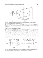
Femoral antetorsion: Anteversion beyond the normal range [2 stan-
dard deviations (SD)].
Femoral anteversion: Angular difference between the forward
inclination of the femoral neck and the transcondylar femoral axis
(Fig. 7.2).
148 Mark D. Bracker et al.
A
CDE
c
B
b
a
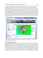
Fig. 7.1. Tests for torsional deformities (see text for full discus-
sion). (A) Foot progression angle (a) is formed by the foot axis (B)
and the line of progression (b). (B) Foot axis. (C) Measurement of
internal femoral rotation. (D) Measurement of external femoral
rotation. (E) Thigh-foot angle (c) is formed by the longitudinal axis
of the femur and the foot axis. (From Lillegard and Kruse,
50
with
permission.)
Foot axis: Imaginary line bisecting the long axis of the foot from the
mid-heel through the middle to the metatarsal heads.
Internal and external femoral rotation: The child lies prone with the
knees flexed to 90 degrees, the pelvis is stabilized, and the angle of
gravity-assisted internal (medial rotation) and external rotation (lateral
rotation) of each leg is measured.
Thigh–foot angle: Measures tibial torsion. The child lies prone and
flexes the knees to 90 degrees; the angle is then placed in neutral
position. Looking down at the sole of the foot, an imaginary line
through the long axis of the foot is measured against the long axis of
the femur. The angle between these two axes is the thigh–foot angle.
Evaluation and Interpretation
The medical history is obtained first and includes the type of defor-
mity, apparent time of onset, amount of progression, family history,
and previous treatment. A complete musculoskeletal and neurological
examination is performed, and finally a torsional (rotational) profile is
generated to determine the severity and level of deformity (Fig. 7.3).
7. Musculoskeletal Problems of Children 149
POSTERIOR
TFA
30 - 35Њ
10 - 15Њ
5 - 10Њ
CHILD
ADULT
TOP VIEWANTERIOR VIEW
INFANT
TFA
TFA
Fig. 7.2. Transcondylar femoral axis (TFA) as it would be meas-
ured radiographically in degrees of rotation.
150 Mark D. Bracker et al.
ROTATIONAL PROFILE
C
MR
girls
B
FPA
R
FPA
MR
LR
TFA
Foot
L
20Њ
10Њ
20Њ
40Њ
60Њ
80Њ
0
20Њ
20Њ
−40Њ
−20Њ
20Њ
40Њ
0
40Њ
60Њ
80Њ
100Њ
40Њ
60Њ
80Њ
0
1
1
3
3
5
5 7 9 11 1315-19 30s 50s70+
79
Age (yrs)
TFA
11 1315-19 30s 50s 70+
1
1 3 5 7 9 11 1315-19 30s 50s70s
3579
Age (yrs)
EFLATERAL ROTATION
THIGH-FOOD ANGLE
LR
Age (yrs) Age (yrs)
11 1315-19 30s 50s 70+
−10Њ
1357911
Age (yrs)
MR boys
D
13 15-19 30s50s 70+
2SD
2SD
2SD
2SD
2SD
2SD
2SD
2SD
2SD
2SD
0
MEDIAL ROTATION
A
FOOD PROGRESSION
ANGLE
MEDIAL ROTATION
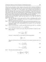
Fig. 7.3. (A) Torsional profile. (B–F) Range of normal values by
age group and sex. (From Engel and Staheli,
2
with permission.)
Foot Progression Angle. It is important to watch the child walk as
naturally as possible. When being observed, children may initially try
to control the amount of in-toeing to please the parent or physician.
Keep in mind also that the amount of in-toeing becomes worse when
a child is fatigued. In-toeing is expressed as a negative value (12–10
degrees) and out-toeing as a positive value. The normal value range
for the foot progression angle is wide, and severe deformity above the
foot may exist with a normal angle.
Hip Rotation. With the child in the prone position, the knees are
flexed to 90 degrees with the pelvis level. The thigh is then rotated
medially (internal rotation of the hip) by gravity alone. Lateral rota-
tion is measured with the child in the same position by allowing the
legs to cross. The diagnosis of medial femoral torsional deformity/
femoral anteversion is made if medial rotation is more than 70
degrees. Total joint laxity must be taken into consideration by con-
current reduction in lateral rotation. Restriction of lateral rotation dur-
ing early infancy is thought to be due to intrauterine position.
Tibial Rotation. Tibial rotation, the most difficult measurement to
make accurately, requires assessment of the thigh–foot angle (TFA).
The TFA increases from early childhood to mid-childhood. Internal
tibial rotation is expressed as a negative angle. A negative value up to
20 degrees is considered normal during infancy. Medial tibial torsion
exists if the TFA is more than 20 degrees. During early childhood the
tibia rotates laterally.
Foot. The sole of the foot is observed to determine its shape; the lat-
eral border is normally straight. Metatarsus adductus is the character-
istic appearance of a “bean-shaped foot” with a wide space between
the first and second toes, prominence at the base of the fifth metatarsal
bone, and convexity at the lateral side of the foot. Metatarsus adduc-
tus is often present in conjunction with tibial torsion.
Clinical Patterns and Management
In-toeing (Metatarsus Adductus). The terms metatarsus adductus
(MA) and metatarsus varus are used interchangeably. MA occurs
when the forefoot bones are deviated medially at the tarsal–metatarsal
junction, causing the foot to appear to curve inward at the midfoot
(bean-shaped foot). It is probably caused by a combination of
intrauterine position and genetic predisposition and can be either flex-
ible or rigid. Studies dispute the belief that hip dysplasia is higher
7. Musculoskeletal Problems of Children 151
among patients with metatarsus varus than in the general population.
3
On physical examination the foot is convex laterally and concave
medially. The lateral border of the base of the fifth metatarsal may
appear prominent. With the heel held in neutral position and pressure
directed laterally at the first metatarsal head, a flexible deformity cor-
rects to neutral but does not overcorrect as do normal feet. One help-
ful test is to stroke the lateral border of the foot, noting if the infant
reflexively corrects the deformity. Treatment for flexible MA involves
having the parents passively correct the range of deformity (as
described above) with each diaper change. Due to the high rate of
spontaneous resolution and the history of natural resolution, no
treatment has been shown to be superior. These treatments vary from
observation to casting to bracing night or day, or both, to orthopedic
shoes. Children with rigid MA require cast correction and are best
treated before 6 months of age and worked up for other neuromuscu-
lar disorders. If begun during the first month of life, correction can
often be obtained within 6 to 8 weeks of casting by a knowledgeable
orthopedist. After age 8 months, cast correction is almost impossible
due to foot stiffness and active kicking by robust toddlers.
The reasons for treating these feet remain controversial, and spe-
cific treatment indications vary among orthopedic surgeons. It is cur-
rently believed that residual MA is not linked to adult degenerative
arthritis.
1
Surgical correction is rarely indicated. When needed,
Heyman–Herndon soft tissue releases are advised for children under
age 4 years, and multiple metatarsal osteotomies are recommended
for older children.
4
In severe cases requiring surgical correction, asso-
ciated heel valgus is common and must be addressed or the child will
be further disabled because correction of the forefoot alone removes
the stable tripod of the foot.
Tibial Torsion. In-toeing can also be due to excessive internal tibial
torsion (medial tibial version). It can be clinically estimated using the
TFA described previously. Normally the tibia is externally rotated 5
degrees at birth and 15 degrees at skeletal maturity. Correction is
almost always spontaneous. Bracing, splints, twister cables, and shoe
modifications have not been shown to be effective and are not recom-
mended, as most of these deformities correct spontaneously by 3 to 4
years of age.
5
Developmental correction may be delayed if the child
sleeps prone with the legs internally rotated or sits with the knees
flexed and feet internally rotated. Although there is no proved benefit
of altering the child’s sitting position, parents may be instructed to
encourage the child to avoid these positions. Derotational osteotomy
is reserved for severe deformity, including significant functional and
152 Mark D. Bracker et al.
cosmetic disability, internal rotation of more than 85 degrees, external
rotation of less than 10 degrees, radiographic anteversion of more
than 45 degrees, or external tibial rotation of less than 35 degrees. The
child must be at least 7 to 8 years old.
6
Femoral Anteversion. The angle between the femoral neck axis and
the transcondylar axis of the distal femur is called femoral version
(Fig. 7.2). Femoral anteversion (FA) decreases from an average of 40
degrees at birth to about 15 degrees at skeletal maturity. Children
commonly sit on their knees with their feet out to the sides in the clas-
sic W position. With femoral anteversion the in-toeing is worse at the
end of the day when compensating muscles fatigue. FA presents by
age 3 to 4 years and resolves slowly over the next 5 years and is more
common in girls than boys.
Infants normally have limited medial rotation due to a tight hip cap-
sule, and external rotation to 90 degrees is common. External rotation
decreases to around 55 degrees by age 3 and slowly decreases there-
after. Internal rotation increases from 35 degrees at birth to 60 degrees
by age 6, at which time it is slightly greater than external rotation.
From birth to 2 years of age the total range should be 120 degrees,
decreasing to 95 to 110 degrees thereafter.
Treatment for an in-toeing gait due to excessive femoral antever-
sion, termed medial femoral torsion, is almost always simple obser-
vation, as 85% resolve with spontaneous derotation of the proximal
femur during normal growth. Bony derotation occurs up to age 8 and
in some cases into adolescence. Surgery is rarely indicated; it is
reserved for severe, uncompensated medial femoral torsion causing
significant functional and cosmetic problems during late childhood.
In rare severe cases, a proximal femoral derotation osteotomy can be
done safely at age 9 or 10.
Angular Abnormalities of the Knee
Newborns generally have a genu varus of approximately 15 degrees
due physiologically to intrauterine positioning. Parents frequently
note bowed legs as their child starts to stand. Children with superim-
posed internal tibial torsion actually look more bowed than they are.
Children progress from genu varus in the newborn until 24 months
and then start to develop genu valgus to about 15 degrees by age 4
years. Then by 6 to 7 years of age, this valgus begins to correct to 5
to 6 degrees, where it essentially remains to adulthood. Pathologic
genu valgum or varus should be evaluated for metabolic disorders,
inflammatory disease, tumors, osteochondrodysplasia, posttraumatic
7. Musculoskeletal Problems of Children 153
conditions, congenital abnormalities, osteogenesis imperfecta, or
Blount’s disease as possible causes.
Bowlegs
Excessive genu varus deformities with a tibial–femoral angle of more
than 20 degrees should be investigated if they have not started correcting
by 2 years of age. Growth charts should be carefully reviewed along with
developmental and family history. Evaluation should include physical
exam, gait observation, knee ligament laxity assessment, rotational
evaluation, and foot position. Appropriate laboratory and standing
radiographic studies should be ordered. The posteroanterior (PA) stand-
ing radiographs must be taken with the child’s feet together or a shoul-
der width apart and neutral rotation with the patella pointing directly
forward. The physis should be carefully examined. A tibiofemoral angle
of more than 20 degrees in toddlers indicates severe physiologic bowing,
or Blount’s disease. Severe physiologic bowing is characterized radi-
ographically as follows.
1. Medial metaphyseal beaking of the proximal fibula and distal
femur
2. Medial cortical thickening
3. Varus angulation of more than 20 degrees based on the metaphy-
seal–diaphyseal angle
4. No pathologic changes in the proximal tibial epiphysis
After other etiologies have been ruled out and severe physiologic
bowing is diagnosed, spontaneous correction can be expected by 7 to
8 years of age. If significant deformity persists past age 8, corrective
tibial osteotomy is necessary in certain cases.
Blount’s Disease
Osteochondrosis deformans tibiae, or Blount’s disease, is due to defec-
tive formation of the posterior medial border of the proximal tibial epi-
physis and may be difficult to distinguish from severe physiologic
bowing. Blount’s disease is more common in blacks than whites and is
associated with obesity and early walking. Radiographic findings after
18 to 24 months are angulation under the posterior medial proximal
epiphysis, metaphyseal irregularity, beaking of the proximal tibia, and
wedging of the proximal epiphysis. Another radiographic sign that has
been found useful to diagnose Blount’s disease is the metaphyseal–
diaphyseal (MD) angle. The angle is derived from drawing a line along
154 Mark D. Bracker et al.
the lateral tibial cortex on a standard PA radiograph, and then drawing
a line perpendicular to the tibial cortex line and one through the epi-
physis. If the angle between the epiphysis and tibial cortex perpendi-
cular line is greater than 11 degrees, Blount’s disease is diagnosed.
7
Most of these children require corrective bracing or surgery and should
be referred as soon as identified.
Knock-Knees
Genu valgus (knock-knees) can be apparent, physiologic, or patho-
logic. Apparent valgus may be due to large thighs, joint laxity, or poor
muscle tone. Most cases are idiopathic or physiologic. Pathologic
causes include juvenile rheumatoid arthritis, rickets, trauma, endocrine
disturbance, and infection. Most children have a slight genu valgus that
generally resolves by 6 years of age; it can become excessive later dur-
ing childhood or early adolescence when the normal valgus fails to
resolve. Genu valgus may represent an acceleration of normal angula-
tion caused by abnormal forces across the knee. Standing PA radi-
ographs with the feet pointing straight ahead may be obtained to
document the tibiofemoral angle and to rule out underlying disease.
Young children with this problem tend toward spontaneous resolution.
With older children, knock-knees is less likely to correct completely.
Surgical correction of severe knock-knees deformity causing sig-
nificant functional or cosmetic problems should be performed 1 year
before the end of physeal growth in the femur (girls, 10–11 years old;
boys, 12–13 years old). A staple encircles the femoral physis, which
continues to grow laterally but not medially.
8
Problems of the Feet
Toe Walking
The tiptoe gait characteristic of beginning toddlers should give way to
an adult-like pattern by 2 years of age. Neuromuscular conditions
such as cerebral palsy or spinal cord lesions such as spina bifida, teth-
ered cord, and diastematomyelia can produce foot deformity, which
can be appropriately evaluated diagnostically or referral made if toe
walking persists beyond age 2.
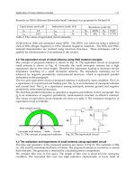
Clubfoot
Talipes equinovarus (clubfoot), which occurs in approximately 1/1000
births,
9
is characterized by talar plantar flexion, hindfoot varus, forefoot
adduction, and soft tissue contractures, resulting in a cavus foot
7. Musculoskeletal Problems of Children 155
deformity (Fig. 7.4).
10
It is thought to be secondary to intrauterine posi-
tion in a genetically predisposed fetus but is also associated with con-
genital hip dislocation, myelomeningocele, and arthrogryposis. The
major deformity of clubfoot is in the subtalar complex, with shortening
and medial deviation of the talus with displacement of the navicular
medially.
11
Radiographs confirm the severity of deformity, allow com-
parisons over time, and are essential for judging the type of surgical cor-
rection needed.
Treatment by an experienced orthopedic surgeon is an acquired
skill that is becoming a lost art. Proper intervention involves reduction
of the displaced navicular on the head of the talus and mobilization of
tight capsules and tendons through manipulation followed by place-
ment in a series of carefully molded corrective casts. The need for
extensive surgery is reduced if casting is early and effective with 30%
to 50% correction obtained.
12
Operative intervention is indicated if
complete correction cannot be obtained or maintained. Recognition
and treatment of clubfoot deformity should be initiated in the new-
born nursery; therefore, recognition and referral of this entity are
imperative. Parents should be reassured it is normal for the affected
foot and calf to be smaller throughout the child’s life.
Cavus Foot
Pes cavus, or cavus foot, is a fixed equinus and pronation deformity
of the forefoot in relation to the hindfoot, usually resulting from an
156 Mark D. Bracker et al.
A) NORMAL
B) METATARSUS VARUS C) CLUB FOOT
25Њ
50Њ
5Њ
60Њ
30Њ
Fig. 7.4. Bone alignment. (A) Normal foot. (B) Metatarsus adduc-
tus (varus). (C) Clubfoot, demonstrating Kite’s angle. Note Kite’s
angle is increased in metatarsus varus and decreased in club
foot.
underlying neuromuscular condition: spinal dysraphism (spina bifida,
lipoma, tethered cord, diastematomyelia), Charcot–Marie–Tooth dis-
ease, Friedreich’s ataxia, or cord tumor. Occasionally, cases are famil-
ial or idiopathic. When unilateral, a spinal disorder is almost always
the cause. All cavus feet demonstrate excessive plantar flexion of the
first ray with pronation of the forefoot in relation to the hindfoot. The
workup includes family and neurological history and exam, weight-
bearing radiographs of the feet, and strong consideration of a referral
to the orthopedist. Corrective shoes and inserts are not effective for
treating cavus feet. Surgical management, best undertaken after age 4
or 5 years, is directed toward medial and plantar release (plantar fas-
cia, short flexors, adductor hallucis) followed by weekly cast changes
to gain full correction.
13
Flatfoot
Flexible Flatfoot
All children have flat feet at birth. Some of these feet remain flat and
asymptomatic and are a normal physiologic variant. The normal foot
may appear flat until the child is 3 to 5 years old. Reasons include lig-
ament laxity, flexibility of cartilage, neuromuscular development,
and the presence of subcutaneous fat that occupies space in the arch.
The support ligaments gradually tighten to form the longitudinal
arch, increasing definition with normal growth. As a result, the true
flexible flatfoot is difficult to diagnose clinically before the child is
2 years old.
The cause is primarily laxity of the ligaments that normally sup-
port the bones forming the arch. The laxity is frequently familial
and is sometimes associated with Down, Marfan, and Ehlers–Danlos
syndromes, all of which include excessive ligament laxity. Testing
is done by having the child dorsiflex the great toe or stand on tiptoe
(looking for the formation of an arch). Observed from the rear, the
patient may have calcaneal valgus when bearing weight, shifting
to varus position when standing tiptoe (a reflection of subtalar
flexibility).
14
Radiographic evaluation aids in confirming the diagnosis, localiz-
ing the malaligned joints, and ruling out other possibilities in the dif-
ferential diagnosis. Anteroposterior and lateral radiographs are
obtained with the patient standing so the feet are in the weight-bearing
position.
No treatment is necessary for the asymptomatic foot, as there is
gradual improvement with growth and development; the greatest
improvement is seen by age 4. Recent studies have shown no greater
7. Musculoskeletal Problems of Children 157
incidence of painful adult feet in children with flexible flat feet.
12
The use of arch supports in asymptomatic children with flexible
flatfoot has not been shown to make a difference in terms of alter-
ing the radiographic or clinical outcome.
14
For the occasional child
who does develop a symptomatic flexible flatfoot, correction with
an orthosis may be indicated. Medial longitudinal arch supports are
helpful, and a medial heel wedge is added if calcaneal valgus is
present.
Rigid Flatfoot
A rigid flatfoot is flat both sitting and standing; it may be due to under-
lying conditions such as infection, old trauma, congenital vertical
talus, or tarsal coalition. Rigid pes planus with normal (nonspastic)
peroneals is usually caused by an old infection of the tarsus, rheuma-
toid arthritis, or injury resulting in ankylosis and deformity that per-
sists after the symptoms of the original pathology have subsided.
15
Rigid pes planus with associated spasm of the peroneus muscles,
termed peroneal spastic flatfoot, is most often secondary to tarsal
coalition or less commonly tarsal joint arthritis, tuberculosis, or old
trauma. The decreased range of motion is due primarily to ankylosis,
and the peroneal spasm is probably secondary to stress from the rigid
tarsus. This stress results in painful strains, which initiate reflux mus-
cle spasms of the peroneals.
15
Deformity of the foot secondary to cere-
bral palsy is common. Typically, the spastic flatfoot occurs in an
ambulatory diplegic individual. In this case contracture of the Achilles
tendon is the primary problem. Tarsal coalitions may be identified on
plain radiographs but are often cartilaginous and best identified with a
computed tomography (CT) scan. Orthopedic surgeons must exercise
care regarding patient selection for surgery. All foot surgery is charac-
terized by several weeks to months of disability during the postopera-
tive period. The adolescent patient is not immune to reflux sympathetic
dystrophy. Therefore, a specific diagnosis is mandatory, and patient
expectations of postsurgical results should be discussed preoperatively.
The patient with diffuse foot pain is a poor surgical candidate.
Elbow–Radial Head Subluxation
Epidemiology
Subluxation of the head of the radius, also known as “pulled elbow”
or “nursemaid’s elbow,” is subluxation of the annular ligament into
the radiohumeral joint. Commonly seen in preschool children 2 to 4
158 Mark D. Bracker et al.
years old, the peak incidence occurs between 1 and 3 years of age.
Injury after 5 years of age is rare and is most likely due to abnormal
anatomic physiology. Salter and Zaltz
16
found that the annular liga-
ment in children older than 5 years of age is thicker and more firmly
attached to the periosteum at the radial neck. Boys are more fre-
quently injured than girls, and the injury is diagnosed more often on
the left side than the right side.
Traction may occur when lifting a child by one arm at the wrist or
hand or swinging a child by both arms. Although this trauma may be
slight, subluxation occurs owing to this longitudinal traction while the
elbow is extended and the forearm pronated, resulting in a transverse
tear of the annular ligament at its distal attachment to the radial neck.
When the forearm is pronated, the radial head has its narrowest diam-
eter in the anteroposterior plane. The radial head protrudes through
the tear and migrates distally with proximal recession of the annular
ligament into the radiocapitellar joint. Once traction is released, the
annular ligament is trapped between the radial head and the capitel-
lum, and full reduction of the radial head is blocked.
Diagnosis
The injured child presents by refusing to use the affected limb but
may not complain of pain. Often the shoulder is suspected to be the
culprit. At presentation, the arm is held at the side with elbow partially
flexed and the forearm pronated. Clinical findings include tenderness
to palpation over the radial head and decreased range of motion at the
elbow. Radiographs may show soft tissue swelling but are usually
negative. Although the elbow is a commonly injured joint in children,
interpretation of the radiograph may be difficult owing to joint
anatomy. Because the radial epiphysis is not ossified, subluxation is
diagnosed on clinical grounds.
Treatment
Reduction of the radial head is possible if the proximal edge of the
annular ligament does not extend beyond the widest part of the radial
head. Reduction of the annular ligaments is achieved by supination of
the forearm, flexion of the elbow, and simultaneous pressure over the
radial head. This maneuver is also achieved when manipulating the
elbow to obtain an anteroposterior roentgenogram. An audible click
may be heard with reduction, associated with significant relief. Often
the arm can be used immediately after reduction. Immobility is not
necessary. The prognosis is excellent after successful reduction,
with only a 5% recurrence rate.
16
On the rare occasion when closed
7. Musculoskeletal Problems of Children 159
reduction is unsuccessful, surgical referral is warranted. After an open
reduction, immobilization of the elbow is recommended in a plaster
splint at 90 degrees of flexion with the forearm in neutral position.
Mobilization can be started within 1 week.
Classification
The traumatic cause of radial head subluxation, as noted above, is
axial traction. In rare cases nontraumatic causes have been identified.
Idiopathic subluxation may be due to congenital conditions. In the
three cases reported by Southmayd and Ehrlich,
17
the radial head was
observed to be enlarged and deformed. Patients presented with no his-
tory of trauma but experienced pain and limitation of the range of
motion at the elbow. The cause of this condition remains unknown.
Other nontraumatic causes of radial head subluxation have been asso-
ciated with Apert syndrome. In such cases subluxation occurs early,
even at birth, and may be the consequence of developmental defor-
mity of abnormal cartilage tissue.
Problems of the Hip and Lower Extremity
Transient Synovitis of the Hip
Transient synovitis of the hip (TSH), a self-limited unilateral disease
of unknown etiology, is the most common disorder causing a limp in
children. TSH is most common between the ages of 2 and 10 years
(average 6 years) and occurs more frequently in boys. The condition
often parallels or follows a viral upper respiratory infection and has
been considered by some to represent a viral or perhaps “viral-immune
response” disorder affecting the hip.
18
The few biopsies reported for
this benign, transitory disease have revealed only nonspecific inflam-
matory congestion and hypertrophy of the synovial membrane.
Children with TSH present with an ill-defined limp, hip or knee
pain, and possibly a low-grade fever. The hip is often held flexed,
abducted, and externally rotated to provide for maximum joint vol-
ume. A complete blood count may show mild leukocytosis without a
left shift. The erythrocyte sedimentation rate (ESR) may be elevated,
exceeding 20 mm/hour in nearly one third of patients.
19
Radiographs
may show capsular swelling characterized by increased distance
between the medial acetabulum and the ossified part of the femoral
head (Fig. 7.5). Ultrasound examination has been used increasingly as
a diagnostic tool to detect hip disorders because of its high sensitivity
for demonstrating effusion in the hip joint.
160 Mark D. Bracker et al.
It may be difficult to differentiate TSH from early septic arthritis;
and if clinical suspicion is high, the hip should be aspirated. Initial
treatment is bed rest, usually at home, but occasionally hospitalization
is required to perform studies needed to rule out sepsis and thus allay
parental and physician concern.
Symptoms may last up to 7 to 10 days but rarely more than 2
weeks. Failure to resolve with rest should lead to a more extensive
workup to exclude juvenile rheumatoid arthritis, sacroiliac joint infec-
tion, osteomyelitis of the ileum, and osteoid osteoma, each of which
may mimic TSH. A few patients with TSH (1–3%) go on to develop
Legg–Calvé–Perthes disease within a year.
20
Therefore, patients with
TSH should have their hips examined once or twice during the year
following acute presentation. Radiographs are unnecessary if hip
motion is full.
Septic Hip
A septic hip is considered a medical emergency, as surgical drainage
of pus soon after onset of symptoms prevents destruction of the
femoral head and neck. Accumulating fluid and pus containing
destructive enzymes rapidly elevate the intraarticular pressure and
permanently injure vessels and articular cartilage. Microorganisms
usually enter the hip joint by bacteremia, the result of distant infection
7. Musculoskeletal Problems of Children 161
Fig. 7.5. Teardrop distance is the interval between the ossified
part of the femoral head or neck and the acetabulum (arrow-
heads). The teardrop distance is a useful criterion for early diag-
nosis of Legg–Calvé–Perthes disease and is also a good indicator
of the presence of excess joint fluid caused by sepsis. In 96% of
normal subjects the teardrop distance in both hips is the same or
differs by only 1 mm or less.
(skin or subcutaneous abscess, otitis media, pharyngitis, pneumonia,
or umbilical infection). In neonates nosocomial infection may occur
via catheters or venipuncture.
In neonates and infants, the early stages of septic hip may be mis-
taken for cellulitis, venous thrombosis, superficial abscess, and sciatic
nerve palsy. Unilateral swelling of the thigh or leg may indicate a rup-
tured septic hip with extravasation of pus into the thigh fascial planes.
Older children usually present as apprehensive, toxic, and experienc-
ing constant hip pain. Typical septic arthritis of the hip in infants and
children can be recognized without difficulty. The child is febrile with
the thigh in a position of flexion, abduction, and external rotation. The
pain is worse with any hip movement. A site of infection and portal of
entry into the bloodstream such as skin abscess, otitis media, or pneu-
monia is usually present.
Laboratory testing may show an elevated complete blood count
(CBC), ESR, and C-reactive protein. C-reactive protein rises within 6
to 8 hours, while the ESR may not rise for 24 to 48 hours. There is
considerable overlap between TSH and septic arthritis. No combina-
tion of physical exam or laboratory findings is 100% sensitive or spe-
cific in diagnosing septic arthritis of the hip.
19
Aspirating pus from the
hip joint remains critical for diagnosis and early decompression.
Blood cultures and cultures from other sites are obtained before initi-
ating antibiotics (see Reference 51, Chapter 43). Staphylococcus and
gram-negative organisms are commonly found in newborns. In chil-
dren 1 to 18 months of age, Haemophilus influenzae is a frequent
cause of septic hip. Salmonella can infect a hip in patients with sickle
cell disease. Intravenous antibiotics should be started following nee-
dle aspiration and culture, but antibiotics alone cannot cure septic hip.
Treatment must include surgical decompression.
Slipped Capital Femoral Epiphysis
Slipped capital femoral epiphysis (SCFE) is the most common serious
disorder of the hip in adolescents. The peak age incidence is 11 years
for girls and 14 years for boys; the incidence in the general population
is approximately 2 per 100,000 with a male to female ratio of
2.5:1.0.
21
SCFE is characterized by sudden or gradual medial dis-
placement of the femoral neck from the capital femoral epiphysis.
The epiphysis remains in the acetabulum, resulting in a retroversion
deformity of the femoral neck. The goals of treatment for a patient
with a SCFE are to stabilize the slip and prevent further displacement
while avoiding the complications of avascular necrosis, chondrolysis,
and early osteoarthritis.
162 Mark D. Bracker et al.
The etiology is multifactorial and ill-defined. Classification of SCFE
has been traditionally based on duration of symptoms. Slips have been
divided into acute (symptoms Ͻ3 weeks), acute-on-chronic (symptoms
of mild pain for Ͼ3 weeks with a recent sudden exacerbation), and
chronic (symptoms Ͼ3 weeks).
22
Newer classification schemes attempt
to address the question of stability because unstable slips have a poorer
prognosis.
23,24
With an acute slip, mild symptoms are present for a short time
before the displacement occurs; minimal trauma may then cause an
acute separation, with pain so severe the child cannot bear weight on
the affected side. Patients with the chronic form have hip pain local-
ized to the groin, buttock, or lateral hip. Occasionally, the child has
only knee pain. There is a decrease in abduction, flexion, and inter-
nal rotation, and as the hip is gently flexed it may roll into external
rotation.
The clinical diagnosis of SCFE requires radiographic confirmation
of femoral head displacement. Radiographic assessment must include
both hips in anteroposterior (AP) and lateral views. Both hips are
included because bilateral disease occurs in one third of cases.
25
The
earliest changes may be subtle, only showing widening or irregularity
of the epiphyseal plate (Fig. 7.6). Since initial displacement occurs
posteriorly, the true lateral or “frog” lateral views are most sensitive to
detect early SCFE. On the AP view the Klein’s line drawn along the
superior femoral neck should intersect 20% of the lateral femoral head
(Fig. 7.6). When the diagnosis is suspected from the clinical findings,
but plain radiographs are not conclusive, magnetic resonance imaging
(MRI) is the best study to demonstrate the subtle widening and irregu-
larity of the physis and even early slippage of the femoral head.
26
Surgery is the only reliable treatment for SCFE. Results are best if
it is performed soon after diagnosis because outcomes depend on
early stabilization. Any attempt to reduce a chronic slip produces
avascular necrosis.
In children who have unilateral disease at diagnosis, nearly 20%
may go on to develop bilateral disease. Most often sequential slips
will occur within 18 months, although reports have documented cases
that occur up to 5 years after initial diagnosis.
25
Frequent follow-up
examination is recommended until definite radiographic evidence of
physeal closure is noted.
Developmental Dysplasia of the Hip
The term developmental dysplasia of the hip (DDH) describes a spec-
trum of disorders: frank dislocation, partial dislocation (subluxation),
7. Musculoskeletal Problems of Children 163
164 Mark D. Bracker et al.
NORMAL
SLIP
Klein's Line
BLURRING
PROXIMAL METAPHYSIS
LATERAL VIEW
Fig. 7.6. Left slipped capital femoral epiphysis. A line drawn
along the superior aspect of the femoral neck (Klein’s line) barely
intersects with the femoral head compared to the normal right
side, a sign of slipping of the left femoral head.
instability, and acetabular dysplasia. Because many of these findings
are not present at birth, the term developmental dysplasia has replaced
the older term congenital hip dislocation. The reported incidence of
all forms of DDH is 2 to 6/1000 and is influenced by genetic and envi-
ronmental factors. The etiology of DDH is multifactorial. The female
to male ratio is 5:1. Hormonal factors play a role in joint laxity.
Mechanical factors increasing the risk of DDH include oligohydram-
nios, primigravida, and breech presentation. Intrauterine positioning
may explain the 3:1 predominance of left hip involvement. One in five
children with DDH has a positive family history.
27,28
In the newborn, Barlow and Ortolani tests (Fig. 7.7) are the most
reliable tests for diagnosis and should be part of every well-baby
examination (see Reference 51, Chapter 17). The infant is examined
relaxed and supine, with one of the examiner’s hands stabilizing the
pelvis. The other hand holds the hip to be examined with the thumb
in the groin and the index or long finger over the greater trochanter.
The hip is flexed to 90 degrees and adducted past the midline while a
gentle outward force is made by the thumb. The hip may be felt to
dislocate during adduction (positive Barlow sign). The hip is then
abducted and gently lifted. Relocation of the dislocated femoral
head may be felt (a pop is not heard), which is a positive Ortolani’s
7. Musculoskeletal Problems of Children 165
Dislocated
Reduced
Fig. 7.7. Barlow and Ortolani tests.
reduction test. A positive test is felt as a “clunk.” The high-pitched
click that is often heard is normal and unrelated to DDH.
In the child over 2 to 3 months of age, muscle tightness may mask
dislocation or reduction. Clinical signs are more subtle as the child
approaches walking age, but the following abnormalities should always
be sought during well-child examinations: an asymmetric hip abduc-
tion, one knee lower than the other (positive Galeazzi’s sign), and asym-
metric thigh creases. Unfortunately, these clinical examinations do not
identify all neonates with DDH, in part because some cases are missed
on initial examination and other children develop instability later.
Standard radiographs are difficult to interpret until the femoral head
begins to ossify at 3 to 6 months of age. During dynamic ultrasonogra-
phy a modified Barlow maneuver is used for the hip evaluation, increas-
ing the accuracy of diagnosing hip instability after 6 weeks of age.
29,30
Neonatal hip instability or dislocation can be treated with a Pavlik-
type harness (Fig. 7.8) with 85% to 90% success in infants up to 6 to
8 months of age.
31,32
This harness holds the infant’s hips in a flexed
and abducted position, directing the femoral head into the developing
acetabulum. Pavlik harness use requires close ultrasound or radi-
ographic monitoring and frequent clinical follow-up. Most hips stabi-
166 Mark D. Bracker et al.
Pavlik harness
Fig. 7.8. Pavlik harness on a newborn. The hips are fully flexed,
then fall out passively into abduction.