Musculoskeletal problems and injuries - part 9 pot
Bạn đang xem bản rút gọn của tài liệu. Xem và tải ngay bản đầy đủ của tài liệu tại đây (548.05 KB, 31 trang )
prohibiting swimming or flying until the tympanic membrane heals
spontaneously. Decongestants and antihistamines are usually recom-
mended. Antibiotics have been suggested but are of uncertain value.
Patients should not dive or fly until they have movement of the tym-
panic membrane on autoinflation during otoscope examination by the
physician. Patients with inner ear barotrauma should be referred to an
otolaryngologist. Sinus barotrauma can be treated with decongestant
nasal sprays, such as phenylephrine 0.5% (Neo-Synephrine), and oral
decongestants, such as pseudoephedrine (Sudafed), which shrink the
nasal mucosa to help open and drain the affected sinuses.
9
Patients
with recurrent sinus barotrauma or sinus barotraumas that is resistant
to medical treatment should be referred to an otolaryngologist.
11
Decompression Sickness/Pulmonary Barotrauma
Decompression sickness (“the bends”) most often occurs after divers
descend and remain deeper than 10 m (33 feet). As divers increase
underwater depth time, nitrogen gradually dissolves in the blood and
tissues. If ascent is rapid, this nitrogen can become insoluble, forming
bubbles in the bloodstream and the tissues. Decompression sickness
usually manifests immediately or shortly after the dive but may occur
as long as 12 hours later. Most commonly, the victim experiences
steady or throbbing pain in the shoulders or elbows with some relief on
“bending” the affected joint. The skin may become pruritic, with rashes
and purplish mottling. Cerebral effects include headache, fatigue, inap-
propriate behavior, seizures, hemiplegia, and visual disturbances.
Pulmonary effects include substernal pain, cough, and dyspnea.
10,12
Pulmonary barotrauma is a risk during SCUBA diving and mechan-
ical ventilation, especially when peak airway pressures are more than
70 cm H
2
O. A scuba diver breathing compressed air who ascends
from depth without exhaling runs a risk of pulmonary trauma as a
result of overdistention of the lungs. Overinflated alveoli can rupture
and allow air to escape into the interstitium, pleural cavity, or pul-
monary vessels. Slow leakage from alveoli may produce subcuta-
neous or mediastinal emphysema. Subcutaneous emphysema may
present as neck fullness and crepitance, dysphagia, and change in
voice quality. Mediastinal emphysema may present with chest pain
and dyspnea. Pneumothorax occurs in as many as 15% of patients on
mechanical ventilators and is difficult to recognize on portable chest
radiographs.
13
If air enters the pulmonary vessels, the symptoms of air embolism
are immediate as bubbles disseminate throughout the circulation. The
CNS is most frequently affected, with neurological manifestations
266 Allan V. Abbott
consistent with acute stroke. Unconsciousness, stupor, focal paralysis,
sensory loss, blindness, and aphasia may be seen. Acute coronary
occlusion and cardiac arrest can occur.
Treatment
Immediate recompression therapy in a compression chamber is essen-
tial for both decompression sickness and air embolization. Family
physicians should know the location of the nearest recompression
chamber. Until recompression is possible, the patient should remain in
a horizontal position breathing oxygen with monitoring of respiratory
and circulatory status, and should receive oral or isotonic intravenous
fluids. The most common treatment error is failure to recompress
mild or questionable cases. Dramatic recoveries from decompression
sickness have occurred even after recompression was delayed for
1 week.
14
Pneumothorax is treated with a chest tube. Subcutaneous
and mediastinal emphysema can be treated symptomatically unless
the emphysema hinders breathing or the circulation.
10
Prevention
To prevent barotitis, scuba divers and individuals flying in aircraft
should have normally functioning eustachian tubes and be able to
“clear their ears” by swallowing, yawning, or performing an autoin-
flation maneuver. The physician can confirm this functioning by
observing each tympanic membrane with an otoscope while the
patient performs an autoinflation maneuver. Each tympanic mem-
brane visibly moves or “pops” as air enters the middle ear through the
eustachian tube. Individuals with a URI who must fly should be
advised to use oral decongestants before flying and a decongestant
nasal spray before descent to help avoid the mild but painful middle
ear barotrauma on descent. Scuba divers undergo thorough training in
the prevention of all types of barotrauma as part of their scuba certi-
fication. Individuals who dive without this training are at great risk for
barotrauma. Pressure-targeted ventilation that limits peak ventilator
pressures to 35 cm H
2
O or less can help prevent barotrauma due to
mechanical ventilation.
13
Burns
Burns are the fifth leading cause of accidental deaths, with 3100
related deaths in the United States annually.
1
Of all age groups,
12. Selected Injuries 267
children have the highest incidence of burn injuries: more than half
occur in preschoolers, with most resulting from hot liquids, especially
hot tap water from heaters set above 54°C (129°F).
15
Most burned patients have minor injuries that can be adequately
treated on an outpatient basis. Family physicians must be able to rec-
ognize and initiate emergency care for more severe burns and inhala-
tion injuries that require hospitalization. Severe burns can cause rapid
derangements of fluids and electrolytes and can lead to sepsis. For
these reasons and for the prevention management of cosmetic and
functional sequelae, surgical consultation is often required.
Pathophysiology
Management of burn injuries requires an understanding of the etiology
and pathophysiology of the injury. In addition to the depth and extent
of the burn, several special conditions may warrant hospitalization.
The skin normally prevents fluid loss, regulates heat, and protects
against infection. As skin is burned it undergoes coagulation necrosis,
with cell death and loss of vascularity. Next to the dead tissues is a
layer of injured cells in which the circulation is impaired. There is
increased capillary permeability and rapid edema development with
rapid loss of fluid and heat. This injured tissue can be damaged fur-
ther by improper care, which may allow drying, trauma, or infection.
Gram-positive and, in a few days, gram-negative bacteria grow rap-
idly on the burned surface.
Partial-thickness burns leak and sequester serous exudate, which
forms a yellow, sticky eschar. During healing, scarring and contrac-
tures occur wherever the dermis is devitalized.
16
Causes
The severity of the burn is determined by the type of burning agent,
the temperature, and the duration of exposure. Temperatures less
than 45°C (113°F) rarely cause cell damage, yet temperatures of
50°C (122°F) can cause burns depending on the duration of expo-
sure. Brief flash burns and scalds tend to cause relatively superficial
injury, yet flash burns can be partial-thickness burns and scalds can
be full-thickness burns. Burns from flames and from adherent sub-
tances cause deeper burns. Electrical injuries may appear to be
minor, yet deep tissue damage may become evident in several days,
often manifesting as red urine caused by the release of myoglobin
from damaged muscle. The skin of elderly patients and the very
young is thin and subject to greater injury.
17
268 Allan V. Abbott
Classification
Treatment and hospitalization decisions depend on classification of
burns according to the extent of the skin burned and the depth and
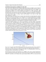
location of the burn. The total area of the burn can be approximated
in adults using the “rule of nines,” although this surface area rule
varies in the young age group (Fig. 12.1).
18
Small burns can be com-
pared to the size of the patient’s hand, which is about 1% of the total
skin area.
Burns are traditionally classified according to depth as first, sec-
ond, or third degree; however, these terms are being replaced by
superficial, superficial partial-thickness, deep partial-thickness, and
full-thickness. Burn depth is rarely uniform and may be difficult to
determine initially and require reevaluation after a few days.
19
Superficial Burns (First Degree)
Superficial burns involve only the superficial epidermis, appear ery-
thematous, and blanch with pressure. Mild sunburn is an example
12. Selected Injuries 269
Age:
15
9.5
13-17
11-13
9-11
7
9.5
9.5
15
17
17
9.5
9.5
9.5
9
32
32
32
36
9
18
18
1-4 years 5-9 years 10-14 years
Adult
(rule of nines)
18
18
Fig. 12.1. Assessment of the percent of the total surface area.
(Lund CC, Browder NC. The estimate of areas of burns. Surg
Gynecol Obstet 1944;79:352–8.)
with uneventful healing and some delayed peeling. The protective
functions of the skin are maintained.
Superficial and Deep Partial-Thickness Burns (Second Degree)
Superficial partial-thickness burns spare the deeper dermis compo-
nents, including hair follicles and the sweat and sebaceous glands, and
are either superficial or deep. These burns form bullae and are red,
painful, and weeping. They blanch with pressure, and the superficial
skin is sometimes wiped away. These burns heal in about 2 to 3 weeks
with little or no scarring. Deep partial-thickness burns are mottled
with red elements (dermal vessels) or are waxy-white and dry and do
not blanch with pressure. They may be nearly painless, with sensation
only to pressure. These burns may take a month or more to heal and
usually form scars. They may progress to full-thickness burns if not
properly treated and take 3 weeks or more to heal.
Full-Thickness Burns (Third Degree)
Full-thickness burns appear dry, white, or charred and inelastic. They
are painless and avascular, and thrombosed vessels may be visible.
A dry eschar covers the burn and may cause constriction of underly-
ing structures. Healing occurs only from the edges by epithelial
migration with scarring and contracture.
15
Hospitalization
Decisions regarding hospitalization can be made according to guide-
lines from the American Burn Association
20
(Table 12.2). Family
physicians should consider surgical consultation anytime there is
doubt about the depth of burns or need for hospitalization. Because
inhalation injury occurs frequently in large fires and is a common
cause of death, the physician must be alert for the presence of associ-
ated signs: facial burns, singed nasal hair, sooty mucus, hoarseness, or
cough. Initial physical examination, chest roentgenograms, and blood
gas measurements may be helpful but may also be normal in the pres-
ence of inhalation injury.
Burn Management
Severe Burns
Immediately after the burn, the victim’s clothing and any hot sub-
stances remaining in contact with the skin are removed, and the victim
270 Allan V. Abbott
is covered with a dry, sterile sheet. Copious irrigation with water is
indicated for chemical injuries. Cool compresses (not ice) can be used
to relieve the pain of small burns but can cause hypothermia if used
for large burns. Breathing is assessed immediately and oxygen admin-
istered if there is any distress or suspicion of carbon monoxide inhala-
tion.
21
Airway. Early endotracheal intubation is warranted at the first indi-
cation of inhalation injury. All patients with inhalation injury should
be placed on humidified oxygen. Steroids are warranted only in the
presence of bronchospasm. Bronchoscopy can confirm large airway
injury, and lung scans can detect small airway damage.
Fluids. Patients with burns of more than 15% to 20% of the surface
area require intravenous fluid replacement. Lactated Ringer’s solution
at a rate of 4 mL/kg per percent of burned area during the first 24
hours is the most common fluid replacement regimen used in the
United States, with half of this amount given during the first 8 hours
after the burn. Many other fluid regimens have been used, but all must
be administered with close monitoring of renal output and cardiovas-
cular status.
12. Selected Injuries 271
Table 12.2. Burns Requiring Hospitalization
20
Moderate burns (require hospitalization)
Partial-thickness burns on 15–25% of total body surface area (2–10%
in children or elderly)
Full-thickness burns on 2–10% of body surface
Suspected inhalation injury
Suspected high-voltage (200 volts) electrical burns (may appear mild
initially)
Circumferential burn (decompressive escharotomy may be needed)
Major burns (consider referral to burn center)
Partial-thickness burns on Ͼ25% body surface (Ͼ20% in children or
elderly)
Full-thickness burns on Ͼ10% of body
Burns with inhalation injury, major trauma, or other poor risk condi-
tion such as diabetes or immunodeficiency that increase risk of
infection
High-voltage (200 volts) electrical burns (may appear mild initially)
All but minimal burns to face, eyes, feet, hands, perineum, or genitalia
where cosmetic or functional impairment is likely
Burns from caustic chemicals such as hydrofluoric acid (may appear
mild initially)
Pain Management. Narcotics and benzodiazepines are used initially
for relief of pain and anxiety with caution because they can exacer-
bate the hypotension that may follow a major burn. Immediate admin-
istration of narcotics may also interfere with evaluation of other
associated trauma. After intravenous fluids have been administered
and fluid status has stabilized, narcotic doses can be increased.
Inhaled or intravenous anesthesia may be needed for the severe pain
of early dressing changes.
22
Consultation. Consultation with a surgical burn specialist is appropri-
ate for all severe burns, small burns that are deep partial-thickness or
deeper, and those located on the face, eyes, ears, or neck or in areas of
critical function including the hands, elbows, popliteal fossae, or feet.
Major complications including sepsis and hypermetabolism, and subse-
quent major burn management is best handled in major burn centers.
23
Minor Burns
Minor burns, those not requiring hospitalization, are by far the most
common type of burn managed by the family physician. Partial-thick-
ness burns contain portions of epithelium that must be protected from
further damage so epithelialization can occur.
Local Care. For all burns, the clothing and any hot or caustic materi-
als are removed immediately; and cool saline-soaked gauze is applied.
The ideal temperature for those compresses is 12°C (54°F), which
avoids hypothermia while relieving pain and increasing circulation for
up to 3 hours after the burn. Burns are cleaned with saline or mild
soapy water; the use of chlorhexidine gluconate (Hibiclens) or half-
strength povidone-iodine (Betadine) is now discouraged because
these agents may inhibit healing. Cytotoxic cleansing agents such as
hydrogen peroxide should be avoided. Necrotic skin is carefully
removed using aseptic technique; whirlpool debridement is often well
tolerated by patients. The yellow eschar of partial-thickness burns
should not be removed initially. Blisters may be left intact but are
removed if they appear to contain cloudy fluid, if broken, or if they
cover possible full-thickness burns.
Topical chemoprophylaxis is used for all but superficial burns to
prevent infection. Silver sulfadiazine (Silvadene) cream, classically
the most commonly used topical agent, is applied to the burn in a
thickness of about 1 to 2 mm and is then covered with a loose-fitting
dressing such as soft gauze. Silver sulfadiazine should not be used on
the face, on patients with sulfonamide sensitivity, or in pregnant
272 Allan V. Abbott
patients. Bacitracin (Baciguent) ointment is a good alternative.
Systemic antibiotics are used only with a proved burn infection. Oral
nonsteroidal antiinflammatory drugs, acetaminophen with codeine,
and rarely narcotics, can be given for pain.
15
An alternative to topical chemoprophylaxis and dressing changes
for superficial partial-thickness burns (not deeper burns) is the use of
synthetic dressings such as Duoderm, Opsite, or Biobrane.
24
These
expensive dressings are applied to fresh, clean, moist burns and are
left in place until the burn heals or until the dressing separates in about
1 to 2 weeks. In many cases these dressings are easy to use, promote
fast healing, decrease infection, do not limit activity, reduce pain, and
are acceptable to the patient overall. Immunity to tetanus should be
ensured, as burns are readily subject to tetanus infections.
25
(See Table
11.3.)
Follow-Up Care. Patients should bathe daily and gently wash off
completely and reapply the silver sulfadiazine. Dressings should
remain intact under any circumstances where the burns might become
dirty but may be removed at home when the burns can be protected.
The physician should recheck partial-thickness burns daily, and
patients should be alert to signs of impaired circulation caused by a
tight dressing and to signs of infection such as chills or fever. The
physician should remain alert for hypertrophic scarring and contrac-
tures and refer these patients to a burn specialist. Depending upon
depth, 6 to 24 months may be required for complete healing; during
this period the healing skin should be protected from sun exposure
and lubricated with moisturizing cream.
26
Sunburn
Superficial burns resulting from sunburn are common in fair-skinned
individuals and frequently come to the attention of the family physi-
cian. The skin appears red, blanches with light pressure, and is tender
and painful. Skin lubricants such as Eucerin may improve comfort.
The use of topical anesthetic sprays should be limited because they
may sensitize the skin to the anesthetics. Topical steroids have little
effect; but with extensive sunburns, constitutional symptoms may be
improved with oral prednisone at a daily dose of 20 mg for 2 to 3 days.
Prevention
Prevention of most burns takes place in the home by the family. Water
heaters should be set to a temperature below 51°C (124°F) to avoid
12. Selected Injuries 273
scalds. Smoke detectors should be installed and checked regularly.
Electrical outlets should be covered to protect children from electrical
injury, and chemicals and caustic agents must be stored away from the
reach of children. In the kitchen, hot pot handles should be turned
away from children, and all foods should be temperature-tested before
being offered to children. Oily rags must be discarded and flammables
stored properly. Finally, sunscreens should be used to prevent sun-
burn, and sun exposure should be avoided between 10 A.M. and 4 P.M.
As many as one in five burns of young children are the result of
abusive acts, so abuse must be considered when a child has more than
two burn sites, burns at various stages of healing, and burns that
follow a particular pattern (e.g., “stocking-glove” distribution).
27
When abuse is suspected, evaluation of previous medical records,
checking with protective services, and hospital admission should be
considered.
Aspirated or Swallowed Foreign Body
Pathophysiology
More deaths in the United States result from suffocation by foreign
bodies than from burns or from firearms accidents. Children younger
than 3 years of age have a natural tendency to place objects in their
mouths, putting them at high risk of choking injury. In children
younger than 1 year, asphyxiation is an important cause of uninten-
tional death. The foreign bodies most often aspirated are food, includ-
ing nuts, vegetable or fruit pieces, seeds, and popcorn. Small items
such as pen caps, beads, or crayons may be aspirated by small children.
Balloons pose a high risk for aspiration and asphyxiation to children of
any age. Items that may become lodged in the cricopharyngeus or
esophagus include coins, pieces of food, pieces of toys or hardware,
batteries, glass, chicken bones, etc.
28
Large objects in the esophagus can cause airway obstruction. The
gastrointestinal (GI) tract can become obstructed or perforated; medi-
astinitis, cardiac tamponade, paraesophageal abscess, or aortotra-
cheoesophageal fistula can occur. Perforation may be the result of
direct mechanical erosion (bones), or chemical corrosion (button
batteries).
29
Most pediatric obstructions occur in the proximal esophagus, and
most obstructions in adults occur at the distal esophagus in those
with a history of esophageal disease. Most swallowed foreign bodies
that pass through the esophagus continue through the entire GI tract
without difficulty, but 10% to 20% require some intervention and
274 Allan V. Abbott
about 1% require surgery. Objects larger than 3 to 5 cm may have
difficulty passing the duodenal loop in the region of the ligament
of Treitz.
Clinical Manifestations
The most frequent symptom of aspirated foreign body is a sudden
onset of choking and intractable cough with or without vomiting.
Other presenting symptoms may be cough, fever, breathlessness, and
wheezing. Some patients will be asymptomatic and many, especially
older adults, are misdiagnosed as having other pulmonary diseases.
On chest radiograph a pneumonic patch or atelectasis may be present
in adults, and air trapping is more common in children. Older adults
predisposed to aspiration include those with stroke or other central
nervous system disease or major underlying lung disease.
30
A swallowed foreign body can be painful and can provoke great
anxiety. Foreign bodies in the esophagus usually cause dysphagia,
especially with solid foods, and occasionally dyspnea due to com-
pression of the larynx. Patients may be unable to swallow their own
secretions. The initial period may be symptom-free, with symptoms
of esophageal obstruction developing later as the result of edema and
inflammation. Increasing pain, fever, and shock suggest perforation.
31
Management
When an aspirated foreign body is suspected or diagnosed on chest
radiograph, bronchoscopy is indicated. Success of foreign body
removal by bronchoscopy depends on the experience of the bron-
choscopist.
Because most ingested foreign bodies pass without problems, eval-
uation and treatment are often expectant. When patients complain of a
sticking sensation in their throat (as is often the case when a fish bone
is swallowed), direct or indirect laryngoscopy permits direct visualiza-
tion and removal with forceps. Esophagogastroscopy is preferred for
removal of most foreign bodies lodged in the esophagus or stomach.
Radiopaque foreign bodies can be easily diagnosed with standard radi-
ographs of the neck, chest, or abdomen. An esophagram can be used
to locate nonopaque objects. The physical examination is repeated to
detect signs of obstruction or early peritonitis with perforation. The
progress of the object through the GI tract can be monitored with
repeat abdominal films. If a foreign body remains in one position dis-
tal to the pylorus for longer than 5 days, surgical removal should be
considered.
12. Selected Injuries 275
Food Impaction
The typical patient with an esophageal food impaction is elderly, usu-
ally a denture wearer. The history is usually that the patient swallowed
a bolus of meat and feels that it is caught “halfway down.” Complete
occlusion is evident when the patient cannot swallow water and
regurgitates. The airway is usually uninvolved, and the patient speaks
and breathes without difficulty. Endoscopic removal is preferred. The
use of proteolytic enzymes, such as aqueous solution of papain (e.g.,
Adolph’s meat tenderizer) is not recommended owing to the risk of
perforation. When endoscopy is not available, intravenous glucagon
(1.0 mg) has been suggested to relax the esophageal smooth muscle.
If the food bolus has not passed in 20 minutes, an additional 2.0 mg
is given intravenously. An esophagram should be obtained to ensure
passage of the impaction.
Coin Ingestion
Half of the children with coins lodged in their esophagus are asympto-
matic; therefore, radiographs are obtained for all children suspected of
swallowing coins. Endoscopy is the preferred and safest method of coin
removal. If endoscopy is not available, a Foley catheter can be passed
down the esophagus beyond the object. The balloon is then inflated, and
as the catheter is slowly withdrawn the coin is withdrawn with it. There
is a high incidence of aspiration with this technique in small children
younger than 5 years of age. Coins have been observed to remain in the
stomach for 2 to 3 months before spontaneous passage.
32
Battery Ingestion
Most batteries pass uneventfully through the GI tract within 48 to 72
hours. However, a button battery lodged in the esophagus is an emer-
gency. These batteries contain 45% potassium hydroxide, which is
erosive to the esophagus and especially hazardous. Button batteries
should be removed endoscopically from the esophagus or if they
remain in the stomach longer than 24 hours.
29
Ingestion of Sharp Objects
Children who have swallowed a sharp object yet are asymptomatic
can be managed on an expectant basis.
31
Progression of the sharp
object should be documented by serial radiographs. If it is not seen
to progress past the stomach and perforation is suspected, a water-
soluble contrast radiograph is obtained. Perforation requires prompt
276 Allan V. Abbott
surgical intervention. Close observation or hospitalization is recom-
mended for children who have swallowed open safety pins or long,
sharp objects such as sewing needles.
Prevention
Young children should not have access to small objects such as toys
with small detachable parts, coins, pins, and the like. Children
younger than 3 years should not be given food in forms that could be
aspirated; nuts, popcorn, vegetable chunks, and so on should be
avoided. Care should be taken to avoid aspiration when feeding older
adults who have stroke or other serious debilitating disease. If
metered dose inhalers are carried in bags or pockets without their
safety caps on, foreign bodies may enter their mechanism and be
expelled forcefully into the bronchial tree. The ensuing symptoms are
often difficult to distinguish from those of an acute attack of asthma.
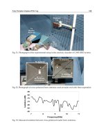
Fishhook Removal
There are four basic strategies for removing a barbed fishhook when
it has accidentally penetrated a person’s skin. Sterile technique, local
cleansing, and local anesthesia are appropriate with all the techniques.
Fishhook injuries are tetanus prone, and antibiotics should be given
when the wound is particularly dirty or when infection is already evi-
dent. Fishhook injuries to the eye or orbit should be referred to an
ophthalmologist.
33
Simple Retrograde Pull
If the hook has a small barb or is not embedded deeply, the hook can
be held close to the skin with a needle holder or hemostat and with-
drawn along its entry path (Fig. 12.2A). A small 1- to 2-mm incision
may be needed to help the barb pass through the dermis.
String-Yank Technique
This technique (Fig. 12.2B) does not involve any incisions or surgical
equipment and may be tried in the field. A strong suture or fishing line
is passed around the bend of the hook, and both ends are held together
with one hand while the hook is stabilized and gently depressed
against the skin with the other. A sharp pull is applied to the suture in
the direction parallel to the shank of the hook.
12. Selected Injuries 277
Needle-Cover Technique
The needle-cover technique (Fig. 12.2C) is often useful when the barb
is large. The hook is held in a needle holder or hemostat, and a 16- or
18-gauge hypodermic needle is introduced through the entry wound
and advanced along the hook’s bend until the barb can be sheathed
within the lumen of the needle. The hook and needle are then gently
withdrawn together. It is my experience that, with practice, this tech-
nique is usually successful.
Advance and Cut Technique
This method is nearly always successful but causes additional trauma
to the surrounding tissue (Fig. 12.2D). The middle of the shank is
firmly grasped with a needle holder and the hook tip is advanced out
through the skin. The exposed point of the hook is removed with wire
cutters, and the hook shank is withdrawn from the wound in a retro-
grade manner.
References
1. Injury Facts, 2000 edition. Itasca, IL: National Safety Council, 2000.
2. Ramesh CS. Near drowning. Crit Care Clin 1999;15:281–96.
3. Levin DL, Morriss FC, Toro LO, Brink LW, Turner GR. Drowning and
near-drowning. Pediatr Clin North Am 1993;40:321–6.
278 Allan V. Abbott
A
B
D
a
b
a
C
b
a
b
b
a
c
Fig. 12.2. Fishhook removal. (A) Simple retrograde pull. (B)
String-yank technique. (C) Needle-cover technique. (D) Push and
cut technique.
4. Christensen DW, Jansen P, Perkin RM. Outcome and acute care hospital
costs after warm water near drowning in children. Pediatrics 1997;99:
715–21.
5. Heimlich H, Hoffman K, Canestri F. Food choking and drowning deaths
prevented by external subdiaphragmatic compression. Ann Thorac Surg
1975;20:188–95.
6. Bratton SL, Jardine DS, Morray JP. Serial neurologic examinations after
near-drowning and outcome. Arch Pediatr Adolesc Med
1994;148:167–70.
7. Lavelle JM, Shaw KN, Seidl T, Ludwig S. Ten-year review of pediatric
near-drownings: evaluation for child abuse and ne-glect. Ann Emerg Med
1995;25:344–8.
8. James JR. Dysbarism: the medical problems from high and low atmos-
pheric pressure. J R Coll Physicians Lond 1993;27:367–74.
9. Jerrard DA. Diving medicine. Emerg Med Clin North Am 1992; 10:
329–38.
10. Moon RE. Treatment of diving emergencies. Crit Care Clin 1999;15:
429–49.
11. Parell JG, Becker GD. Neurological consequences of scuba diving with
chronic sinusitis. Laryngoscope 2000;110:1358–60.
12. Melamed Y, Shupak A, Bitterman H. Current concepts: medical prob-
lems associated with underwater diving. N Engl J Med 1992;326:30–5.
13. Marcy TW. Barotrauma: detection, recognition, and management. Chest
1993;104:578–84.
14. Boettger ML. Scuba diving emergencies: pulmonary overpressure acci-
dents and decompression sickness. Ann Emerg Med 1983;12:563–7.
15. Feller I. Burn epidemiology: focus on youngsters and the aged. Burn
Care Rehabil 1982;3:285.
16. Griglak MJ. Thermal injury. Emerg Med Clin North Am 1992;10:
369–83.
17. Carvajal HF. Burns in children and adolescents: initial management as
the first step in successful rehabilitation. Pediatrician 1991;17:237–43.
18. Lund CC, Browder NC. The estimate of areas of burns. Surg Gynecol
Obstet 1944;79:352–8.
19. Clark J. Burns. Br Med Bull 1999;55:885–94.
20. Joint Committee of the American Burn Association and the American
College of Surgeons Committee on Trauma. Assessment and initial care
of burn patients. Chicago: ACS, 1986.
21. Robertson C, Fenton O. ABC of major trauma: management of severe
burns. BMJ 1990;301:282–6.
22. Henry DB, Foster RL. Burn pain management in children. Pediatr Clin
North Am 2000;47:681–98.
23. Nguyen TT, Gilpin DA, Meyer NA, Herndon DN. Current treatment of
severely burned patients. Ann Surg 1996;223:14–25.
24. Wyatt D, McGowan DS, Najarian MP. Comparison of a hydrocolloid
dressing and silver sulfadiazine in the outpatient management of second-
degree burns. J Trauma 1990;30:857–65.
25. Smith DJ. Burn wounds: infection and healing. Am J Surg 1994;167:
46S–8S.
12. Selected Injuries 279
26. Morgan ED, Scott CB, Barker J. Ambulatory management of burns. Am
Fam Physician 2000;62:2016–26.
27. Rosenberg NM, Marino D. Frequency of suspected abuse/
neglect in burn patients. Pediatr Emerg Care 1989;5:219–21.
28. Rimell FL, Thome A, Stoll S, et al. Characteristics of objects that cause
choking in children. JAMA 1995;274:1763–6.
29. Litovitz T, Schmitz BE. Ingestion of cylindrical and button batteries, an
analysis of 2382 cases. Pediatrics 1992;89:727.
30. Baharloo F, Veyckemans F, Francis C, et al. Tracheobronchial foreign
bodies: presentation and management in children and adults. Chest 1999;
115:1357–62.
31. Paul RI, Jaffe DM. Sharp object ingestions in children: illustrative case
and literature review. Pediatr Emerg Care 1988;4:245.
32. Caravati EM, Bennett DL, McElwee NE. Pediatric coin ingestion: a
prospective study on the utility of routine roentgenograms. Am J Dis
Child 1989;143:549.
33. Gammons M, Jackson S. Fishhook removal. Am Fam Physician 2001;
63:2231–6.
280 Allan V. Abbott
INDEX
A
ABC of emergency care, 215
Abdominal aortic aneurysm, 12
Acetaminophen, 92–93
with codeine, 273
Achilles tendonitis, 68
ACL, see Anterior cruciate ligament
Acromioclavicular separation
diagnosis of, 220
management of, 220
Activities of daily living (ADL), 181
Acute disc herniation, clinical
diagnosis of, 6
Acute phase of CRPS, 133
Adam’s testing, 175
Adaptive mobility aids, 92
Addison’s disease, 182
Adhesives, 254–255
β-Adrenergic aerosols, 263
Adrenocorticotropic hormone
(ACTH), 201
Advanced cardiopulmonary life
support (ACLS), 262–263
Alcohol, 182, 186
Alendronate (Fosamax), 190–191
Allopurinol, 201–202
side effects of, 202
Ambien (Zolpidem), 130
American Association of Clinical
Endocrinologists Clinical
Practice Guidelines, see Bone
mineral density (BMD)
screening
American Burn Association
guidelines, 270
Amitriptyline (Elavil), 130
Amoxicillin-clavulanate, 257
ANA, see Antinuclear antibody
Ancillary tests, 8
Anesthetic methods
field blocks, 237
infiltration blocks, 237
nerve blocks, 238
Anesthetization, of wounds
injectable agents, 236–237
methods in, 237–238
topical agents, 235–236
Angle of gait, 148, 150
Angular abnormalities, of
knee
Blount’s disease, 154
bowlegs, 154
knock knees, 155
Ankle injuries
diagnosis of, 210
management of, 210–211
orthopedic referral, 211
Ankle
Achilles tendon ruptures, 76–77
sprains, 76
talar dome fractures, 77–78
tibial plafond fractures, 77
Ankylosing spondylitis, 11–12,
121
treatment of, 122
Anterior axillary nerve, 38
Page numbers with t and f indicate table and figure respectively
Anterior cruciate ligament (ACL)
injury
diagnosis of, 213–214
management of, 214
Anterior cruciate ligament (ACL),
68, 70
Anterior drawer test, 213
Antibiotics, 256–257, 266
Antidepressant(s), 188
low-dose tricyclic, 134
therapy, 20
Anti–double-stranded DNA
antigens, 113
Antihistamines, 266
Antimalarials
for psoriatic arthritis treatment,
122
for SLE treatment, 118
Antinuclear antibody (ANA), 102
Anti-Ro (Robert antigen)
antibodies, 113
Anti-Sm (Smith antigen) antigens,
113
Apert syndrome, 160
Aphasia, 267
Apophyseal injuries
Osgood–Schlatter disease, 170
Osteochondritis dissecans (OCD),
170–172
Sever’s disease, 170
Sinding–Larsen–Johansson
syndrome, 168
Apophysitis, of hip, 168
Aristocort (triamcinolone diacetate),
107
Arthralgias, 114
Arthritis Self-Help Course, 105
Arthritis, 114
psoriatic, 122
rheumatoid, see Rheumatoid
arthritis
Arthroplasty patients, 94
Aspirated or swallowed foreign
body
clinical manifestations in, 275
management of, 275–277
pathophysiology, 274–275
prevention of, 277
Aspirin, 139, 201–202
Assessments, of osteoporosis
bone densitometry assessment,
183–185
bone remodeling assessment,
185–186
laboratory assessment, 183
risk factor assessment, 182–183
Asymmetric polyarticular
arthritides, 97–98
Athletic activity, effects of excess,
see Apophyseal injuries; Spine,
problems of
Athletic injuries
at risk population, 205, 207
common injuries and injury rates,
206
mechanisms of, 205–207
overuse injuries, see Specific
overuse injuries, in athletes
prevention of, 229
traumatic injuries, see Traumatic
injuries, in athletes
Atlantoaxial (C1-2) subluxation,
101
Atrophic phase of CRPS, 133
Avascular necrosis, 163
B
Baciguent, see Bacitracin ointment
Bacitracin ointment, 273
Back disorders
background, 1–4
chronic low back pain, 20
clinical presentation, 4–6
diagnosis, 6–8
differential diagnosis, 9–13
herniated intervertebral disc,
16–19
management, 13–16
prevention, 20–21
Balloons, 274
282 Index
Barlow maneuver, 166
Barotitis, 267
Battery ingestion, see Aspirated or
swallowed foreign body
“Bean-shaped foot”, 151
Behavior modification in FM
treatment, 130
Benadryl, see Diphenhydramine
Benemid, see Probenecid
Benign tumors, of bone
chondromas, 139
giant cell tumor, 139–141
osteochondroma, 138–139
osteoid osteomas, 139
osteomas, 139
Benzodiazepines, 188, 239
Betadine, see Povidone-iodine
Biceps tendon, 40
Bicipital tendonitis, 40–41
Biobrane, 273
Biofeedback theory, 132
Biomechanical factors of OA,
89–80
Bisphosphonates
Alendronate (Fosamax), 190–191
Risedronate (Actonel), 191
Blount’s disease, see Angular
abnormalities, of knee
BMD, see Bone mineral density
Bone
benign tumors of, 138–141
densitometry assessment, for
osteoporosis, 183–185
malignant tumors of, 141–142
mineral density, see Bone mineral
density (BMD)
miscellaneous conditions of, see
Bone conditions, miscellaneous
stimulators for formation of, see
Bone formation stimulators
Bone conditions, miscellaneous
nonossifying fibroma, 142–143
Paget’s disease of bone, 143–145
Bone formation stimulators
fluoride, 191–192
parathyroid hormone (PTH), 191
Bone mineral density (BMD), 61,
183–184
assessment indications, 185
screening, 186
Bone remodeling assessment, for
osteoporosis, 185–186
Bony hypertrophy, 90
Boston-type spinal orthosis, 174
Bowlegs, see Angular
abnormalities, of knee
“Boxer’s fracture,” 48
Brachial plexus injury
diagnosis of, 221
management of, 221
“Bridge therapy,” 107–108
Bronchoscopy, 271, 275
Bupivacaine, 237
Burns
causes, 268
classification, 269–270
hospitalization, 270
management of, 270–273
pathophysiology, 268
prevention of, 273–274
Bursitis, 67–68
C
C- telopeptide, 185
Caffeine, 120
Calcific tendonitis, 40, 222–223
Calcitonin, 191
for Paget’s disease treatment,
143, 145
Calcium, 183, 186, 189
optimal intake of, 187
pyrophosphate deposition disease
(CPDD), see Pseudogout
pyrophosphate dihydrate, 203
Canard’s test, 223
Capsaicin cream, 134
Carbocaine, see Mepivacaine
Carpal bones, 47
Cartilages in OA, degeneration of,
90
Index 283
Cauda equina syndrome, 4, 12
Cavus foot, 156
Ceftriaxone, 257
Central hip dislocations, 64
Cerebral edema, 262
Cerebral hypoxia, in liquid medium,
see Near drowning
Cervical collar, 27
Cervical myelopathy
clinical presentation, 24
diagnosis, 24
management, 24
Cervical radiculopathy, 21
clinical presentation, 22
diagnosis, 22
management, 22–23
Cervical spine injury, 215
Cervical spondylosis, radiologic
diagnosis in, 24
Cervical whiplash, 25
clinical presentation, 26
diagnosis, 26–27
management, 26–28
prognosis, 28–29
Charcot–Marie–Tooth disease, 156
Chem panel, 183
Chemonucleolysis, 18–19
Chemoprophylaxis, 272
Chemo-therapeutic agents, 118
Chlorhexidine gluconate, 241, 272
Cholestyramine, 111
Chondrocalcinosis, 203
Chondroitin sulfate, 93
Chondromas, 139
Chordoma, 142
Chronic impingement, 39
Circulatory shock, 12
Clavicle
AC joint arthritis, 36
AC joint dislocations, 36
clavicular fractures, 35–36
Closed head injuries
diagnosis of, 216
grading of, 217
management of, 216, 218
Club foot, see Feet problems,
pediatric
Cobb method, 174–175
Coin ingestion, see Aspirated or
swallowed foreign body
Colchicine, 200–202
Colles’ fracture, 46
Collision sports, 206
Complete blood count (CBC), 183
Complex regional pain syndrome
(CRPS)
clinical criteria for diagnosis of,
133
family issues for, treatment, 135
invasive treatments for, 134
noninvasive medications for,
treatment, 134
phases in, 133
physical therapy for, treatment,
134
prevention for, 134–135
psychotherapy for, treatment, 134
Compression fracture, plain
radiograph demonstrating, 7f
Computed axial tomography (CAT)
scan, 216
Computed tomography (CT), 62, 91
for low back pain, 8
in SLE treatment, 117
Concomitant anxiety, 104
Concurrent therapy, 256–257
Contusions, 66
Cord tumor, 157
Corticosteroids, 183
injection, 48
Cortisol, 183
Cosmetic deformity, 176
COX-1, 107
COX-2, see Cyclooxygenase-2
inhibitors
C-reactive protein, 162
Crepitance, 90
CREST syndrome, 120
CRPS, see Complex regional pain
syndrome
284 Index
Cushing’s syndrome, 182
Cyclooxygenase-2 (COX-2)
inhibitors, 92, 107
Cyclophosphamide, for SLE
treatment, 118
Cyclosporine, 201
Cytokines, 90
D
DC, see Dupuytren’s contracture
De Quervain’s tenosynovitis, 48
Debridement process, 241
Decadron-LA (dexamethasone
acetate), 107
Decompression sickness
prevention, 267
treatment of, 267
Decompressive laminectomy, 9–10
Decongestants, 266
Deep venous thrombosis (DVT), 75,
227
Depo-triamcinolone, 107, 201
Dermabond, see Histoacryl Blue
Derotational osteotomy, 152
Developmental dysplasia, of hip
(DDH)
clinical signs, 166
diagnosis of, 163
etiology of, 163
treatment of, 163
Dexa scanning, 61
Dexamethasone acetate (Decadron-
LA), 107
Dietary therapy, for RA treatment,
104
Digital nerve block, 238f
DIP, see Distal interphalangeal
joints
Diphenhydramine, 237
Diphosphonates, for Paget’s disease
treatment, 143, 145
Disease-modifying antirheumatic
drugs (DMARDs), 108–110
Disorders of the back
background, 1–4
chronic low back pain, 20
clinical presentation, 4–6
diagnosis, 6–8
differential diagnosis, 9–13
herniated intervertebral disc, 16–19
management, 13–16
prevention, 20–21
Disorders of the neck
cervical myelopathy, 23–25
cervical radiculopathy, 21–23
cervical whiplash, 25–29
Distal interphalangeal (DIP) joints,
51–52, 101, 221
Diuretics, 201
DMARD, see Disease-modifying
antirheumatic drugs
Dog ear, 251
Doxycycline, 257
Dual-energy x-ray absortiometry
(DEXA), 184
Duoderm, 273
Dupuytren’s contracture (DC), 50,
136–137
Dystrophic phase of CRPS, 133
E
Ear and sinus barotrauma
barotisis media, 265
external barotitis, 264–265
inner ear barotrauma, 265
management of, 265–266
sinus barotrauma, 265
Early menopause, 182
Ehlers–Danlos syndromes, 157
Elavil (amitriptyline), 130
Elbow
epicondylitis, 43
fractures of radial head, 43
olecranon bursitis, 44–45
radial head subluxation, 43–44
Elbow-radial head subluxation
classification, 160
diagnosis, 159
epidemiology, 158–159
treatment, 159–160
Index 285
Electromyography (EMG), 9, 26
Ely’s test, 66
EMLA, 236
Endometrial cancer, risk of, 190
Endotracheal intubation, in
hospitalized patients, 263
Epidemiology of OA, 89
Epidural corticosteroid injection, 17
Epidural hematoma, 216
Erythrocyte sedimentation rate
(ESR), 91, 102–103, 160, 183,
199
Esophagogastroscopy, 275
ESR, see Erythrocyte sedimentation
rate
Estrogen, 182, 190
Ethyl chloride, 236
Ewing’s sarcoma, 141
Exercise
for FM treatment, 130–131
importance of, 188
Exogenous thyroid hormone, 183
Exostosis, see Osteochondroma
Experimental therapies for RA
treatment, 111
F
Factor V Leiden mutation, 166
Feet problems, pediatric
cavus foot, 156–157
clubfoot, 155–156
toe walking, 155
Femoral anteversion, 147–148, 152
Femoral head, superoanterior
dislocation of, 64
Fentanyl, 240
Fibrocartilaginous glenoid labrum,
37
Fibromyalgia (FM)
behavior modification for
treatment of, 130
characteristics of, 127
clinical features of, 129
diagnosis of, 130
diagnostic criteria of, 128
differential diagnosis of, 129
exercise for treatment of,
130–131
medication for treatment of, 130
prognosis of, patients, 131
Fibrous cortical defect, see
Nonossifying fibroma
Finger
fractures, 50–51
gamekeeper’s thumb, 54–55
infections, 55–56
PIP joint dislocations, 51
tendon injuries, 51–54
trigger fingers, 54
Finkelstein test, 48
Fish hook injuries
advance and cut technique, 278
needle cover technique, 278
simple retrograde pull, 277
string-yank technique, 277
Flash burns, 268
Flat foot
flexible, 157
rigid, 158
Flexeril (cyclobenzaprine), 130
Flexion of elbow, 159
Flexor retinaculum, 81
Fluoride, 191–192
FM, see Fibromyalgia
Food and Drug Administration
(FDA), 184, 190–192
Food impaction, see Aspirated or
swallowed foreign body
Foot, 151
anterior tarsal tunnel syndrome,
82
axis, 149
Forefoot injuries, 84
Haglund’s deformity, 81
interdigital neuritis, 86
metatarsals, 84–86
midfoot injuries, 82–83
plantar fasciitis, 78–81
progression angle, see Angle of
gait
286 Index
Tarsal tunnel syndrome, 81–82
Forefoot injuries
sesamoids, 84
turf toe, 84
Fractures, in fingers
distal tip fractures, 50
middle and proximal phalangeal
fractures, 51
Friedreich’s ataxia, 157
Full-thickness burns, 270
G
Gait abnormalities
clinical patterns and management
of, 151–153
evaluation and interpretation of,
149–151
terminology of, 148–149
Gamekeeper’s thumb, 221–222
Ganglion cysts, 137–138
Gastrointestinal (GI) toxicity,
107
Gastroprotective therapy, 92
Genu valgus, see Knock knees
Giant cell arteritis, see Temporal
arteritis
Giant cell tumor, 139–141
Glenohumeral disorders
adhesive capsulitis, 41
osteonecrosis, 41
Glenohumeral joint, 37, 39–40
Glucocorticoids, 107
long-term systemic, for JRA
treatment, 112
for SLE treatment, 118
systemic, for RA treatment,
107–108
Glucosamine sulfate, 93
Gonadotropin releasing hormone
(GnRH) analogues, 183
Gout disorder
diagnosis, 198–199
diagnostic studies, 199
management of, 201
treatment of, 199–200
H
Haemophilus influenzae, 162
Hamstring, 65–66
reflex, 6
strain, 67
Hand
infections, 49–50
metacarpal fractures, 48–49
OA, 91
Heberden’s nodes, 91
Hematologic disorder in SLE
patients, 117–118
Hemogram, 91
Hemostasis, 242, 250, 254
Herniated lumbar disc, 3
Hexachlorophene (pHisoHex), 241
Heyman–Herndon soft tissue
releases, 152
Hibiclens, see Chlorhexidine
gluconate
Hip
dislocation, types of, 62–63
fractures, risk factors, 60–62
fractures aging, 59–60
and lower extremity, problems of,
see Hip and lower extremity,
problems of
OA, 90–91
pain, 4
rotation, 150–151
Hip and lower extremity, problems
of
apophyseal injuries, 168–172
developmental dysplasia of hip,
163–167
Legg–Calvé–Perthes disease
(LCPD), 166–168
septic hip, 161–162
slipped capital femoral epiphysis,
162–163
transient synovitis of hip (TSH),
160–161
Histoacryl Blue, 254
Hormone Replacement Therapy
(HRT), 187, 189–190
Index 287
Human leukocyte antigen (HLA)-
B27, 12
Human tetanus immune globulin
(TIg), 257
Humerus, 42
Hyaline cartilages, 139
Hypercortisolism, 182
Hyperextension lateral films, use of,
176
Hyperparathyroidism, 182, 185
Hyperreflexia, 24
Hyperthyroidism, 182
Hyperuricemia, 197
causes of, 198
treatment of, 201–203
Hypothermia, 262
Hypoxia, 262
I
Idiopathic scoliosis, 174–175
Imaging studies for RA, 103
Immersion syndrome, 261
Immunosuppressive agents, 121
Indocin, see Indomethacin
Indomethacin, 200
Infections, in fingers
felon, 56
paronychia, 55–56
tenosynovitis, 56
Infections, in hand
dorsal hand infections, 49–50
palmar hand infections, 49
Inflammatory arthritis, see Psoriatic
arthritis; Rheumatoid arthritis
Interdigital neuritis, 86
Internal and external femoral
rotation, 149
Interphalangeal (IP) joint, 54
Intervertebal discs, degenerative
changes in, 3
In-toeing, see Metatarsus adductus
Intraarticular corticosteroid
injection, 28
Intravenous antibiotics, 162
Invasive treatments, for CRPS, 134
IP, see Interphalangeal joint
Isolated low back pain, 6
J
Joint deformity, 90
Joint instability, 98
Joint pain
differential diagnosis of, 97–98
physical examination in, patients,
98–99
Joint stiffness, 98
Joints in OA, symptoms of affected,
90–91
Jones’ and sesamoid bone fractures
diagnosis of, 227, 229
management of, 229
JRA, see Juvenile rheumatoid
arthritis
Juvenile kyphosis, see
Scheuermann’s disease
Juvenile rheumatoid arthritis (JRA),
111
clinical manifestations of, 112
NSAIDs for treatment of,
112–113
K
Kenalog, see Depo-triamcinolone
Ketamine, 239
Kite’s angle, 156f
Knee braces, 211
Knee extension, 5
Knee pain
anterior cruciate ligament tear,
70–72
causes of, 69
chondral fractures, 74
knee dislocation, 69–70
ligamentous knee injuries, 72–73
meniscal injuries, 68–69
osteochondral fractures, 74
osteochondritis dissecans (OCD),
73–74
patellar dislocation, 74–75
posterior cruciate ligament tear, 72
288 Index
Knee radiographs, 211
Knock knees, see Angular
abnormalities, of knee
Kyphosis, 176
L
Lacerations
beveled, 250
care of, see Laceration care
complex, 251–252
dog ears, 251
finger injuries, 252–253
across landmark, 250
Laceration care
alternative techniques, to sutures,
253–255
anesthetization of wounds,
235–238
concurrent therapy, 256–257
instructions for patients, 256
postrepair management of,
255–256
sedation, 238–240
skin anatomy of, 233–234
specific circumstances in,
250–253
suture techniques, 245–250
wound closure, 243–245
wound healing process, 234–235
wound preparation, 240–243
Lachman test, 213
Lactated Ringer’s solution, 271
Lateral collateral ligament (LCL),
70
Lateral epicondylitis, see Tennis
elbow
Leflunomide, 111
Left slipped capital femoral
epiphysis, 164f
Legg–Calvé–Perthes disease
(LCPD), 161
Catterall classification of, 167
classification of, 167
diagnosis of, 167
early stages, 167
treatment of, 167–168
Lidocaine, 107, 134, 236
Light touch, sensation to, 5
Lisfranc injury, 82–83
Low back injury, prevention of,
20–21
Low back pain
with cough, 4
demographic characteristics, 3
epidemiology, 1–2
natural history, 2–4
psychosocial factors, 3–4
risk factors, 2
Lower extremity variations, in
pediatric groups; see also Hip
and lower extremity, problems
of
angular abnormalities, of knee,
153–155
feet problems, 155–157
flat foot, 157–158
gait abnormalities, 147–153
Lower leg
plantaris tendon, 75
stress fractures, 75
Lumbar spine
surgery, 17
symptomatic cancer of, 11
Lupus anticoagulant antigens, 113
Lymphomas, 142
M
Magnetic resonance imaging (MRI),
91, 163, 216
for cyst detection, 138
for low back pain, 8
in SLE treatment, 117
Malar rash, 115
Malignant bone tumors
chordoma, 142
Ewing’s sarcoma, 141
metastatic malignant tumors,
142
osteosarcoma, 141–142
primary chondrosarcoma, 141
Index 289
Management of FM
behavior modification, 130
medication, 130
patient education, 130
water exercise, 130–131
Management of OA
goals in, 91
nonpharmacologic management
strategies for, 91–92
pharmacologic approaches in,
92–93
surgical approaches for, 93–94
Management, back pain
acupuncture, 16
back school, 16
bed rest, 13
exercise, 16
medications, 13
nonspecific low back pain, 13
spinal manipulation, 16
unproven treatment, 13–16
Marcaine, see Bupivacaine
Marginal osteophyte formation, 91
McMurray’s test, 212
MCP, see Metacarpophalangeal
joints
Medial collateral ligament (MCL)
sprain
diagnosis of, 212
management of, 213
Medial femoral torsion, 153
Medial knee taping, 92
Mediastinal emphysema, 266–267
Meniscus flaps, 211
Meniscus injuries
diagnosis of, 212
management of, 212
Mepivacaine, 237
Mesenchymal cells, 141
Metacarpophalangeal (MCP) joints,
49, 101
Metastatic malignant tumors, 142
Metatarsals
bunion, 85
fracture of the fifth metatarsal, 85
metatarsalgia, 84
Metatarsophalangeal (MTP) joint,
84
Metatarsus adductus, 147
and femoral anteversion, 153
and tibial torsion, 152–153
treatment of flexible, 151–152
treatment of rigid, 152
Metatarsus varus, 151
Microdiscectomy, 18
Midazolam, 239–240
Milwaukee brace, 176
Moll’s lateral flexion test, 121
Monsel’s solution, 254
Morning stiffness, 98, 100–101
Morton’s neuroma (MN), see
Interdigital neurities; Neuroma
MPS, see Myofascial pain
syndrome
Multimodal treatments, 28
Multiple myeloma, 10
Muscle
relaxants, 13, 22
strain, 65–67
Musculoligamentous injuries, 3
sprains, 25
Myelogram for low back pain, 8
Myelography, 8
Myofascial pain syndrome (MPS),
131
clinical features of, 131–132
treatment of, 132
N
N- telopeptide, 185
Nail bed injuries, 252–253
Narcotic analgesics, 16–17
National Health and Nutrition
Examination Survey
(NHANES I) study on OA, 89
National Osteoporosis Foundation
guidelines, see Bone mineral
density (BMD), screening
Near drowning
clinical presentation, 261–262
290 Index