Biochemical, Genetic, and Molecular Interactions in Development - part 4 ppt
Bạn đang xem bản rút gọn của tài liệu. Xem và tải ngay bản đầy đủ của tài liệu tại đây (864.64 KB, 45 trang )
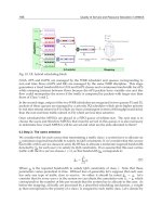
Regulation of BMP, Wnt, and Hh Signaling 117
mature Nodal cleaved from its native precursor protein is highly unstable whereas that cleaved from
a chimeric precursor containing the BMP-4 prodomain is stable (16).
The requirement for proteolytic removal of the prodomain for activity is supported by the finding
that cleavage mutant forms of BMPs in which the -RXXR- motif has been disrupted are inactive and
can dimerize with and inhibit the cleavage, secretion and bioactivity of native BMPs (23). A few
exceptions to this rule do exist, however, in that precursor forms of inhibin A (24), lefty (25), and
Xenopus nodal related-2 (26) possess some bioactivity.
The mechanism(s) by which the prodomain regulates the activity of mature BMPs is unknown and
is likely to vary between individual family members. In the case of TGF-`, which has been better
studied than BMPs, the prodomain remains noncovalently associated with the mature ligand, form-
ing an inactive, latent complex that is stored in the extracellular matrix (ECM) in association with the
latent TGF-` binding protein. The major regulatory step controlling TGF-` activity takes place out-
side of the cell when proteases or other agents either release the prodomain or induce a conformational
change that exposes the receptor binding sites on TGF-` (27). Analogous to TGF-`, the prodomain
of BMP-7 remains noncovalently associated with the mature region after cleavage but, unlike TGF-`,
this complex can bind to and activate BMP receptors without further processing or alteration (28).
Recent genetic data support a functional interaction between BMP-7 and the latent TGF-` binding
protein family member Fibrillin-2 and suggest that the bioactivity or availability of BMP-7, like that
of TGF-`, may be regulated by interactions with the ECM (29). Processing of BMP-4 is more com-
plex than that of BMP-7 in that the precursor is sequentially cleaved by furin at two sites and this
ordered proteolysis regulates the activity and signaling range of mature BMP-4 (14,15). Specifically,
proBMP-4 is initially cleaved at a consensus furin motif adjacent to the mature ligand domain and
this allows for subsequent cleavage at an upstream nonconsensus furin motif within the prodomain.
Failure to cleave at the upstream site generates a ligand that is targeted for rapid degradation, leading
to lower bioactivity and signaling distance in vivo. Conversely, a mutant form of the precursor that is
rapidly cleaved at both sites generates ligand that is more active and signals over a greater range. An
intriguing possibility is that the upstream site is cleaved in a tissue-specific fashion, thereby provid-
ing a mechanism to spatially regulate the levels and distance of BMP signaling in vivo. This same
mechanism may operate for the closely related family member BMP-2 because the two cleavage sites
are conserved in BMP-4 and BMP-2 from all species, but not in other family members.
Role of Homo- vs Heterodimerization
Closely related members of the BMP family, for example BMP-2-4 and/or -7, BMP-2 and GDF-6,
or different nodal-related proteins, can form heterodimers within the secretory pathway before pro-
teolytic processing and in some cases the heterodimers are more potent signaling molecules than are
homodimers (30–33). Recent studies have shown that more distantly related family members can also
heterodimerize. BMP-4, for example, forms heterodimers with Xenopus derriere or nodal-related pro-
teins (26) and BMP-7 forms heterodimers with nodal (34). BMP-4 and -7 bind to a distinct class of
receptors and activate a different intracellular signal transduction pathway than do derriere or nodals,
raising the questions of whether these heterodimers are active and, if so, which class of receptors and
signaling pathways are activated. An alternate possibility is that this class of heterodimer blocks acti-
vation of both signaling pathways as has been suggested for BMP-7/nodal heterodimers (34).
Processing of Wnts
Regulated Glycosylation
Unlike Hh and BMPs, Wnts are subject only to regulated glycosylation and not cleavage. In trans-
fected tissue culture cells, most Wnt protein is retained as an unglyosylated form in the endoplasmic
reticulum associated with an HSP70 protein (35). This inefficient processing suggests that generation
118 Hackenmiller et al.
of active Wnt protein is a complex process and may require tissue specific accessory proteins. Consis-
tent with this, genetic studies identified Porcupine (Porc) as a member of an evolutionarily conserved
family of multipass transmembrane ER proteins, which is required for processing the Drosophila Wnt
family member, Wg (36,37). Porc was recently shown to bind an N-terminal region of Wg that is highly
conserved among all Wnts and to stimulate glycosylation of nearby sites. In addition, Porc was shown
to be dispensable for N-glycosylation in the presence of dithiothreitol (DTT), suggesting that the cotrans-
lational formation of intramolecular disulfide bonds in Wnt proteins normally inhibits efficient glyco-
sylation. Based on these studies, a model has been proposed in which Porc tethers Wg to the ER membrane
bringing it into close proximity with the oligosaccharyl transferase complex, thereby accelerating
glycosylation and minimizing competition with cotranslational disulfide bond formation. Porc shares
homology with a family of acetyltransferases, raising the possibility that it may anchor Wg to the ER
membrane via acetylation (38).
Processing of Hedgehog
Autoproteolysis and Cholesterol Attachment
Hh is synthesized as a 45-kDa precursor that is autoprocessed to generate a 20-kDa N-terminal
fragment (Hh-N) that possesses all known signaling activity and a 25-kDa C-terminal domain (Hh-C)
that catalzyes intramolecular cleavage of the precursor (39–41). Cleavage occurs through the for-
mation of a thioester intermediate that undergoes nucleophilic attack by cholesterol, resulting in the
covalent attachment of cholesterol to the C-terminus of Hh-N (42). This yields the mature signaling
form of Hh, which is denoted Hh-Np.
The addition of cholesterol to Hh-N initially was thought to be essential for ligand function, possi-
bly by mediating binding to the Hh receptor, Ptc (reviewed in ref. 43), but is now known to be dis-
pensable for activity and receptor binding. This was demonstrated with a truncated form of Hh lacking
the cholesterol modification, which retains full signaling activity both in vitro and in vivo (41,44)
and binds to Ptc with similar affinity as does Hh-Np (45).
In Drosophila, the cholesterol adduct can limit the range over which Hh signals, as evidenced by
the finding that overexpressed Hh-N signals over a much greater distance than does Hh-Np. This
restriction is caused by the ability of Ptc to sequester and thereby limit the travels of Hh-Np, but not
Hh-N. This presents an unresolved paradox, however, because earlier studies have shown that Ptc binds
to Hh-N and Hh-Np with equal affinity. The difference in receptor interactions in vivo may be medi-
ated by differential association of Hh-N and Hh-Np with HSPGs, as described in the Activity Regula-
tion by HSPGs section.
Curiously, the cholesterol moiety not only restricts the range over which Hh can signal but also
enables Hh to signal beyond producing cells. Hh-Np can signal across several cell diameters whereas
a membrane tethered form of Hh can signal only to adjacent cells, thereby demonstrating that choles-
terol does not function as a simple membrane anchor. Release of Hh-Np from producing cells is depen-
dent on the function of yet-to-be identified HSPGs, which is discussed in the next section on extracellular
regulation of activity, and a novel transmembrane protein, Dispatched (Disp).
Disp is a 12 pass transmembrane protein with a sterol sensing group that was identified by genetic
studies as being required in Hh-producing cells for release of Hh-Np but not Hh-N (46). In the absence
of functional Disp, Hh-Np is synthesized, processed, reaches the cell surface, and can signal (47) but
is not released from the cell. The mechanism by which Disp regulates Hh release is unknown.
Most of what is known about the role of cholesterol in modulating the range of Hh signaling has
come from genetic studies in Drosophila. Recent studies in mice led to the surprising conclusions
that, unlike in the fly, addition of cholesterol to vertebrate Hh is essential for long range activity but
is dispensable for short-range signaling and sequestration by Ptc (48). Specifically, mice were gener-
ated in which a stop codon was introduced into the Sonic Hh (Shh) gene such that only a truncated
Regulation of BMP, Wnt, and Hh Signaling 119
form of Shh analogous to Hh-N was expressed. This unprocessed, unmodified form of Shh protein
was expressed at normal levels, interacted genetically with Ptc, and was able to signal to nearby cells
but was not distributed to distal cells that normally receive Shh. The observed differences in the signal-
ing range of Hh-N in the fly vs the mouse may be caused by the use of overexpression approaches in
Drosophila vs knock-in mutations in the mouse, the use of different accessory proteins to regulate
Hh signaling in each species (e.g., Disp in flies, HIP in mouse, see below), or differences in cellular
context.
Palmitoylation
In addition to cholesterol modification, Hh undergoes an additional posttranslational lipid modifi-
cation, the palmitoylation of its most N-terminal cysteine via an acylation intermediate (45). Studies
in tissue culture suggest that palmitoylation, like cholesterol coupling, can anchor Hh to the mem-
brane (45), but a variety of indirect evidence suggests that acylation alone is not sufficient to restrict
the range of action of Hh in vivo. This issue remains to be resolved, but what is clear is that palmitoy-
lation is essential to generate a fully active ligand. In Drosophila, acylation is catalyzed by a transmem-
brane acyltransferase encoded by the skinny hedgehog (ski) gene (49), also referred to as sightless (sit;
ref. 50), central missing (cmn; ref. 47), or Rasp (51). The activity of Hh-N and Hh-Np is abolished in
embryos mutant for this gene. Further evidence that acylation is required to generate functional Hh is
provided by studies in which the N-terminal cysteine to which palmitate is attached was mutated. This
mutation inactivates the protein and generates a dominant mutant form that interferes with endog-
enous Hh activity (52). In vertebrates, palmitoylation is not absolutely essential for Hh activity but
generates a more potent signaling molecule in cell culture (45) and tissue assays. Specifically, although
unacylated recombinant Shh can induce formation of ventral cell types in chick forebrain explant cul-
tures, it is much less potent on mouse forebrain explants than is acylated protein (53). In addition, muta-
tion of the N-terminal cysteine residue to serine generates a signaling molecule with reduced patterning
activity in a mouse limb bud assay relative to the wild-type Shh (52).
The mechanism by which acylation potentiates the signaling activity of Hh is unclear. Addition of
hydrophobic amino acids or other hydrophobic moieties to the N-terminus of Shh enhances the potency
of the ligand but does not alter binding affinity for Ptc and has no apparent effect on structure (54).
Although these modifications do not appear to restrict the range of Hh, they may localize the protein
to specific membrane domains and/or alter its affinity for cofactors or other proteins involved in sig-
naling and transport.
ACTIVITY REGULATION BY EXTRACELLULAR MODES
In addition to the posttranslational modifications that impact on the action of BMP, Wnt, and Hh,
there are a large number of extracellular proteins that regulate ligand activity and/or availability. In this
section we focus on two extracellular regulatory mechanisms: secreted extracellular binding proteins
and cell surface HSPGs. These diverse extracellular modulators either facilitate or inhibit the signal-
ing activities of BMP, Wnt, and Hh by a variety of molecular mechanisms.
Sequestration of BMPs and Wnts by Secreted Extracellular Binding Proteins
In general, the soluble extracellular binding proteins described below affect the concentrations of
BMPs and Wnts (no secreted extracellular regulators have been identified for Hh) that signal at the
surface of responding cells. These interactions serve to regulate the amount of a particular ligand that
a cell “sees,” thus indicating its position within the morphogen gradient. Most of these extracellular
regulators are high-affinity secreted binding proteins that prevent receptor activation by binding to the
ligand, thereby acting as antagonists. Interestingly, there is little or no sequence similarity between
the different classes discussed below.
120 Hackenmiller et al.
BMP-Secreted Extracellular Regulators
Noggin
Noggin is a small glycoprotein (32 kDa) that was originally identified as a molecular component
of Spemann’s organizer, a specialized signaling center located on the dorsal side of gastrulating
Xenopus embryos. Noggin functions as a homodimer that binds specifically to BMPs secreted by ven-
tral cells and antagonizes BMP signaling by blocking interaction with its receptors (55). These inter-
actions are critical for normal dorsoventral patterning in Xenopus embryos. Noggin can also bind to
and inhibit Xenopus GDF-6 (a TGF-` family member), preventing its ability to induce epidermis and
blocking neural tissue formation (56). Additional biochemical studies have shown that noggin binds
to BMP-2, BMP-4 and GDF-6 with high affinity, but to BMP-7 with low affinity (55,56).
Noggin-null mice demonstrate that antagonism of BMP activity by noggin is critical for proper
skeletal development. In addition to defects in neural tube and somite development noggin-null mice
have excess cartilage and fail to initiate joint formation (57). Two human genetic disorders, proximal
symphalangism and multiple synostoses syndrome, which are characterized by bony fusions of joints,
have been shown to be caused by dominant mutations in noggin (58), further underscoring the impor-
tance of noggin in joint development.
Chordin/Short Gastrulation (Sog)
Chordin is a 120-kDa protein secreted from the Spemann’s organizer. In the same manner as noggin,
chordin, and its Drosophila ortholog, short gastrulation (Sog) antagonizes BMP signaling by binding
the ligand and preventing it from interacting with its receptor (59). Because it is much larger than other
BMP antagonists, chordin may diffuse less efficiently in tissues, altering its ability to function as a
BMP inhibitor.
In both vertebrates and invertebrates, the activity of chordin orthologs is negatively regulated by a
family of secreted zinc metalloproteases, including Drosophila Tolloid, Xenopus Xolloid, and human
BMP-1. Biochemical studies have shown that Tolloid cleaves chordin and decreases its affinity for
BMP ligands, thus functioning as a BMP agonist (60–62). The activity of Drosophila Tolloid appears
to be different than that of the other Tolloid orthologs. Drosophila Tolloid cleavage activity is depen-
dent on the formation of the Dpp–Sog complex, whereas in Xenopus and zebrafish, chordin cleavage
is independent of BMP binding (60,61,63). Nonetheless, Tolloid orthologs can regulate the availabil-
ity of BMP signals by regulating the amount of BMP bound by chordin.
Paradoxically, in Drosophila, whereas Dpp is inhibited by high levels of Sog, it appears to be
enhanced by low levels of Sog, and this process requires Tolloid (64). Sog may facilitate diffusion of
Dpp, allowing the inactive complex to accumulate and then be activated by tolloid-mediated cleav-
age at sites distant from the Sog source.
Adding complexity, it has recently been shown that the secreted protein Twisted gastrulation (Tsg)
acts as a BMP antagonist when complexed with chordin and BMP (65–68). Tsg promotes the binding
of chordin to BMP and together the three form a ternary complex that inactivates BMP signaling
more efficiently than chordin alone. Additionally, Tsg enhances tolloid cleavage of chordin. It is not
clear whether this generates “supersog-like molecules,” that can inhibit additional members of the
BMP family not inhibited by unprocessed Sog (69) or whether it inactivates chordin, freeing BMP to
signal (70). One possibility is that the chordin/Tsg/BMP complex helps BMP diffuse through the
embryo, in part by preventing its association with cell surface receptors along the way. This would
allow for high levels of BMP signaling at a distance from the chordin source (see above and ref. 71).
Follistatin
Follistatin is a soluble secreted glycoprotein with cysteine-rich modules originally identified as a
protein that binds and inhibits activin (72). When follistatin is overexpressed in ventral blastomeres
of a Xenopus embryo, it can induce a secondary body axis (73) and when overexpressed in Xenopus
ectoderm, it can induce neural tissue (74). These results suggest that follistatin might inhibit the
Regulation of BMP, Wnt, and Hh Signaling 121
action of proteins in addition to activin, namely BMPs. Additionally, follistatin has been shown to
co-immunoprecipitate with BMP-4 in tissue culture (75), indicating a direct interaction between BMPs
and follistatin. In contrast to the mode of action of noggin and chordin, follistatin does not compete
with the type I receptor for BMP-4 binding. Instead, it forms a tetrameric complex with BMP and the
type I and type II BMP receptor to block receptor activation (73).
DAN Family
DAN, Cerberus, Gremlin, Caronte, and other structurally related proteins are collectively called
the DAN family (76). All members of this family characterized to date have been shown to antago-
nize BMP signaling by preventing BMP–receptor interaction. Unrelated to other BMP antagonists,
all DAN family members have a conserved 90 amino-acid cystine-knot motif that at least in Cerberus
and Caronte includes the BMP-binding region (77,78).
DAN
DAN, originally isolated as a putative zinc-finger protein that has tumor-suppressor activity
(79,80) was later shown to be a secreted factor that like other BMP antagonist can neutralize ectoder-
mal explants from Xenopus embryos and convert ventral mesoderm to more dorsal fates (76). DAN
directly binds to BMP-2 in vitro (76) but experimental evidence suggests it may be a more potent
inhibitor of the GDF class of BMPs in vivo (81). The exact role of DAN in developmental processes
is unclear because DAN mutant mice have no obvious abnormalities (81). In developing mouse
neurons dan mRNA is localized to axons, suggesting a potential role for DAN in axonal outgrowth or
guidance.
Cerberus
The Xenopus cerberus gene was identified as a Spemann organizer-associated transcript that encodes
a secreted protein able to induce ectopic heads when injected into Xenopus embryos (82). Cerberus is
a multidimensional antagonist: it has been shown to bind and inhibit BMPs, Wnts, Nodals, and Acti-
vin, but the binding sites are independent (77). BMP-4 and Xnr1 (nodal family member) bind in the
cystine-knot region, whereas Xenopus wnt-8 (Xwnt-8) binds to the unique amino terminal half of
cerberus. Cerberus appears to restrict trunk formation to the posterior part of the body by coordinately
antagonizing three trunk-forming pathways—the BMP, Nodal, and Wnt pathways—in the anterior
part of the developing embryo.
Gremlin
Gremlin was isolated in studies to identify dorsalizing factors that can induce a secondary axis in
the Xenopus embryo (76). In addition to antagonizing BMP activity, Gremlin also blocks signaling of
Activin and Nodal-like members of TGF-` superfamily. Gremlin is expressed in cells of the neural
crest lineage, suggesting it may have a role in neural crest induction and later patterning events. Grem-
lin has also been shown to be a central player in the outgrowth and patterning of the vertebrate limb (83).
Wnt-Secreted Extracellular Regulators
The sFRP Family
The Wnt antagonists known as secreted frizzled-related proteins (sFRP) are a large family of
secreted proteins that share homology to the putative Wnt-binding region of the Frizzled (Fz) family
of transmembrane receptors (84,85). Frzb-1 is the founding family member, and it was identified by
researchers two ways: in a screen while looking for cDNAs enriched in the Xenopus Spemann’s orga-
nizer (84,85) and in articular cartilage extracts while looking for in vivo chondrogenic activity (86).
Frzb-1 coimmunopreciptates with Xwnt-8, showing a direct interaction between Frzb and Wnts (84),
and Frzb blocks the axis-inducing activity of Xwnt-8 and mouse Wnt-1 when coinjected on the ven-
tral side of cleaving embryos, demonstrating that Frzb is an antagonist of Wnt signaling. Additional
122 Hackenmiller et al.
experiments have demonstrated that the antagonistic effects of Frzb and Wnt take place in the extra-
cellular space where the two proteins are secreted (87), preventing productive interactions between
Wnt and the Fz receptor.
All sFRP family members have been shown to have dorsalizing activities in Xenopus whole embryo
assays, but the various family members have diverse expression patterns and different affinities for
specific Wnts (88). This suggests that particular sFRPs are required at specific times and in specific
tissues to antagonize signaling of specific Wnts. Biochemical data regarding the target Wnt protein
for the various sFRPs has been inconclusive. For example, Frzb1 can bind to Xwnt-3a, Xwnt-5, and
Xwnt-8 in vitro but only interacts with Xwnt-8 in the embryo (89). Similar results have been obtained
for Frzb2 and Sizzled 2 (90), making the in vivo requirement for the different sFRPs unclear.
A simple interaction between sFRP and Wnt proteins may not be able to fully explain the mecha-
nism by which FRPs act. Recent data have demonstrated that sFRPs interact not only with Wnt pro-
teins but also with other FRPs and with Fz receptors (91), leaving open an alternative mode of action
for sFRP-mediated antagonism of Wnt signaling.
Wnt Inhibitory Factor-1
Wnt inhibitory factor-1 (WIF-1) is another secreted Wnt antagonist that binds to Wnt proteins and
blocks their interaction with the Fz receptors (92). Its earliest expression is seen at neurula stages in the
somitic mesoderm and anterior forebrain of mice (92), and WIF-1 has been shown to bind to Xwnt-8
and Wg in vitro. WIF-1 has an N-terminal signal sequence, a domain of approx 150 amino acids termed
the WIF domain that binds to Wnt/Wg, five epidermal growth factor-like repeats, and a hydrophobic
domain of approx 45 amino acids at the C-terminus. The WIF domain partially overlaps with the Wnt
binding domain in Fz-2.
Xenopus studies demonstrate that the action of WIF-1 is different than that of the Frzb family mem-
bers. Coinjection of the BMP antagonist chordin with Frzb leads to a low frequency of secondary
axis formation and when formed, the ectopic heads are always cyclopic. By contrast, co-injection of
WIF-1 and chordin promotes complete secondary axes and no cyclopic eyes. The WIF domain alone
is able to synergize with chordin to give secondary axes, but the heads are always cyclopic, suggest-
ing that the epidermal growth factor-like repeats are necessary for full activity of WIF-1 (92).
Cerberus
As discussed above, cerberus is a multivalent inhibitor that can block BMP, Wnt, Nodal, and Acti-
vin signaling. Cerberus directly binds to Xwnt-8, inhibiting its interaction with the Fz receptors. It is
expressed in the Xenopus Spemann’s organizer and is thought to have a role in head induction, a pro-
cess inhibited by ectopic Xwnt-8 signaling in the gastrula dorsal mesoderm (93).
Dickkopf
Dickkopf (Dkk-1) encodes a member of a novel protein family of secreted Wnt antagonists. Dkk-1
is expressed in the anterior mesentoderm and is proposed to function in head induction (94). Dick-
kopf’s mode of antagonism is different than previously described antagonistic proteins. Dkk-1 antag-
onizes Wnt signaling by binding to and inactivating the Wnt co-receptor LRP (arrow in Drosophila;
refs. 95–98) but does not directly bind to Wnt. Dkk regulates coreceptor availability rather than ligand
availability. It has recently been demonstrated that the membrane-anchored molecule Kremen binds
to Dkk and triggers internalization and clearing of the Dkk-LRP complex from the cell surface (99).
This renders Wnt unable to activate the intracellular pathway necessary for target gene expression. It
remains to be determined how Kremen triggers internalization of the Dkk-LRP complex.
Activity Regulation by HSPGs
HSPGs are large macromolecules found abundantly on the cell surface that modulate the function
of intracellular signaling molecules in many ways (100). BMPs, Wnts, and Hh have been shown to
Regulation of BMP, Wnt, and Hh Signaling 123
interact with components of the ECM, such as HSPGs, and it is becoming clear that these interactions
play an important role in modulating the levels, facilitating the movement, and/or acting as corecep-
tors for these ligands (101).
BMP
In Drosophila, genetic analysis of a mutation in the glypican gene dally (division abnormally delayed)
has implicated this protein in both Wg (discussed below) and Dpp signaling (102,103). Reducing
Dpp levels in a dally mutant background enhances defects in the eye, antenna and genitalia, and over-
expression of Dpp can rescue the defects in these tissues (104). Interestingly, although these genetic
interactions indicate that Dally regulates Dpp activity (103), the requirement for Dally in Dpp signal-
ing appears to be restricted to the imaginal disks.
Several studies on mouse glypican-3 (gpc-3) knockouts have provided evidence that BMP/HSPG
interactions are important in mouse embryogenesis. When gpc-3-deficient animals are mated to BMP-
4 haploinsufficient mice, the offspring display a high penetrance of postaxial polydactyly and rib
malformations not seen in either parent strain (105). Additional studies show that Gpc-3 modulates
BMP-7 activity during embryogenic kidney morphogenesis (106).
Work in Xenopus has identified a basic core of amino acids in the N-terminal region of BMP-4
necessary for BMP binding to HSPGs (107). Mutating these three amino acids does not alter receptor
binding or induction of target genes but does increase the effective range of BMP signaling, indicat-
ing that HSPGs restrict the diffusion of BMPs in vivo. Together, these results demonstrate that HSPGs
are important regulators of BMP function and signaling range during both Drosophila and vertebrate
development.
Wnt/Wg
Genetic studies in Drosophila confirm a role for HSPGs in Wg signaling. Sugarless (sgl/kiwi) encodes
an uridine diphosphate (UDP)-glucuronate involved in the biosynthesis of heparin, heparan sulfate
(HS), chondroitin sulfate, and hyaluronic acid. Mutations of sgl demonstrate a noncell autonomous
defect in Wg-receiving cells (102,108), which is mediated by loss of HS. Exogenous HS can rescue sgl
mutants whereas overexpression of HS in wild-type embryos gives rise to excess Wg signaling (102).
Wg signaling is also impaired in sulfateless (slf) mutants, which lack an enzyme involved in the modi-
fication of HS. Together, these studies suggest that proteoglycans and specifically HSPGs interact
with Wg in receiving cells either to stabilize the ligand, limit its diffusion, increase the effective local
concentration of the ligand (102), or to act as a low-affinity co-receptor (108).
As discussed above, Dally is a GPI-linked glypican that is modified by Sfl. Dally protein is expressed
in the same cells as the Wg receptor, Dfz2 where it may act as a co-receptor with Dfz2 to generate a
high-affinity binding site for Wg (103,109).
A second glypican molecule involved in reception of Wg signaling is Dally-like (Dly). Overex-
pression of Dly leads to an accumulation of extracellular Wg and generates a wg phenotype. This sug-
gests Dly acts to sequester Wg and acts as an antagonist, preventing access to or activation of Dfz2
(110). In contrast to the apical localization of Wg mRNA, association of Wg with glycosylphosphatidyl-
inositol (GPI)-linked HSPG targets it to the basolateral surface of cells (111), contributing to the poste-
rior spread of Wg signaling.
QSulf1, a sulfatase family member, is another genetically linked enzyme in the Wg pathway (112)
necessary for the degradation of HSPGs (113). Disruption of QSulf1 specifically inhibits expression
of MyoD, a Wnt-responsive gene, suggesting that breakdown of HSPGs is integral to Wnt signaling.
In transient transfection assays, addition of QSulf1 enhances Wnt signaling, whereas addition of hep-
arin or chlorate antagonizes QSulf1, abrogating Wnt signaling (112). One explanation for how Qsulf1
alters Wnt signaling is that QSulf1 desulfates HS to locally release Wnt-bound HSPG, enabling the
ligand to bind its cognate receptor and initiate signaling.
124 Hackenmiller et al.
Hh
Genetic evidence that HSPGs are essential for trafficking of Hh was provided by the identification
of tout velu (ttv) as a gene that is required for movement of Hh-Np, but not Hh-N, in Drosophila (114,
115). Ttv is a homolog of the human EXT genes that were identified through their association with
the bone disorder multiple exostoses (116). These genes encode enzymes essential for heparan sul-
fate glycosaminoglycan biosynthesis (117). Glycosaminoglycan have also been shown to be impor-
tant for movement of vertebrate Hh away from its source (118). Several models have been proposed
for the role of HSPGs in Hh-Np movement or receptor binding. It is possible, for example, that
association of Hh-Np, but not Hh-N, with HSPGs increases its local concentration, thereby enabling
it to bind to and be sequestered by Ptc. Alternatively, or in addition, binding to a specific class of
HSPGs, such as the GPI-linked glypicans, might enable transport of Hh from cell to cell directly (119)
or via transcytosis (120) as has been observed for other GPI-linked proteins. Association with glypi-
cans might also function to promote localization of Hh-Np to lipid raft microdomains within the mem-
brane through which transport can occur. Rafts are microdomains rich in cholesterol, sphingolipids,
and GPI-anchored proteins and Hh-Np is associated with this membrane fraction, either by virtue of
its sterol modification alone, or perhaps by association with a glypican molecule (121).
REGULATION OF RECEPTOR ACTIVATION: FEEDBACK LOOPS
Research in recent years has shown that the BMP-, Wnt-, and Hh-signaling pathways are often
subjected to regulation by autofeedback loops in addition to the action of extracellular regulators.
Most of these feedback loops consist of transcriptional targets of the pathways that once activated
turn off or downregulate BMP, Wnt, or Hh activity by interfering with future signaling events. Intra-
cellular targets, such as inhibitory SMADs, which block intracellular events in the BMP pathway, are
not discussed, although these are an important component of feedback loops that are further described
in several recent reviews (8–10). Instead, we highlight feedback loops that alter receptor activation
or accessibility.
BMP Feedback Loops
BAMBI
BMP and activin membrane-bound inhibitor) (Bambi; ref. 122) is a transmembrane protein related
to TGF-`-family type I receptors that lacks an intracellular kinase domain. In all species examined,
embryonic expression of Bambi overlaps that of BMPs and is induced by BMP ligands. Bambi acts
as a pseudoreceptor by intercalating in the TGF-` complex and disrupting receptor signaling, thus func-
tioning as a naturally occurring dominant mutant of BMP signaling.
Tkv
In the developing wing disk of Drosophila, Dpp negatively regulates expression of its own type I
receptor thickveins (Tkv; ref. 123). This results in Tkv levels being lowest in Dpp-expressing cells
and highest in cells furthest from the source of Dpp (123,124). Low levels of Tkv enable Dpp to
spread over long distances, in part generating the Dpp morphogen gradient. High levels of Tkv pre-
sumably limit the spread of Dpp. Hh also represses tkv expression in dpp-expressing cells (125), add-
ing an additional level of regulation.
Noggin
Noggin expression in chondrocyte and osteoblast cultures is increased by BMP signaling and
noggin in turn abolishes the bioactivity of BMPs (see Regulation of Receptor Activation: Feedback
Loops section and refs. 126,127). This suggests that noggin may participate in a BMP-negative feed-
back loop.
Regulation of BMP, Wnt, and Hh Signaling 125
Wnt Feedback Loops
Binding of Wg to its receptor, Dfz2, has been shown to stabilize Wg in the wing imaginal disk (128).
This stabilization allows Wg to diffuse further from its source at the dorsoventral boundary of the
imaginal disk. Wg signaling represses dfz2 transcription, resulting in dfz2 expression being low near
secreting cells and increasing distally. This sets up an inverted gradient of wg/dfz2 expression, which
promotes ligand stability at a distance (129). Conversely, early in embryogenesis, overexpression of
Dfz2 acts to restrict distribution of Wg, suggesting the receptor can also act to sequester ligand (87).
Hedgehog Feedback Loops
ptc Upregulation
The ptc gene is a transcriptional target of the Hh-signaling pathway. In Drosophila and mouse, ptc
upregulation in response to Hh signaling is responsible for the sequestration of Hh and restriction of
Hh movement (130,131). Hh upregulation of ptc is a self-limiting mechanism by which Hh attenu-
ates its own movement through responsive tissues. In addition, high levels of Ptc block the intrinsic
activity of Smo. As discussed above, Ptc-mediated sequestration of Hh is dependent on cholesterol
modification of Hh.
HIP
Hedgehog-interacting protein (HIP) is a membrane glycoprotein that binds to all three mammalian
Hh proteins with an affinity similar to Ptc (132). HIP was the only protein identified in an expression
screen for Hh-interacting proteins that promoted cell surface binding of Hh. Binding of Hh to HIP
most likely regulates the availability of ligand, resulting in signal attenuation (10). An example of
HIP-negative regulation of Hh signaling is seen in cartilage where Indian hedgehog (Ihh) controls
growth, and overexpression of HIP leads to a shortened skeleton similar to that observed in ihh knock-
out mice (132). Hip, like ptc, is a transcriptional target of Hh signaling. HIP expression is induced by
ectopic Hh expression and is absent in Hh-responsive cells in Hh mutants. Interestingly, no HIP
othologs have been identified in Drosophila, providing a possible molecular mechanism to explain
the different actions of Hh in the mouse vs the fly.
CONCLUSION
mRNA expression patterns alone do not describe the activities and interactions of BMPs, Wnt, and
Hh as mediators of many fundamental processes in embryonic development. As we have described,
these proteins are regulated at multiple levels beyond transcription. They are regulated posttransla-
tionally via covalent modifications, proteolytic processing, and regulated secretion; within the extra-
cellular space by secreted binding proteins and HSPGs; and via autoregulartory feedback loops. These
modifications and interactions result in a complex pattern of ligand activity that cannot be achieved
by transcriptional regulation alone.
Although we have tried to highlight some of the modes of regulating the activity of BMP, Wnt,
and Hh signaling, there has been a large amount of recent work on how ligands move from cell to cell.
Passive diffusion, long thought to be the way morphogen gradients were generated, is now viewed as
only one of a handful of ways that a tissue/organism traffics its morphogens. Movement by carrier
molecules, endocytosis, argosomes (vesicle-mediated transport), transcytosis (sequential endocyto-
sis and exocytosis), and cytonemes (threads of cytoplasm connecting distant cells) are additional mech-
anisms used to generate morphogens gradients (for recent reviews, see refs. 133–136). It is becoming
apparent that depending on the time in development the tissue, and even the organism, many different
tools can be used establish the necessary distribution of particular morphogens. Future studies will
likely show that differently modified forms of the ligands have different affinities for antagonistic
proteins and HSPG molecules and that these associations in turn regulate how, when, and where the
126 Hackenmiller et al.
ligand is transported. Although many of the specifics of the BMP, Wnt, and Hh pathways have been
worked out, understanding how these pathways (and others) are integrated to form complex organisms
remains a critical problem in developmental biology.
REFERENCES
1. Wozney, J. M., Rosen, V., Celeste, A., et al. (1988) Novel regulators of bone formation: molecular clones and activi-
ties. Science 242, 1528–1534.
2. Nusse, R. and Varmus, H. E. (1982) Many tumors induced by the mouse mammary tumor virus contain a provirus
integrated in the same region of the host genome. Cell 31, 99–109.
3. Rijsewijk, F., Schuermann, M., Wagenaar, E., et al. (1987) The Drosophila homolog of the mouse mammary oncogene
int-1 is identical to the segment polarity gene wingless. Cell 50, 649–657.
4. Nusslein-Volhard, C. and Wieschaus, E. (1980) Mutations affecting segment number and polarity in Drosophila.
Nature 287, 795–801.
5. Neumann, C. and Cohen, S. (1997) Morphogens and pattern formation. Bioessays 19, 721–729.
6. Gonzalez, F., Swales, L., Bejsovec, A., et al. (1991) Secretion and movement of wingless protein in the epidermis of
the Drosophila embryo. Mech. Dev. 35, 43–54.
7. Johnson, R. L. and Tabin, C. (1995) The long and short of hedgehog signaling. Cell 81, 313–316.
8. Nakayama, T., Cui, Y., and Christian, J. L. (2000) Regulation of BMP/Dpp signaling during embryonic development.
Cell Mol. Life Sci. 57, 943–956.
9. Huelsken, J. and Birchmeier, W. (2001) New aspects of Wnt signaling pathways in higher vertebrates. Curr. Opin.
Genet. Dev. 11, 547–553.
10. Ingham, W. and McMahon, A. (2001) Hedgehog signaling in animal development: paradigms and principles. Genes
Dev. 15, 3059–3087.
11. Hammonds, R. G. Jr., Schwall, R., Dudley, A., et al. (1991) Bone-inducing activity of mature BMP-2b produced from
a hybrid BMP-2a/2b precursor. Mol. Endocrinol 5, 149–155.
12. Massague, J. (1990) The transforming growth factor-beta family. Annu. Rev. Cell Biol. 6, 597–641.
13. Steiner, D. F. (1998) The proprotein convertases. Curr. Opin. Chem. Biol. 2, 31–39.
14. Cui, Y., Jean, F., Thomas, G., et al. (1998) BMP-4 is proteolytically activated by furin and/or PC6 during vertebrate
embryonic development. EMBO J. 17, 4735–4743.
15. Cui, Y., Hackenmiller, R., Berg, L., et al. (2001) The activity and signaling range of mature BMP-4 is regulated by
sequential cleavage at two sites within the prodomain of the precursor. Genes Dev. 15, 2797–2802.
16. Constam, D. B. and Robertson, E. J. (1999) Regulation of bone morphogenetic protein activity by pro domains and
proprotein convertases. J. Cell Biol. 144, 139–149.
17. Constam, D. B. and Robertson, E. J. (2000) SPC4/PACE4 regulates a TGFbeta signaling network during axis forma-
tion. Genes Dev. 14, 1146–1155.
18. Roebroek, A. J., Umans, L., Pauli, I. G., et al. (1998) Failure of ventral closure and axial rotation in embryos lacking
the proprotein convertase Furin. Development 125, 4863–4876.
19. Gray, A. M. and Mason, A. J. (1990) Requirement for activin A and transforming growth factor—beta 1 pro-regions
in homodimer assembly. Science 247, 1328–1330.
20. Shinde, U. and Inouye, M. (2000) Intramolecular chaperones: polypeptide extensions that modulate protein folding.
Semin. Cell Dev. Biol. 11, 35–44.
21. Kessler, D. S. and Melton, D. A. (1995) Induction of dorsal mesoderm by soluble, mature Vg1 protein. Development
121, 2155–2164.
22. Jones, C. M., Armes, N., and Smith, J. C. (1996) Signalling by TGF-beta family members: short-range effects of Xnr-
2 and BMP-4 contrast with the long-range effects of activin. Curr. Biol. 6, 1468–1475.
23. Hawley, S. H., et al. (1995) Disruption of BMP signals in embryonic Xenopus ectoderm leads to direct neural induc-
tion. Genes Dev. 9, 2923–2935.
24. Mason, A. J., Farnworth, G., and Sullivan, J. (1996) Characterization and determination of the biological activities of
noncleavable high molecular weight forms of inhibin A and activin A. Mol. Endocrinol 10, 1055–1065.
25. Ulloa, L. and Tabibzadeh, S. (2001) Lefty inhibits receptor-regulated Smad phosphorylation induced by the activated
transforming growth factor-beta receptor. J. Biol. Chem. 276, 21397–2404.
26. Eimon, M. and Harland, R. M. (2002) Effects of heterodimerization and proteolytic processing on Derriere and Nodal
activity: implications for mesoderm induction in Xenopus. Development 129, 3089–3103.
27. Khalil, N. (2001) Post translational activation of latent transforming growth factor beta (L-TGF-beta): clinical impli-
cations. Histol. Histopathol. 16, 541–551.
28. Jones, W. K., Richmond, E. A., White, K., et al. (1994) Osteogenic protein-1 (OP-1) expression and processing in
Chinese hamster ovary cells: isolation of a soluble complex containing the mature and pro-domains of OP-1. Growth
Factors 11, 215–225.
29. Arteaga-Solis, E., Gayraud, B., Lee, S. Y., et al. (2001) Regulation of limb patterning by extracellular microfibrils. J.
Cell Biol. 154, 275–281.
30. Aono, A., Hazama, M., Notoya, K., et al. (1995) Potent ectopic bone-inducing activity of bone morphogenetic pro-
tein-4/7 heterodimer. Biochem. Biophys. Res. Commun. 210, 670–677.
Regulation of BMP, Wnt, and Hh Signaling 127
31. Hazama, M., Aono, A., Ueno, N., et al. (1995) Efficient expression of a heterodimer of bone morphogenetic protein
subunits using a baculo-virus expression system. Biochem. Biophys. Res. Commun. 209, 859–866.
32. Suzuki, A., Kaneko, E., Maeda, J., et al. (1997) Mesoderm induction by BMP-4 and -7 heterodimers. Biochem. Biophys.
Res. Commun. 232, 153–156.
33. Nishimatsu, S. and Thomsen, G. H. (1998) Ventral mesoderm induction and patterning by bone morphogenetic pro-
tein heterodimers in Xenopus embryos. Mech. Dev. 74, 75–88.
34. Yeo, C. and Whitman, M. (2001) Nodal signals to Smads through Cripto-dependent and Cripto-independent mecha-
nisms. Mol. Cell 7, 949–957.
35. Kitajewski, J., Mason, J. O., and Varmus, H. E. (1992) Interaction of Wnt-1 proteins with the binding protein Bi. Mol.
Cell Biol. 12, 784–790.
36. Kadowaki, T., Wilder, E., Klingensmith, J., et al. (1996) The segment polarity gene porcupine encodes a putative
multitransmembrane protein involved in Wingless processing. Genes Dev. 10, 3116–3128.
37. Tanaka, K., Okabayashi, K., Asashima, M., et al. (2000) The evolutionarily conserved porcupine gene family is involved
in the processing of the Wnt family. Eur. J. Biochem. 267, 4300–4311.
38. Tanaka, K., Kitagawa, Y., and Kadowaki, T. (2002) Drosophila segment polarity gene product porcupine stimulates
the posttranslational N-glycosylation of wingless in the endoplasmic reticulum. J. Biol. Chem. 277, 12816–12823.
39. Bumcrot, D. A., Takada, R., and McMahon, A. (1995) Proteolytic processing yields two secreted forms of sonic
hedgehog. Mol. Cell Biol. 15, 2294–2303.
40. Lee, J. J., Ekker, S. C., von Kessler, D. P., et al. (1994) Autoproteolysis in hedgehog protein biogenesis. Science 266,
1528–1537.
41. Porter, J. A., von Kessler, D. P., Ekker, S. C., et al. (1995) The product of hedgehog autoproteolytic cleavage active
in local and long-range signalling. Nature 374, 363–366.
42. Porter, J. A., Young, K. E., and Beachy, A. (1996) Cholesterol modification of hedgehog signaling proteins in animal
development. Science 274, 255–259.
43. Osborne, T. F. and Rosenfeld, J. M. (1998) Related membrane domains in proteins of sterol sensing and cell signaling
provide a glimpse of treasures still buried within the dynamic realm of intracellular metabolic regulation. Curr. Opin.
Lipidol. 9, 137–140.
44. Marti, E., Bumcrot, D. A., Takada, R., et al. (1995) Requirement of 19K form of Sonic hedgehog for induction of
distinct ventral cell types in CNS explants. Nature 375, 322–325.
45. Pepinsky, R. B., Zeng, C., Wen, D., et al. (1998) Identification of a palmitic acid-modified form of human Sonic
hedgehog. J. Biol. Chem. 273, 14037–14045.
46. Burke, R., Nellen, D., Bellotto, M., et al. (1999) Dispatched, a novel sterol-sensing domain protein dedicated to the
release of cholesterol-modified hedgehog from signaling cells. Cell 99, 803–815.
47. Amanai, K. and Jiang, J. (2001) Distinct roles of central missing and dispatched in sending the Hedgehog signal.
Development 128, 5119–5127.
48. Lewis, M., Dunn, M. P., McMahon, J. A., et al. (2001) Cholesterol modification of sonic hedgehog is required for
long-range signaling activity and effective modulation of signaling by Ptc1. Cell 105, 599–612.
49. Chamoun, Z., Mann, R. K., Nellen, D., et al. (2001) Skinny hedgehog, an acyltransferase required for palmitoylation
and activity of the hedgehog signal. Science 293, 2080–2084.
50. Lee, J. D. and Treisman, J. E. (2001) Sightless has homology to transmembrane acyltransferases and is required to
generate active Hedgehog protein. Curr. Biol. 11, 1147–1152.
51. Micchelli, C. A., The, I., Selva, E., et al. (2002) Rasp, a putative transmembrane acyltransferase, is required for
Hedgehog signaling. Development 129, 843–851.
52. Lee, J. D., Kraus, P., Gaiano, N., et al. (2001) An acylatable residue of Hedgehog is differentially required in Droso-
phila and mouse limb development. Dev. Biol. 233, 122–136.
53. Kohtz, J. D., Lee, H. Y., Gaiano, N., et al. (2001) N-terminal fatty-acylation of sonic hedgehog enhances the induc-
tion of rodent ventral forebrain neurons. Development 128, 2351–2363.
54. Taylor, F. R., Wen, D., Garber, E. A., et al. (2001) Enhanced potency of human Sonic hedgehog by hydrophobic mod-
ification. Biochemistry 40, 4359–4371.
55. Zimmerman, L. B., De Jesus-Escobar, J. M., and Harland, R. M. (1996) The Spemann organizer signal noggin binds
and inactivates bone morphogenetic protein 4. Cell 86, 599–606.
56. Chang, C. and Hemmati-Brivanlou, A. (1998) Neural crest induction by Xwnt7B in Xenopus. Dev. Biol. 194, 129–134.
57. Brunet, L. J., McMahon, J. A., McMahon, A. P., et al. (1998) Noggin, cartilage morphogenesis, and joint formation
in the mammalian skeleton. Science 280, 1455–1457.
58. Gong, Y., Krakow, D., Marcelino, J., et al. (1999) Heterozygous mutations in the gene encoding noggin affect human
joint morphogenesis. Nat. Genet. 21, 302–304.
59. Piccolo, S., Sasai, Y., Lu, B., et al. (1996) Dorsoventral patterning in Xenopus: inhibition of ventral signals by direct
binding of chordin to BMP-4. Cell 86, 589–598.
60. Piccolo, S., Agius, E., Lu, B., et al. (1997), Cleavage of chordin by Xolloid metalloprotease suggests a role for pro-
teolytic processing in the regulation of Spemann organizer activity. Cell 91, 407–416.
61. Marques, G., Musacchio, M., Shimell, M. J., et al. (1997) Production of a DPP activity gradient in the early Droso-
phila embryo through the opposing actions of the SOG and TLD proteins. Cell 91, 417–426.
62. Larrain, J., Bachiller, D., Lu, B., et al. (2000) BMP-binding modules in chordin: a model for signalling regulation in
the extracellular space. Development 127, 821–830.
128 Hackenmiller et al.
63. Blader, P, Rastegar, S., Fischer, N., et al. (1997) Cleavage of the BMP-4 antagonist chordin by zebrafish tolloid. Science
278, 1937–1940.
64. Ashe, H. L. and Levine, M. (1999) Local inhibition and long-range enhancement of Dpp signal transduction by Sog
[see comments]. Nature 398, 427–431.
65. Scott, I. C., Blitz, I. L., Pappano, W. N., et al. (2001) Homologues of Twisted gastrulation are extracellular cofactors
in antagonism of BMP signalling. Nature 410, 475–478.
66. Ross, J. J., Shimmi, O., Vilmos, P., et al. (2001) Twisted gastrulation is a conserved extracellular BMP antagonist.
Nature 410, 479–483.
67. Chang, C., Holtzman, D. A., Chau, S., et al. (2001) Twisted gastrulation can function as a BMP antagonist. Nature 410,
483–487.
68. Ray, R. and Wharton, K. A. (2001) Twisted perspective: new insights into extracellular modulation of BMP signaling
during development. Cell 104, 801–804.
69. Yu, K., Srinivasan, S., Shimmi, O., et al. (2000) Processing of the Drosophila Sog protein creates a novel BMP inhib-
itory activity. Development 127, 2143–2154.
70. Oelgeschlager, M., Larrain, J., Geissert, D., et al. (2000) The evolutionarily conserved BMP-binding protein Twisted
gastrulation promotes BMP signalling. Nature 405, 757–763.
71. Harland, R. M. (2001) Developmental biology. A twist on embryonic signalling. Nature 410, 423–424.
72. de Winter, J., ten Dijke, P., de Vries, C. J., et al. (1996) Follistatins neutralize activin bioactivity by inhibition of
activin binding to its type II receptors. Mol. Cell Endocrinol. 116, 105–114.
73. Iemura S, Y. T., Takagi, C., Uchiyama, H., Natsume, T., Shimasaki, S., Sugino, H., et al. (1998) Direct binding of
follistatin to a complex of bone-morphogenetic protein and its receptor inhibits ventral and epidermal cell fates in
early Xenopus embryo. Proc. Natl. Acad. Sci. USA 95, 9337–9342.
74. Hemmati-Brivanlou, A., Kelly, O. G., and Melton, D. A. (1994) Follistatin, an antagonist of activin, is expressed in
the Spemann organizer and displays direct neuralizing activity. Cell 77, 283–295.
75. Fainsod, A., Deissler, K., Yelin, R., et al. (1997) The dorsalizing and neural inducing gene follistatin is an antagonist
of BMP-4. Mech. Dev. 63, 39–50.
76. Hsu, D.R., Economides, A. N., Wang, X., et al. (1998) The Xenopus dorsalizing factor Gremlin identifies a novel
family of secreted proteins that antagonize BMP activities. Mol. Cell 1, 673–683.
77. Piccolo, S., Agius, E., Leyns, L., et al. (1999) The head inducer Cerberus is a multifunctional antagonist of Nodal,
BMP and Wnt signals. Nature 397, 707–710.
78. Yokouchi, Y., Vogan, K. J., Pearse, R. V., 2nd, et al. (1999) Antagonistic signaling by Caronte, a novel Cerberus-
related gene, establishes left-right asymmetric gene expression. Cell 98, 573–583.
79. Ozaki, T. and Sakiyama, S. (1993) Molecular cloning and characterization of a cDNA showing negative regulation in
v-src-transformed 3Y1 rat fibroblasts. Proc. Natl. Acad. Sci. USA 90, 2593–2597.
80. Ozaki, T. and Sakiyama, S. (1994) Tumor-suppressive activity of N03 gene product in v-src-transformed rat 3Y1
fibroblasts. Cancer Res. 54, 646–648.
81. Dionne, M. S., Skarnes, W. C., and Harland, R. M. (2001) Mutation and analysis of Dan, the founding member of the
Dan family of transforming growth factor beta antagonists. Mol. Cell Biol. 21, 636–643.
82. Bouwmeester, T., Kim, S., Sasai, Y., et al. (1996) Cerberus is a head-inducing secreted factor expressed in the ante-
rior endoderm of Spemann’s organizer. Nature 382, 595–601.
83. Zuniga, A., Haramis, A. P., McMahon, A. P., et al. (1999) Signal relay by BMP antagonism controls the SHH/FGF4
feedback loop in vertebrate limb buds. Nature 401, 598–602.
84. Wang, S., Krinks, M., Lin, K., et al. (1997) Frzb, a secreted protein expressed in the Spemann organizer, binds and
inhibits Wnt-8. Cell 88, 757–766.
85. Leyns, L., Bouwmeester, T., Kim, S. H., et al. (1997) Frzb-1 is a secreted antagonist of Wnt signaling expressed in the
Spemann organizer. Cell 88, 747–756.
86. Hoang, B., Moos, M., Jr., Vukicevic, S., et al. (1996) Primary structure and tissue distribution of FRZB, a novel
protein related to Drosophila frizzled, suggest a role in skeletal morphogenesis. J. Biol. Chem. 271, 26131–26137.
87. Reichsman, F., Smith, L., and Cumberledge, S. (1996) Glycosaminoglycans can modulate extracellular localization
of the wingless protein and promote signal transduction. J. Cell Biol. 135, 819–827.
88. Wang, S., Krinks, M., and Moos, M. Jr. (1997) Frzb-1, an antagonist of Wnt-1 and Wnt-8, does not block signaling by
Wnts -3A, -5A, or -11. Biochem. Biophys. Res. Commun. 236, 502–504.
89. Lin, K., Wang, S., Julius, M. A., et al. (1997) The cysteine-rich frizzled domain of Frzb-1 is required and sufficient
for modulation of Wnt signaling. Proc. Natl. Acad. Sci. USA 94, 11196–11200.
90. Bradley, L., Sun, B., Collins-Racie, L., et al. (2000) Different activities of the frizzled-related proteins frzb2 and
sizzled2 during Xenopus anteroposterior patterning. Dev. Biol. 227, 118–132.
91. Bafico, A., Gazit, A., Pramila, T., et al. (1999) Interaction of frizzled related protein (FRP) with Wnt ligands
and the frizzled receptor suggests alternative mechanisms for FRP inhibition of Wnt signaling. J. Biol. Chem. 274,
16180–16187.
92. Hsieh, J. C., Kodjabachian, L., Rebbert, M. L., et al. (1999) A new secreted protein that binds to Wnt proteins and
inhibits their activities. Nature 398, 431–436.
93. Christian, J. L. and Moon, R. T. (1993) Interactions between Xwnt-8 and Spemann organizer signaling pathways
generate dorsoventral pattern in the embryonic mesoderm of Xenopus. Genes Dev. 7, 13–28.
94. Glinka, A., Wu, W., Delius, H., et al. (1998) Dickkopf-1 is a member of a new family of secreted proteins and func-
tions in head induction. Nature 391, 357–362.
Regulation of BMP, Wnt, and Hh Signaling 129
95. Wehrli, M., Dougan, S. T., Caldwell, K., et al. (2000) arrow encodes an LDL-receptor-related protein essential for
Wingless signalling. Nature 407, 527–530.
96. Mao, B., Wu, W., Li, Y., et al. (2001) LDL-receptor-related protein 6 is a receptor for Dickkopf proteins. Nature 411,
321–325.
97. Semenov, M. V., Tamai, K., Brott, B. K., et al. (2001) Head inducer Dickkopf-1 is a ligand for Wnt coreceptor LRP6.
Curr. Biol. 11, 951–961.
98. Bafico, A., Liu, G., Yaniv, A., et al. (2001) Novel mechanism of Wnt signalling inhibition mediated by Dickkopf-1
interaction with LRP6/Arrow. Nat. Cell Biol. 3, 683–686.
99. Mao, B., Wu, W., Davidson, G., et al. (2002) Kremen proteins are Dickkopf receptors that regulate Wnt/beta-catenin
signalling. Nature 417, 664–667.
100. Selleck, S. B. (1999) Overgrowth syndromes and the regulation of signaling complexes by proteoglycans. Am. J. Hum.
Genet. 64, 372–377.
101. Perrimon, N. and Bernfield, M. (2000) Specificities of heparan sulphate proteoglycans in developmental processes.
Nature 404, 725–728.
102. Binari, R. C., Staveley, B. E., Johnson, W.A., et al. (1997) Genetic evidence that heparin-like glycosaminoglycans
are involved in wingless signaling. Development 124, 2623–2632.
103. Tsuda, M., Kamimura, K., Nakato, H., et al. (1999) The cell-surface proteoglycan Dally regulates Wingless signalling
in Drosophila. Nature 400, 276–280.
104. Jackson, S. M., Nakato, H., Sugiura, M., et al. (1997) Dally, a Drosophila glypican, controls cellular responses to the
TGF-beta-related morphogen, Dp. Development 124, 4113–4120.
105. Paine-Saunders, S., Viviano, B. L., Zupicich, J., et al. (2000) glypican-3 controls cellular responses to Bmp4 in limb
patterning and skeletal development. Dev. Biol. 225, 179–187.
106. Grisaru, S., Cano-Gauci, D., Tee, J., et al. (2001) Glypican-3 modulates BMP- and FGF-mediated effects during renal
branching morphogenesis. Dev. Biol. 231, 31–46.
107. Ohkawara, B., Iemura, S., ten Dijke, P., et al. (2002) Action range of BMP is defined by its N-terminal basic amino
acid core. Curr. Biol. 12, 205–209.
108. Haerry, T. E., Heslip, T. R., Marsh, J. L., et al. (1997) Defects in glucuronate biosynthesis disrupt Wingless signaling
in Drosophila. Development 124, 3055–3064.
109. Lin, X. and Perrimon, N. (1999) Dally cooperates with Drosophila Frizzled 2 to transduce Wingless signalling. Nature
400, 281–284.
110. Baeg, G. H., Lin, X., Khare, N., et al. (2001) Heparan sulfate proteoglycans are critical for the organization of the
extracellular distribution of Wingless. Development 128, 87–94.
111. Strigini, M. and Cohen, S. M. (2000) Wingless gradient formation in the Drosophila wing. Curr. Biol. 10, 293–300.
112. Dhoot, G. K., Gustafsson, M. K., Ai, X., et al. (2001) Regulation of Wnt signaling and embryo patterning by an
extracellular sulfatase. Science 293, 1663–1666.
113. Robertson, D. A., Freeman, C., Morris, C. P., et al. (1992) A cDNA clone for human glucosamine-6-sulphatase
reveals differences between arylsulphatases and non-arylsulphatases. Biochem. J. 288, 539–544.
114. Bellaiche, Y., The, I., and Perrimon, N. (1998) Tout-velu is a Drosophila homologue of the putative tumour suppres-
sor EXT-1 and is needed for Hh diffusion. Nature 394, 85–88.
115. The, I., Bellaiche, Y., and Perrimon, N. (1999) Hedgehog movement is regulated through tout velu-dependent syn-
thesis of a heparan sulfate proteoglycan. Mol. Cell. 4, 633–639.
116. Stickens, D., Clines, G., Burbee, D., et al. (1996) The EXT2 multiple exostoses gene defines a family of putative
tumour suppressor genes. Nat. Genet. 14, 25–32.
117. Lind, T., Tufaro, F., McCormick, C., et al. (1998) The putative tumor suppressors EXT1 and EXT2 are glyco-
syltransferases required for the biosynthesis of heparan sulfate. J. Biol. Chem. 273, 26265–26268.
118. Gritli-Linde, A., Lewis, P., McMahon, A. P., et al. (2001) The whereabouts of a morphogen: direct evidence for
short- and graded long-range activity of hedgehog signaling peptides. Dev. Biol. 236, 364–386.
119. Kooyman, D. L., Byrne, G. W., McClellan, S., et al. (1995) In vivo transfer of GPI-linked complement restriction
factors from erythrocytes to the endothelium. Science 269, 89–92.
120. Dierick, H. and Bejsovec, A. (1999) Cellular mechanisms of wingless/Wnt signal transduction. Curr. Top. Dev. Biol.
43, 153–190.
121. Rietveld, A., Neutz, S., Simons, K., et al. (1999) Association of sterol- and glycosylphosphatidylinositol-linked
proteins with Drosophila raft lipid microdomains. J. Biol. Chem. 274, 12049–12054.
122. Onichtchouk, D., Chen, Y G., Dosch, R., et al. (1999) Silencing of TGF-` signalling by the pseudoreceptor BAMBI.
Nature 401, 480–485.
123. Lecuit, T. and Cohen, S. M. (1998) Dpp receptor levels contribute to shaping the Dpp morphogen gradient in the
Drosophila wing imaginal disc. Development 125, 4901–4907.
124. Haerry, T. E., Khalsa, O., O’Connor, M. B., et al. (1998) Synergistic signaling by two BMP ligands through the SAX
and TKV receptors controls wing growth and patterning in Drosophila. Development 125, 3977–3987.
125. Tanimoto, H., Itoh, S., ten Dijke, P., et al. (2000) Hedgehog creates a gradient of DPP activity in Drosophila wing
imaginal discs. Mol. Cell 5, 59–71.
126. Gazzerro, E., Gangji, V., and Canalis, E. (1998) Bone morphogenetic proteins induce the expression of noggin, which
limits their activity in cultured rat osteoblasts. J. Clin. Invest. 102, 2106–2114.
127. Kameda, T., Koike, C., Saitoh, K., et al. (1999) Developmental patterning in chondrocytic cultures by morphogenic
gradients: BMP induces expression of indian hedgehog and noggin. Genes Cells 4, 175–184.
130 Hackenmiller et al.
128. Zhang, J. and Carthew, R. W. (1998) Interactions between Wingless and DFz2 during Drosophila wing development.
Development 125, 3075–3085.
129. Cadigan, K. M., Fish, M. P., Rulifson, E. J., et al. (1998) Wingless repression of Drosophila frizzled 2 expression
shapes the Wingless morphogen gradient in the wing. Cell 93, 767–77.
130. Chen, Y. and Struhl, G. (1996) Dual roles for patched in sequestering and transducing Hedgehog. Cell 87, 553–563.
131. Briscoe, J., Chen, Y., Jessell, T. M., et al. (2001) A hedgehog-insensitive form of patched provides evidence for
direct long-range morphogen activity of sonic hedgehog in the neural tube. Mol. Cell 7, 1279–1291.
132. Chuang, T. and McMahon, A. (1999) Vertebrate Hedgehog signalling modulated by induction of a Hedgehog-bind-
ing protein. Nature 397, 617–621.
133. Christian, J. L. (2002) Argosomes: intracellular transport vehicles for intercellular signals? Sci. STKE 2002, E13.
134. Teleman, A. A., Strigini, M., and Cohen, S. M. (2001) Shaping morphogen gradients. Cell 105, 559–562.
135. Seto, E. S., Bellen, H. J., and Lloyd, T. E. (2002) When cell biology meets development: endocytic regulation of
signaling pathways. Genes Devel. 16, 1314–1336.
136. Tabata, T. (2001) Genetics of morphogen gradients. Nat. Rev. 2, 620–630.
FGF4 and Skeletal Morphogenesis 131
131
From: The Skeleton: Biochemical, Genetic, and Molecular Interactions in Development and Homeostasis
Edited by: E. J. Massaro and J. M. Rogers © Humana Press Inc., Totowa, NJ
9
FGF4 and Skeletal Morphogenesis
Valerie Ngo-Muller, Shaoguang Li, Scott A. Schaller,
Manjong Han, Jennifer Farrington, Minoru Omi,
Rosalie Anderson, and Ken Muneoka
INTRODUCTION
Of vertebrate organ systems, the developing limb has been especially well characterized. Embryo-
logical studies combined with molecular manipulations have yielded a wealth of information about the
control of pattern formation during limb outgrowth. A number of key signaling pathways have been
implicated in the control of numerous aspects of limb development, including the establishment of
the early limb field, determination of limb identity, elongation of the limb bud, specification of digit
pattern, and sculpting of the digits. Although there is clear evidence that specific signaling pathways
that operate in the limb field and early limb bud control the specification of pattern, little is known
about how these signals interface with the cell biology of limb development (1). One instance where
some progress has been made concerns the role of FGF4 signaling by the apical ectodermal ridge
(AER) in the limb bud.
The AER is a developmentally transient ectodermal specialization at the distal tip of the limb bud,
where it runs along the distal boundary separating the dorsal and ventral ectodermal surfaces. It is typi-
fied by closely grouped, pseudostratified columnar epithelial cells that are linked by gap junctions
(2) and separated from underlying mesenchymal cells by a basement membrane (3). The AER is indis-
pensable for limb outgrowth (4,5) and achieves its function by maintaining underlying mesenchymal
cells in an undifferentiated, proliferative state known collectively as the progress zone (6). Pattern
specification occurs within the progress zone, and its importance is indicated by the distally localized
expression of a number of developmentally important genes, many of which are regulated by the AER.
Among these are the 5' members of the Hoxa and Hoxd gene clusters, which play roles in the regional
specification of the limb skeletal pattern (reviewed in Chapter 7). As the limb bud grows, cells leave
the progress zone, and differentiation is initiated at proximal levels of the limb bud.
Limb patterning is most frequently related to the pattern of differentiated skeletal elements that
can be described along its three primary axes: proximal–distal, anterior–posterior, and dorsal–ven-
tral. The early skeletal pattern is useful for morphological studies because of clear anatomical differ-
ences between the various skeletal components that make up the proximal–distal axis. Additionally,
the general organization of tissue types is highly conserved among tetrapod vertebrates, even though
there is considerable diversity of final morphology (7). The anterior–posterior limb pattern is assessed
based on the digit sequence, and there is firm evidence that digit identity is controlled by the production
of sonic hedgehog protein (SHH) by the zone of polarizing activity (ZPA) located in the posterior limb
bud (8). Digit identity is defined in the early limb bud long before the initiation of differentiation (9),
132 Ngo-Muller et al.
but it can be modified even at relatively late stages of limb outgrowth (10). Thus, the developmental
window for digit specification is open for a relatively long time. The limb skeleton is first established
as a chondrogenic template that is later replaced by bone tissue during endochondrial ossification. In
all vertebrates, the pattern of chondrogenesis occurs in a proximal to distal sequence.
The interface between the specification of cell fate in the progress zone and the actual differentia-
tion of limb structures at more proximal levels represents an important area of limb development that
is almost completely unexplored. In this review, we provide a model for skeletal morphogenesis that
bridges this interface by linking the control of cell movements within the progress zone by the AER
to the onset of chondrogenic differentiation at levels proximal to the progress zone.
Fibroblast Growth Factor (FGF) Signaling in the Limb Field
Before the appearance of a limb bud, a field of cells along the embryonic flank acquires the capac-
ity to develop into a limb. In the chick embryo, the limb bud is apparent by stage 17, but explant
studies indicate that the wing-forming region has the capacity to form limb structures by stage 12 (11).
The stage 12 prebud region has been mapped to an area adjacent to somites 15 to 20 and is approx
480 µm along the anterior–posterior axis, 200 µm along the dorsal–ventral axis, and 120 µm along
the prospective proximal–distal axis. Fate mapping studies suggest that this prebud region expands
in an organized manner (Fig. 1). During stages 12 to 14, the anterior–posterior dimension more than
doubles whereas the dorsal–ventral and proximal–distal dimensions remain constant (12). From stage
14, the anterior–posterior dimension remains relatively constant whereas the dorsal–ventral compo-
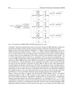
Fig. 1. Schematic illustration depicting changes in limb field size as determined by fate mapping studies. The
limb field increases in size in a highly organized manner before the appearance of the limb bud. The size of the
limb field changes in only the anterior–posterior dimension between stages 12 and 14; the dorsal–ventral and
proximal–distal dimensions remain constant. Between stage 14 and 16, the expansion of the anterior–posterior
dimension declines and the dorsal–ventral dimension increases in size. After stage 16, the proximal–distal dimen-
sion grows in a disproportionate manner in comparison with the other axes.
FGF4 and Skeletal Morphogenesis 133
nent increases (13). The proximal–distal dimension expands as the limb bud forms between stages 16
and 17, and this expansion continues with bud elongation (12). Thus, changes in the size of the pre-
bud region and the limb bud itself indicates highly coordinated patterns of growth and expansion.
The FGF family of signaling proteins play an important role in setting up the prebud field. FGFs
are intercellular signaling molecules that display a strong binding affinity for the extracellular matrix
and signal via the FGF receptor (FGFR), a member of the tyrosine kinase superfamily of cell surface
receptors (14). The Fgf gene family is very large and includes at least seven members expressed
during limb development, Fgf2, Fgf4, Fgf8, Fgf9, Fgf10, Fgf17, and Fgf18 (15,16). Of these Fgf10
and Fgf18 are expressed only in mesenchymal cells, Fgf4, Fgf8, Fgf9, and Fgf17 are expressed only
in the ectoderm, specifically the AER, and Fgf2 is expressed in both the ectoderm and the mesen-
chyme. The FGFr gene family includes four members, of which three, FGFr1, FGFr2, and FGFr3,
are expressed during limb development. FGFr1 is expressed predominately in undifferentiated mes-
enchyme (17–19). There are two isoforms of FGFr2 expressed in the limb bud; FGFr2b is expressed
in the limb ectoderm, including the AER, and FGFr2c is expressed in the ectoderm and in prechon-
drogenic condensations (18–20). FGFr3 is expressed late in skeletogenesis and is associated with dif-
ferentiating cartilage (18,19).
Fgf10 loss-of-function studies in the mouse result in a limbless phenotype, indicating that FGF10
is required for limb outgrowth (21,22). Similarly, interrupting the action of FGF10 either by over-
expressing a soluble, dominant-negative derivative of the FGFr2B gene or by the deletion of the FGF
binding domain of the FGFr2 gene results in a limbless or distally truncated phenotype (23,24). In the
chick, Fgf10 is expressed in lateral plate mesoderm at stage 12 when the limb field becomes tissue auton-
omous (25). At this stage, Fgf10 is expressed beyond the mapped boundary of the limb; however, it is
downregulated in the surrounding tissue so that by stage 15 it is expressed only in the prebud mesoderm.
One downstream target of FGF10 signaling is the AER-specific gene Fgf8. Fgf8 expression in the
prebud ectoderm in first observed at stage 16, some 3 h after localization of Fgf10 expression to the
prelimb mesenchymal tissue (26–30). The initial Fgf8 expression domain encompasses a broad band
of ectodermal cells that includes the future AER, and once the bud forms, Fgf8 expression is exclu-
sively restricted to the AER. Expression of Fgf8 in the limb ectoderm is FGF10 dependent (21,22)
and can be induced by ectopic FGF10 application (25,31). FGF8 application in the limb bud induces
an expansion of the Fgf10 expression domain, thus suggesting a reciprocal regulatory loop between
mesenchymal FGF10 and ectodermal FGF8 (14,25). The absence of FGF8 during limb outgrowth
results in relatively normal limb limbs that display reduced skeletal elements at all levels (32,33).
The absence of FGF8 in the limb bud results in the anterior expansion of the Fgf4 expression domain,
thus suggesting that Fgf4 expression in the AER is negatively regulated by FGF8.
Limb defects are not observed in loss of function studies targeting Fgf2, Fgf4, Fgf9, or Fgf17
genes (34–37); however, gain of function studies in which purified FGF proteins are delivered on
slow-release microcarrier beads into the limb-forming region provide evidence that these factors play
key roles in the regulation of limb outgrowth. In the chick, nonlimb, embryonic flank tissue (stages
13–17) responds to an ectopic source of FGFs by initially forming an ectopic limb bud that later
develops into identifiable limb structures (38,39). The ectopic limb is always of reverse handedness
in comparison with the neighboring, endogenous forelimbs and hindlimbs, and the ectopic limb is
generally a chimera of both tissues types (40). A number of FGFs have been tested using this assay,
including FGF1, FGF2, FGF4, FGF7, FGF8, and FGF10. Of these, only FGF7 failed to induce the
formation of ectopic limb structures (29,30,38,39). Ectopic limbs are generally induced by implants
of microcarrier beads loaded with purified FGF protein, although implantation of cells expressing dif-
ferent Fgfs can induce a similar response (39). Ectopic expression of Fgf4 or Fgf8 in flank cells through
retroviral infection (30,41) or ubiquitous expression of Fgf2 or Fgf4 in transgenic models (42,43) do
not result in ectopic limb formation, thus suggesting that the spatial distribution of FGF is important
for this response.
134 Ngo-Muller et al.
CELL MIGRATION AND A DYNAMIC PROGRESS ZONE
In the chick, the transition between prebud stages to limb bud stages is marked by the lateral bulg-
ing of the limb mesenchyme to form the limb bud, a homogeneous population of mesenchymal cells
covered by ectoderm. The AER is a prominent ectodermal structure that rims the distal tip of the limb
bud in all amniote vertebrates. In the chick, the AER forms soon after the bud is visible, and in the
mouse, the AER does not form until limb bud outgrowth is well underway (44). The late appearance
of the mouse AER as well as studies of the limbless mutation in the chick shows that initial formation
of the limb bud is an AER-independent event (45).
As with limb initiation, the dependency of mesenchymal outgrowth on the AER is known to be a
function of FGF activity. Numerous studies have shown that outgrowth can proceed after AER removal
in the presence of ectopically applied FGF; thus, FGF signaling is linked to the maintenance of the
progress zone. Although this function can be provided for by either FGF2, FGF4, or FGF8 (30,46–
48), FGF8 is assumed to be physiologically relevant because it is expressed throughout the AER with
no axial bias (26,27). Fgf2 is present in the dorsal ectoderm and peripheral mesenchyme in addition
to the AER (49,50), and Fgf4 transcripts are restricted to the posterior AER in the early limb bud (51,
52) but are expressed distally as bud outgrowth proceeds. Both the AER and ectopically applied FGF
also induce distal outgrowth of amputated limb buds, thus indicating that FGF signaling is involved
in the reformation of the progress zone associated with a regeneration response (53–55).
The outgrowth-promoting properties of FGFs in the limb bud is contrasted by studies showing
that ectopic FGF application in the presence of the AER has an inhibitory effect on limb outgrowth
(56,57). Studies with ectopic FGF2 bead implantation into the ZPA of an otherwise-normal chick limb
bud inhibits limb outgrowth in a dose-dependent manner (Fig. 2A-E). This FGF2 response is position
specific in that a similar response is not observed after ectopic application of FGF-2 into the anterior
limb bud (56,58). Outgrowth inhibition by FGF2 is associated with dramatic changes in limb bud shape
and with the expansion and bifurcation of the Shh and HoxD13 expression domains. Cell marking stud-
ies show that ectopic FGF-2 modifies the normal distalward movement of ZPA cells, but not anterior
cells, during limb outgrowth. Thus, understanding the role of FGF2 signaling in the limb bud is com-
plicated by the apparent paradoxical result that FGF2 promotes limb outgrowth but also inhibits limb
outgrowth (56). A similar set of paradoxical findings are known for both FGF4 and FGF8. Application
of FGF4 to the limb bud after AER removal or bud amputation replaces AER function by inducing
distal outgrowth (47,55); however, application of FGF4 to a subdistal location of an otherwise-intact
limb bud causes localized shortening of the limb bud and reductions in the length of skeletal elements,
thus FGF4 inhibits bud outgrowth (Fig. 2F-H). As mentioned above, FGF8 application to the flank of
the embryo results in the induction of supernumerary limbs from flank tissues; however, the inhibition of
limb bud outgrowth is observed when FGF8 beads are implanted near the endogenous limb field (30).
As a solution to these paradoxical effects of FGFs on limb formation, we have proposed that a
major role of FGF signaling by the AER is to control patterns of cell movements important for mor-
phogenesis and pattern formation (1,57). In our in vivo studies, we have found that FGF4 acts as a
potent and specific chemoattractive agent for mesenchymal cells of the limb bud (Fig. 3). Thus, an
ectopic source of FGF4 can induce posterior limb bud cells to migrate in either an anterior or proxi-
mal direction. The in vivo migration response to FGF-4 is dose dependent both in the number of cells
stimulated to migrate and the distance migrated. The AER was also found to be a potent chemo-
attractant, directing the migration of mesenchymal cells within 75 µm of the AER to make contact
with the AER within a 24-h period and mesenchymal cells within at least 150 µm to migrate toward
the AER. These studies indicate that FGF4 produced by the AER has a long-range chemoattractive
function and regulates proximal–distal patterns of cell migration during limb outgrowth. In experi-
ments that result in the inhibition of limb bud outgrowth, we propose that altering the normal migra-
tion of these cells results in dramatic and rapid changes in limb bud shape and alters morphogenesis
FGF4 and Skeletal Morphogenesis 135
Fig. 2. FGF bead implantation studies demonstrate that FGF2 and FGF4 cause a dramatic alteration of limb
bud shape, inhibiting outgrowth and modifying skeletal morphogenesis. Affi-Gel Blue beads containing FGF-2
implanted into the posterior mesenchyme of an otherwise normal wing bud (A) induced dramatic alterations of
limb bud morphology 18 and 40 h (B) after implantation. Distal outgrowth (arrow) of the posterior region of the
bud was inhibited as compared with the nonoperated bud on the same embryo (shown on the left). Skeletal
morphogenesis is modified from control limbs (untreated bead implantation; C), displaying severe loss of digits
(D) or truncation (E) . The arrows in C–E identify implanted beads. Taken from Li et al., 1996 (56). FGF4 bead
implantation also inhibits limb outgrowth and skeletal morphogenesis. Two FGF-4 beads (*) implanted into the
subapical region of a stage 24 limb bud (F) locally inhibits outgrowth (arrows) 24 h later, giving the distal limb
bud an “arrowhead” appearance. Skeletal preparation of the resulting limb shows a complete skeletal pattern in
which proximal-distal elongation of many skeletal elements is inhibited. Taken from Li and Muneoka, 1999 (57).
136 Ngo-Muller et al.
of the skeleton (57). FGF-4 has also been shown to direct the migration of nonlimb cells into the limb
bud (59). These findings indicate that the progress zone is a dynamic region of the limb bud where
differential cell migration toward the AER results in continuous changes in the type of cell–cell inter-
actions that can occur.
There are a number of implications important for our understanding of limb morphogenesis that
result from this dynamic description of the progress zone. Based on direct measurements, we are able
to estimate that migrating mesenchymal cells move at a maximum rate of about 50 µm/h (Li and
Muneoka, unpublished data). At that rate, migrating cells encounter and move pass about five cells
Fig. 3. FGF-4 is a chemoattractant for limb bud cells. A, The in vivo assay for migration consisted of DiI label-
ing of posterior–distal cells of a stage 24 limb bud (arrowhead) and implantation of a carrier bead (*) containing
FGF-4 into the central–distal region of the bud. Figures B–F are computer overlays of whole-mount limb buds
(dorsal view, distal is to the right and posterior is at the bottom) imaged in bright field and also with fluorescence
microscopy to identify DiI-labeled cells. B, In phosphate-buffered saline-treated bead implantation control limb
buds, DiI-labeled cells after 12 h of incubation expanded distally (arrowhead) but did not migrate toward the
implanted bead (*). C, 12 h after implantation of a FGF-4-treated bead, two clusters of DiI-labeled cells are appar-
ent: one associated with the FGF-4 bead (*) located centrally in the limb bud (arrow) and a second at the posterior
injection site (arrowhead). D, In a minority of cases, DiI-labeled cells were scattered along a trail that extended
from the posterior injection site (arrowhead) to the FGF-4 bead (*). E, 6 h after implantation of a FGF-4 treated
bead, DiI-labeled cells are observed migrating anteriorly toward the FGF-4 bead (*) and a few cells can be seen
making contact with the bead (arrow). F, Experiments in which a FGF-4 bead (*) was implanted proximal to the
posterior injection site (stage 24 limb bud) resulted in the migration of labeled cells in a proximal direction. After
12 h of incubation, the majority response was the formation of two clusters of DiI-labeled cells: one associated
with the FGF-4 bead (arrow) located at the base of the limb bud and a second at the posterior injection site
(arrowhead).
FGF4 and Skeletal Morphogenesis 137
each hour, or one cell every 12 min; thus the nature of cell–cell interactions occurring within the limb
bud will be influenced by these cell movements. Because only a subset of cells are migrating to pop-
ulate the progress zone, it is reasonable to speculate that these cells are uniquely different from their
nonmigrating counterparts. The recent demonstration that FGF1-induced migration in NBT-II rat
bladder carcinoma cells in vitro is cell cycle dependent (60) raises the possibility that a similar cell
cycle-specific response occurs in migrating limb bud cells. In the distal limb bud, all cells are prolif-
erating; thus, it is possible that the response to FGF4 signaling could vary in a cell cycle-dependent
manner. This possibility can account for our observation that cells that failed to migrate to the FGF4
bead were later found to migrate distally during limb outgrowth (57). One consequence of a cell cycle–
dependent migration response in the limb bud is that there will be a tendency for both migrating and
nonmigrating cells to become synchronized, and reports of unexplainable regions of synchronized cells
in the limb bud have been reported (61). In addition, if G
1
is the migration-responsive phase, as has
been shown for cultured cells, then the migration event would also cause an artificial depletion of S-
phase cells (low apparent proliferation rate) immediately subjacent to the AER, and an artificial enrich-
ment of S-phase cells (high apparent proliferation rate) at more proximal levels. This unusual and
unexplained observation has been noted multiple times in studies characterizing the growth dynamics
of the early limb bud (62–64).
It is generally assumed that the AER provides a mitogenic signal that maintains cell proliferation
within the progress zone. The AER and FGFs have been shown to be mitogenic for limb bud cells in
vitro (65–68); however, the endogenous patterns of cell proliferation in the limb bud do not support
the conclusion that the AER produces a unique mitogenic signal. Mesenchymal cell proliferation in
the early limb bud is initially uniform, and only after significant elongation and the onset of proximal
chondrogenesis are gradients of proliferation evident (69). In the chick limb bud, a distal-to-proxi-
mal gradient of cell proliferation is discernible by stage 24, but this gradient is associated with a prox-
imal decline in mitotic rate associated with chondrogenesis in the center of the limb bud. At this same
stage, the dorsal–ventral axis also displays a gradient of mesenchymal cell proliferation that is high-
est at the dorsal and ventral surfaces and lowest at the center of the limb bud where chondrogenesis
is commencing. Importantly, growth differences are not apparent when comparing mesenchymal cells
at the distal tip to cells at either the ventral or dorsal periphery at proximal levels. Thus, the mitotic
gradients in the limb bud are linked to the onset of differentiation and not to specific mitogenic sig-
naling associated with the AER. In support of this conclusion, after AER removal, the rate of
3
H-thy-
midine incorporation in subridge mesoderm is not changed, and the mitotic index is only transiently
depressed (70,71). Thus, there is no in vivo evidence for an AER-specific mitogenic signal. Because
AER removal inhibits limb outgrowth without modifying cell proliferation rates, the data indicate
that cell proliferation and the control of limb elongation by the AER are independent events. How-
ever, these data are consistent with a cell migration model in which limb bud outgrowth is driven by
distalward cell movements and a relatively uniform rate of cell proliferation. We have proposed that
the mitogenic effects of the AER and FGFs in vitro is an indirect consequence of FGF regulated cell
migration; limb cell proliferation can be either stimulated or inhibited by FGFs depending on where
they are directed to migrate (57).
FGF SIGNALING AND BRANCHING MORPHOGENESIS
The elongating limb bud is characterized by an apical progress zone where pattern specification
occurs, a subapical zone of proliferating undifferentiated cells, and a proximal differentiation zone
where the onset of differentiation is associated with a decline in cell proliferation. The entire inter-
face between the events important for the specification of patterns that are occurring in the progress
zone and the events regulating the differentiation of limb structures is largely a mystery. In the limb,
patterning studies have focused almost exclusively on the skeletal pattern, and therefore it is appropri-
ate to target skeletal formation in considering the interact between patterning events and differentiation
138 Ngo-Muller et al.
events. The onset of chondrogenic differentiation is marked by the condensation of mesenchymal cells.
These condensations are characterized by an increased packing density, the associated expression of
a number of surface proteins (e.g., fibronectin, tenascin, neural cell adhesion molecule, and N-cadhe-
rin), and a change in the extracellular matrix composition. Differential adhesion between cells plays a
critical role in the initiation of mesenchymal condensations (see Chapter 1 by Tuan). Shubin and Alberch
(72) have proposed that the morphogenesis of the vertebrate limb skeletal pattern occurs through a
hierarchical sequence of de novo condensation followed by elongation, branching, and segmentation
of chondrogenic rudiment. Thus, once initiated, condensations grow by cell recruitment and the skel-
etal pattern emerges as these condensations elongate, bifurcate, and segment. For example, the proxi-
mal long bones of the forelimb, that is, humerus, radius, and ulna, form as a result of elongation with
a single bifurcation and segmentation event, and the short bones of the carpal/tarsal region form as a
result of multiple bifurcation and segmentation events. Although the actual skeletal pattern is specified
early in limb development, the pattern itself is laid down much later by regulating these morphoge-
netic processes.
The development of the limb skeleton and the evolution of diverse tetrapod limb morphologies
can be explained as a result of controlling the spatial–temporal pattern of branching and segmentation
events. The axis from which branching events arise is called the metapterygial axis, and it is gener-
ally accepted that this axis runs along the proximal–distal axis on the posterior side of the limb and
curves from posterior to anterior in congruence with the digital arch (72,73). Skeletal elements proxi-
mal to the digits arise from a segmentation/bifurcation mechanism and the digits themselves form by
a bifurcation from the digital arch followed by elongation and segmentation without bifurcation. This
model of skeletal morphogenesis proposes that alteration in skeletal pattern emerges as a result of
physiochemical interactions that regulate whether or not a bifurcation response occurs. During skele-
tal morphogenesis, the expansion of the prechondrogenic domain reaches a critical mass and induces
a mathematical bifurcation as has been proposed in mechanochemical models of skeletal pattern for-
mation (74). In the developing limb bud, these morphogenetic events are occurring at the interface
between the undifferentiated subapical zone and the differentiation zone where mesenchyme conden-
sation is initiated.
Ectopic application of FGF4 modifies patterns of cell migration that are associated with changes in
limb bud shape and the pattern of chondrogenesis, and we have proposed that these events are causally
linked. One obvious way that FGF4-modified cell migration patterns can result in changes in skeletal
patterning is by modifying, either directly or indirectly, the processes controlling skeletal morpho-
genesis. During normal limb outgrowth, we have shown that the AER can influence the migration of
cells in the subapical zone; thus, we propose that apical cell migration plays a role in controlling the
pattern of skeletal morphogenesis (Fig. 4). One way that this might occur is if the interaction between
chondrogenic cells is favored or enhanced by the distal emigration of nonchondrogenic cells toward the
AER. Differential cell migration toward the AER in the dynamic progress zone model makes two clear
predictions about limb outgrowth. First, undifferentiated cells in the progress zone and the subapical
zone migrate distally and remain undifferentiated. Second, nonmigrating cells remain at a proximal
location, enter the differentiation zone, and initiate chondrogenesis. One consequence of these dif-
ferential cell movements is that there will be a reorganization of cells within the subapical zone. Thus,
the emigration of distally migrating cells out of this zone results in a concentration of non-migrating
cells. We propose that this reorganization of cells in conjunction with the expression of cell adhesion
molecules facilitates adhesive interactions between prechondrogenic cells that trigger mesenchymal
condensation. In this model, patterns of cell migration that is, in part, under the control of the AER
provide the interface between the specification of skeletal pattern and the actual regulation of mesen-
chymal condensation associated with the establishment of the pattern. This model is supported by the
results of fate mapping studies that show anterior or posterior shifts in the migration of mesenchymal
cells that are associated with the branching of specific skeletal elements (75).
FGF4 and Skeletal Morphogenesis 139
The hypothesis that FGF signaling is controlling branching morphogenesis of the limb skeletal pat-
tern is supported by studies in other developing organ systems that link the regulation of cell migra-
tion by FGF to the control of branching morphogenesis. Organs such as the lung, kidney, and salivary
gland in vertebrates develop abnormally or fail to develop at all in mice carrying null mutations for
either an FGF family member or an FGF receptor (21–23,76–78). For example, lung development is
inhibited in mice carrying a null mutation for Fgf10 (21,22) or FgfR2 (76–78), the putative receptor
for FGF10. In addition, in vitro experiments indicate that FGF10 functions as a chemotactic signal
important for branching during lung formation (79); thus, FGF10 in the developing lung and FGF4 in
the developing limb appear to play similar roles. In Drosophila, the FGF homolog branchless acts via
the FGF receptor, breathless, in controlling branching of the developing tracheal system, and here the
evidence also indicates that branchless acts as a chemotactic signal (80).
Beyond the link between FGF signaling and branching morphogenesis, there is considerable evi-
dence that FGF signaling control specific episodes of cell migration in many developing organs. Sex
cell migration is controlled in Caenorhabditis elegans by the FGF-like protein, EGL-17, which is secreted
by the target gonadal cell (81). FGF signaling is required for vertebrate myoblast migration (82), and
in vitro experiments indicate that FGF2 and FGF4 act as a chemotactic signals (83). FGF-8 and/or
FGF4 have also been shown to be critical for cell migration in gastrulating mouse embryos, and it is
required for successful gastrulation (84). Thus, vertebrate FGFs may have evolved from an ancestral
gene that functioned in the control of cell movements, and the extensive duplication of Fgf genes
correlates with the extensive use of cell migration in vertebrate development.
Fig. 4. The dynamic progress zone model for FGF4 directed cell migration and skeletogenesis. A, FGF-4
produced by the AER stimulates limb bud outgrowth by selectively directing cell migration distally (arrows),
thereby maintaining a progress zone at the tip of the bud and enhancing interactions between nonmigrating (<)
chondrogenic cells proximally. Light stippling indicates cells in the undifferentiation zone and dark stippling
indicates cells in the differentiation zone. B–D, Skeletogenesis is influenced by distal cell migration as a result of
emigration, promoting cell–cell interactions between chondrogenic cells. Modifying the direction of cell emigra-
tion can influence branching morphogenesis by modulating the size of the mesenchymal condensation.
140 Ngo-Muller et al.
FGF4 AND SKELETAL BIFURCATIONS
Later stages of limb development are characterized by the formation and separation of the digits.
The autopodial plate is dorsal-ventrally flattened and fans out with chondrogenic rays that represent
the forming digits. Fate maps of these digit rays indicate that they primarily form the metatarsal/meta-
carpal of the mature digit, with the phalangeal elements forming from only the distal-most region of
each ray (85). Individual digit rudiments can develop at ectopic sites, indicating that at this stage
each digit rudiment is an autonomously developing morphogenetic field (86). The primary events of
digit morphogenesis are distal elongation of the digit blastema, segmentation of the chondrogenic
digit ray, and programmed cell death within the interdigital regions that separate the digits. The digits
themselves do not normally bifurcate, thus making their formation a potential model system for study-
ing the induction of bifurcation events. Digit formation is also characterized by the downregulation
of many of the Fgf genes that are expressed during earlier stages of limb development, providing a
temporal relationship with bifurcation events.
When prechondrogenic cells of the autopod aggregate to form the digit rays, the AER and the ZPA
are no longer present; however, many patterning genes that were expressed in the progress zone of
the early limb bud (such as the Hoxa and Hoxd gene clusters and the Msx genes) remain expressed in
the digit rudiments, suggesting that the patterning of individual digit rudiments is still incomplete.
Furthermore, a number of experimental observations indicating that active patterning of digit rudi-
ments continues into later limb bud stages. First, digit tip amputations result in a rapid level-specific
regeneration response that includes re-expression of the AER-dependent Msx genes (86). Second, the
digit rudiments of mouse limb buds (stage 7/8, E12.5) respond to implantation of a FGF4-releasing
microcarrier bead by inducing specific bifurcation of digit IV, indicating that patterning of digit IV is
incomplete (87). Third, interdigital cells of the chick autopod are able to form digits in response to
wounding or to implantation of transforming growth factor-`-releasing microcarrier beads (88).
Fourth, digit identity in the chick hind limb can be modified by interdigital tissues and ectopic BMP
application after the formation of the digit ray (10). These studies support the view that patterning of
individual digit rudiments continues during later stages of limb development, although endogenous
patterning signals that act during digit formation are largely unknown.
Using exo utero surgery, we have used ectopic application of FGF4 into the autopod of the mouse
hindlimb at a stage shortly after AER regression to study the influence of FGF4 on digit formation (87).
Although our studies uncovered a number of FGF4-induced effects on digit formation, two observa-
tions are directly relevant for skeletal morphogenesis. First, FGF4 causes a local and transient inhibi-
tion of chondrogenesis involving the formed autopodial digit ray. A similar response has been reported
in the chick limb (89,90), and FGF signaling has been shown to inhibit chondrogenesis in high density
cultures of limb bud cells (91,92). Second, FGF4 induces a bifurcation response by prechondrogenic
cells at the tip of digit IV (Fig. 5A). We demonstrate with cell-marking studies that this bifurcation
response is associated with an FGF4-induced migration response that results in a reorganization of
prechondrogenic cells at the distal tip of the digit ray (Fig. 5B). This response does not involve inter-
digital cells known to have chondrogenic potential. Consistent with models that predict mathematical
bifurcation responses, the FGF4 response is associated with an expansion of the prechondrogenic zone
at the digit tip (Fig. 5C). Gene expression studies indicate that this response is associated with the mod-
ified expression of number of genes, including Msx1, Igf2, and the posterior members of the HoxD
cluster. These findings support our branching morphogenesis model for skeletogenesis by showing that
FGF4-induced cell migration is associated with the expansion of the distal prechondrogenic tissue
and a skeletal bifurcation response.
SUMMARY
Classical embryological and recent molecular studies have combined to increase our understand-
ing of the complex process of limb development. Although recent years have unarguably broadened
FGF4 and Skeletal Morphogenesis 141
Fig. 5. A, FGF4 bead implantation in E12.5 mouse left hind limb bud causes inhibition of cartilage growth and
differentiation (arrow) and the distal bifurcation of digit IV. B, FGF4 influences cell migration of digit cells. Cells
located at the anterior or posterior digit tip (digit III and IV) were labeled with DiI, and an FGF4 bead was simul-
taneously implanted in the interdigit III-IV (top left). Anterior is to the left, and distal is toward the top. After 72 h,
posterior digit IV cells (away from the bead) were not influenced by the FGF4 bead and migrated distally (lower
right), whereas anterior digit IV cells (closest to the bead) migrated toward the bead and established a secondary
digit tip (*). A summary map of cell migration in digits III and IV is presented (lower right): red dots represent the
location of posterior digit III and anterior digit IV injection sites and green dots represent the location of anterior
digit III and posterior digit IV injection sites. C, FGF4 influences gene expression and the pattern of condensation
in digit tips. Igf2 gene expression is used as a marker for chondrogenesis and identifies digit tip condensations in
control E13.5 day digits (left panel); 48 h after FGF4 bead implantation, Igf2 expression is distally expanded,
indicating a widening of the chondrogenic condensation associated with a bifurcation response (center panel).
The widening of Igf2 expression domain is associated with a Msx1-negative domain at the tip of digit IV (right
panel).