The Tourniquet Manual: Principles and Practice - part 6 pdf
Bạn đang xem bản rút gọn của tài liệu. Xem và tải ngay bản đầy đủ của tài liệu tại đây (339.44 KB, 12 trang )
This page intentionally left blank
B
EFORE THE TOURNIQUET
is inflated, it is essential to empty the limb of as much blood
as possible. This prevents unnecessary oozing and helps precise anatomical dissec-
tion.
When Lister used a tourniquet in 1879 to help him radically excise tuberculous wrist
joints, he was aware of the need for preliminary elevation of the limb.
1
He was fasci-
nated by the mechanism of blanching of the hand when it was elevated to 90
degrees, and he was convinced that this was brought about by the nervous system.
He described simple experiments in a volunteer, and he also experimented on an
anaesthetised horse. In a lecture to the Harveian Society, Lister described how his
attention had been drawn to the problem about 15 years previously. He advocated
preliminary elevation for a few minutes before the application of a tourniquet.
Distefano and colleagues, using impedance plethysmography, found that the
maximal decrease in volume by elevation alone, with the limb at 45 degrees, occurred
after 15–20 seconds, with no noticeable change thereafter.
2
While any changes in
impedance theoretically represent changes in intravascular and extravascular
compartments, in this study it would reflect only the former. Warren and colleagues,
measuring changes of circumference of the limb with mercury in silastic strain
gauges, found that the optimal time for elevation was five minutes.
3
For the maximal
effect, they suggested that the upper limb should be elevated at 90 degrees; for
the lower limb, they suggested 45 degrees of elevation, since further elevation was
likely to kink the femoral vein due to the flexion of the hip.
Using a gamma-camera technique and the injection of autologous 99m technetium-
labelled erythrocytes, Blond and colleagues showed that there was little change in
the reduction of blood volume of the lower limb at 60 degrees with an increase of
the duration of elevation.
4, 5
The results after half a minute were 45%, one minute
45%, two minutes 43%, four minutes 44%, six minutes 43%, and ten minutes 44%.
This pattern was also seen in the upper limb.
4.1 External Compression
External compression in addition to elevation has been shown to improve the degree
of exsanguination However, it is contraindicated in patients who have a suspected
infected or malignant lesion. Use of an Esmarch bandage or hand-over-hand manual
exsanguination
6
are more effective than elevation alone.
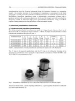
Use of an Esmarch bandage is time-consuming and can damage the skin over a
fracture or the atrophic skin of a patient with rheumatoid arthritis (Figure 4.1). It can
also detach pre-existing venous thromboses and produce pulmonary emboli.
7–9
53
Furthermore, there is no effective control of the pressure with which an Esmarch
bandage is applied. Undue stretching as each turn is applied results in increased
pressure. The average tension produced on routine application is 125 N; 175 N is
near the tensile limit of the bandage.
10
Martin’s bandage made of cream-coloured
latex rubber is used in a similar manner.
Sterilisation of an Esmarch bandages requires care but is effective if the bandage is
rolled loosely with a gauze bandage between the layers and placed in an autoclave.
11
For knee surgery, the adequacy of exsanguination produced by an Esmarch bandage
has been compared with the effect of elevation for two minutes.
12
A blinded,
randomised, prospective trial was undertaken in 50 patients having total knee
replacement and 50 patients having arthroscopy. The mean blood loss during total
knee replacement was significantly greater in the group that was elevated. The
haematocrit of samples of arthroscopy drainage was consistently less than 1%, irre-
spective of the method of exsanguination. None of the operating surgeons reported
that they considered that the surgery had been made more difficult by the use of
elevation alone. With elevation, the skin and superficial tissues are not cleared of
blood as effectively as with an Esmarch bandage. Minimal superficial bleeding did
not interfere with the surgical procedure at a deeper level. It was concluded that
considering the established risks of Esmarch bandages and the adequacy of the field
provided by elevation, the latter method was preferable. In contrast, Strover, in a
large personal series, did not use a tourniquet or mention exsanguination for either
arthroscopies or total knee replacements.
13
1111
2
3
4
5
611
7
8
9
1011
11
2
3111
4
5
6
7
8
9
2011
1
1
2
3
4
5
6
7
8
9
3011
1
1
2
3
4
5
6
7
8
9
4011
1
211
54
The Tourniquet Manual ➀➁➂➍➄➅➆
Figure 4.1 Application of an Esmarch bandage. By keeping close to the limb, one can avoid undue tension.
A technique described by Burchell and Stack
14
was later modified in the form of the
Northwick Park Hospital Exsanguinator.
15
Use of this apparatus did not require eleva-
tion of the limb and thus could be used single-handedly. The apparatus consisted
of a plastic cover applied to the limb distal to the tourniquet and inflated to systolic
pressure for one minute for exsanguination, before the tourniquet was inflated. The
splint was then deflated and removed.
In a similar manner, an arm splint used for the stabilisation of fractures for trans-
port was inflated by compressed air to a pressure of 200 mm Hg. The tourniquet
was then inflated. The splint was deflated, unzipped and removed before intra-
venous regional anaesthesia was given (see Chapter 6).
16
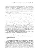
The need for control of the pressure that is applied has led to the development of
appliances such as the Rhys-Davies Exsanguinator.
17
This is an inflated elastic cylinder
that is rolled on to the limb (Figure 4.2). As the exsanguinator is applied, the pres-
sure within the sleeve increases. With small limbs, this is not marked and the degree
of exsanguination is less. On a very large limb, the maximum pressure generated is
55
➀➁➂➍➄➅➆ Exsanguination of the Limb
Figure 4.2 Use of the Rhys-Davies Exsanguinator: (a) Preliminary grip.
1111
2
3
4
5
611
7
8
9
1011
11
2
3111
4
5
6
7
8
9
2011
1
1
2
3
4
5
6
7
8
9
3011
1
1
2
3
4
5
6
7
8
9
4011
1
211
56
The Tourniquet Manual ➀➁➂➍➄➅➆
Figure 4.2 (c) Completion.
Figure 4.2 (b) Starting on the limb.
only about 150 mm Hg, which is distributed uniformly over the whole exposed
surface of the limb. The exsanguinator does not produce localised ridges of high
pressure or distort superficial tissues. Reinflation and direct measurement of the
inflation pressure are done through a valve in the wall of the cylinder, and a sphyg-
momanometer cuff is rolled up inside it. The exsanguinator requires regular
maintenance, and the manufacturer recommends that it be replaced annually.
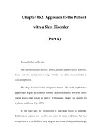
External methods of exsanguination reduce limb volume by forcing blood from it.
Using a water-displacement method, Silver and colleagues showed that a limb would
swell immediately by approximately 10% of its original volume after release of a
pneumatic tourniquet (Figure 4.3).
18
About half of the swelling is due to the return
of the exsanguinated blood to the limb. Further swelling is an effect of reactive
hyperaemia, and additional swelling can occur following both haematoma forma-
tion due to surgery and the accumulation of oedema from anoxia. Therefore, the
postoperative dressing must allow for this inevitable swelling (Figure 4.4). A plaster
should never be completely circumferential; instead, it should be split in the midline
or applied only as a backslab, or there should be a well-padded dressing.
In summary, the combination of elevation and the use of a Rhys-Davies
Exsanguinator is a safe and easy method for daily practice and can be used without
fear of complications.
57
➀➁➂➍➄➅➆ Exsanguination of the Limb
Figure 4.3 To show changes in
volume occurring after
exsanguination.
Reproduced with
permission of Lippincott, Williams &
Wilkins from Silver, R, de la Garza, J,
Koreska, J, Rang, M (1986). Limb
swelling after release of tourniquets.
Clinical Orthopaedics
206: 86–89.
Figure 4.4 Effect on the volume
of the limb, as seen in a rabbit’s
hind limb, following deflation
after application of a tourniquet
for three hours.
4.2 Sickle Cell Disease
The role of exsanguination and the use of tourniquets are controversial in patients
with sickle cell disease. Sickle cell disease is common in the West Indies and West
Africa. The erythrocytes assume a crescent-like or sickle shape when deprived of
oxygen. Blood that remains in a limb distal to a tourniquet may sickle. Tourniquets
induce the three most critical conditions known to produce sickling: circulatory stasis,
acidosis and hypoxia. Homozygous patients with erythrocytes containing the
abnormal haemoglobin S are at risk, but heterozygous patients who have the sickle
cell trait but also normal haemoglobin A are not at risk. Sickle cell trait is the result
of inheritance of normal haemoglobin from one parent and haemoglobin S from
the other. The small proportion of haemoglobin S is of less clinical significance,
although it is not completely innocuous. The erythrocytes contain enough haemo-
globin S to sickle in the laboratory preparation. Ludham and Jellis, working in Zambia,
pointed out that a bloodless field may shorten the time taken for surgery, make
surgery safer, and minimise blood loss, especially if no blood is available for trans-
fusion.
19
Conversely, blood that remains in the limb may sickle, with potentially
serious effects. With careful monitoring of systemic pO
2
by pulse oximetry, Ludham
and Jellis have not seen any marked lowering after deflation of the tourniquet in
patients with sickle cell disease. The benefits afforded by the use of a tourniquet
need to be balanced against the dangers, and a decision should be made for each
individual operation. Careful exsanguination is the key to safety. This approach
accords with the report of Stein and Urbaniak, who found in a series of 21 patients
carrying the sickle cell gene and who underwent 29 operations under tourniquet
that there was no statistically increased incidence of complications when compared
with a control group of black patients without the sickle cell trait and who had
similar operations.
20
References
1 Godlee, RJ (1924). Lord Lister, 3rd edn. Oxford: Clarendon Press. p. 632.
2 Distefano, V, Nixon, JE, Stone, RH (1974). Bioelectric impedance plethysmography as an investigative tool
in orthopaedic surgery – a comparative study of limb exsanguination techniques. Clinical Orthopaedics 99:
203–206.
3 Warren, PJ, Hardman, PJ, Woolf, VJ (1992). Limb exsanguination. i. The arm: effects of angle of elevation
and arterial compression. ii. The leg: effects of angle of elevation. Annals of the Royal College of Surgeons
of England 74: 320–322, 323–325.
4 Blond, L, Kirketorp-Moller, K, Sonne-Holm, S, Madsen, JL (2002). Exsanguination of lower limb in healthy
male subjects. Acta Orthopaedica Scandinavica 73: 89–92.
5 Blond, L, Madsen, JL (2002). Exsanguination of the upper limb in healthy young volunteers. Journal of Bone
and Joint Surgery 84B: 489–491.
6 Colville, J, Small, JO (1986). Exsanguination of the upper limb in hand surgery – comparison of four
methods. The Hand 11B: 469–470.
7 Austin, M (1963). The Esmarch bandage and pulmonary embolism. Journal of Bone and Joint Surgery 45B:
384–385.
8 Pollard, BJ, Lovelock, HA, Jones, RM (1983). Fatal pulmonary embolism secondary to limb exsanguination.
Anaesthesiology 58: 373–374.
1111
2
3
4
5
611
7
8
9
1011
11
2
3111
4
5
6
7
8
9
2011
1
1
2
3
4
5
6
7
8
9
3011
1
1
2
3
4
5
6
7
8
9
4011
1
211
58
The Tourniquet Manual ➀➁➂➍➄➅➆
9 Hoffman, A, Wyatt, RWB (1985). Fatal pulmonary embolism following tourniquet inflation. Journal of Bone
and Joint Surgery 67A: 633–634.
10 McClaren, AC, Rorabeck, CH (1985). The pressure distribution under tourniquets. Journal of Bone and Joint
Surgery 67A: 433–438.
11 O’Hara, JN, Coleman, M, Hutton, RM (1991). A simple and effective method of sterilizing Esmarch bandages.
Journal of Arthroplasty 6: 95–96.
12 Marshall, PD, Patel, M, Fairclough, JA (1994). Should Esmarch bandages be used for exsanguination in
knee arthroscopy and knee replacement surgery? A prospective trial of Esmarch exsanguination versus
simple elevation. Journal of the Royal College of Surgeons of Edinburgh; 38: 189–190.
13 Strover, A (1996). Are tourniquets in total knee replacement and arthroscopy necessary? The Knee 3:
115–119.
14 Burchell, G, Stack, G (1993). Exsanguination of the arm and hand. The Hand 5: 124–126.
15 Klenerman, L (1978). A modified tourniquet: preliminary communication. Journal of the Royal Society of
Medicine 71: 121–122.
16 Winnie, AP, Ramamurthy, S (1970). Pneumatic exsanguination for intravenous regional anaesthesia.
Anaesthesiology 33: 664–665.
17 Rhys-Davies, NC, Stotter, AT (1985). The Rhys-Davies Exsanguinator. Annals of the Royal College of Surgeons
of England 67: 193–195.
18 Silver, R, de la Garza, J, Koreska, J, Rang, M (1986). Limb swelling after release of tourniquets. Clinical
Orthopaedics 206: 86–89.
19 Ludham, CA, Jellis, J (2002). Blood disorders and AIDS. In Benson, M, Fixsen, JA, MacNicol, M, Parch, K, eds.
Children’s Clinical Orthopaedics and Fractures, 2nd edn. London: Churchill Livingstone, p. 116.
20 Stein, RE, Urbaniak, J (1980). Use of tourniquet during surgery in patients with sickle cell haemoglo-
binopathies. Clinical Orthopaedics 151: 231–233.
59
➀➁➂➍➄➅➆ Exsanguination of the Limb
This page intentionally left blank
Chapter 5
Complications
This page intentionally left blank
A
TOURNIQUET CAN
damage any of the tissues of a limb. Complications arise because of
failure to treat the limb physiologically and should be preventable. In addition, there
is an inevitable complication that always follows the use of a tourniquet to some
extent: swelling. Swelling is related directly to the duration of ischaemia, and it must
be anticipated. Release of the tourniquet and haemostasis before the wound is closed
will help to reduce swelling. There should be space available in well-padded postop-
erative dressings, and the limb should be elevated for the first few hours after opera-
tion. The effects of swelling must not be aggravated by constrictive dressings or
plaster casts. Complete plasters must never be applied unless they are split immedi-
ately. Backslabs are preferred, since these avoid the possibility of compartment
syndrome due to external compression.
Complications often have medicolegal implications. The most common problems,
in my experience, are nerve lesions, burns following spirit-based antiseptic solutions
seeping beneath the cuff, and failure to recognise peripheral vascular disease before
the operation, which may lead to delayed wound healing or even amputation.
5.1 Damage to Nerves
Harvey Cushing introduced the use of a pneumatic tourniquet because of the prob-
lems of nerve injuries produced by Esmarch bandages and solid rubber tourniquets.
Speigel and Lewin
1
claim that the first available reports of tourniquet paralysis are
those of Montes, recorded in a Mexican journal, and Putnam, recorded in a report
to the Boston Society for Medical Improvement.
2
The largest series of similar cases
is that collected by Eckhoff, who described 14 patients.
3
Eckhoff stated: “no effort
is too great in the prevention of this condition”. He hoped his article would stimu-
late the routine use of a pneumatic tourniquet. In his series, most lesions recovered
within three months.
Middleton and Varian investigated the number of neurological complications after
the use of a tourniquet by means of a questionnaire sent to 151 members of the
Australian Orthopaedic Association.
4
The incidence of peripheral nerve lesions was
one in 5000 for the arm and one in 13 000 for the leg. The arm palsies fell into
two main groups: the largest group involved median, ulnar and radial nerves below
the tourniquet, while the slightly smaller group comprised isolated radial nerve
lesions. The lesions occurred with both Esmarch bandages and pneumatic cuffs. All
except one patient made a full recovery; the exception developed a complete radial
nerve injury, which persisted. The approximate time for recovery was four to five
months, although some palsies were transient and others required up to 12 months
to disappear.
63