Báo cáo y học: " Pigmented villonodular synovitis of the knee in a patient on oral anticoagulation therapy: a case report" potx
Bạn đang xem bản rút gọn của tài liệu. Xem và tải ngay bản đầy đủ của tài liệu tại đây (1.1 MB, 3 trang )
BioMed Central
Page 1 of 3
(page number not for citation purposes)
Journal of Medical Case Reports
Open Access
Case report
Pigmented villonodular synovitis of the knee in a patient on oral
anticoagulation therapy: a case report
Balasundaram Ramesh*, Sanathkumar Shetty and Salah S Bastawrous
Address: Department of Trauma and Orthopaedics, Glan Clwyd Hospital, Sarn Lane, Bodelwyddan, Rhyl, North Wales, LL18 5UJ, UK
Email: Balasundaram Ramesh* - ; Sanathkumar Shetty - ;
Salah S Bastawrous -
* Corresponding author
Abstract
Introduction: Pigmented villonodular synovitis is a disease which affects the synovial joints and
tendon sheaths. Although the exact aetiological factors are not known, we believe that recurrent
haemarthrosis has a role in the aetiology of this condition.
Case presentation: A 62-year-old Caucasian man presented with gradually worsening pain and
stiffness in his right knee. The patient was on anticoagulation therapy and had been treated for
recurrent episodes of spontaneous haemarthrosis of the knee. The International Normalized Ratio
on each occasion suggested poor control of the anticoagulation therapy. A diagnosis of pigmented
villonodular synovitis was made based on intra-operative findings and was further confirmed by a
histopathological examination.
Conclusion: This report is presented to highlight the unusual association of haemarthrosis and
pigmented villonodular synovitis.
Introduction
Pigmented villonodular synovitis (PVNS) is a disease of
unknown aetiology affecting the synovial joints. The aeti-
ology of PVNS remains controversial and a number of the-
ories have been postulated. Haemarthrosis has been
suggested as a possible aetiological factor. Only one
description of PVNS of the ankle in a patient on anticoag-
ulation therapy has been previously reported [1]; we
describe the second known case of PVNS of the knee joint
in a patient on anticoagulation therapy.
Case presentation
A 62-year-old Caucasian man had an uneventful total left
knee replacement five years prior to presentation. Two
years following the total knee replacement, the patient
was diagnosed with dilated cardiomyopathy and was
started on warfarin. Following this, he had recurrent epi-
sodes of sudden pain and swelling on his right knee. Dur-
ing each of these episodes, there was no history of trauma
and the patient was systemically well. Although his Inter-
national Normalized Ratio (INR) was high, his blood tests
for full blood count and C-reactive protein were within
the normal range. All of these episodes were in the initial
phase of his warfarin therapy and they ceased once his
INR was stabilized.
The condition of the patient's right knee gradually deteri-
orated. Clinically, the knee joint was diffusely swollen
and tender but stable. His active range of movement was
from neutral to 100 degrees of flexion and any further
flexion was painful. Radiographs of the knee showed
advanced arthritic changes and he was admitted for a total
Published: 13 November 2009
Journal of Medical Case Reports 2009, 3:121 doi:10.1186/1752-1947-3-121
Received: 7 January 2008
Accepted: 13 November 2009
This article is available from: />© 2009 Ramesh et al; licensee BioMed Central Ltd.
This is an Open Access article distributed under the terms of the Creative Commons Attribution License ( />),
which permits unrestricted use, distribution, and reproduction in any medium, provided the original work is properly cited.
Journal of Medical Case Reports 2009, 3:121 />Page 2 of 3
(page number not for citation purposes)
knee replacement. Intraoperatively, the synovium was
found to be hypertrophic and stained reddish orange and
the synovial fluid was reddish-orange in colour (Figure 1).
These appearances suggested a diagnosis of PVNS. A syn-
ovectomy was performed, which was then followed by a
total knee replacement. A synovial specimen was sent for
histopathological examination. The microscopic features
were consistent with a diagnosis of PVNS (Figure 2). The
postoperative period was uneventful and the patient was
asymptomatic after three years of follow-up treatment.
Discussion
PVNS typically occurs in adults in their third or fourth
decade of life, with a male-to-female ratio of 1.9 to 1.3.
Involvement is usually monoarticular [2]. The knee joint
is the most frequently affected site, followed by the fin-
gers, feet, ankles, hips, wrists and shoulders in a decreas-
ing order of frequency [2,3].
Since the first description of this condition by Jaffe et al. in
1941 [4], the aetiology of this benign tumour involving
the synovial membrane has remained unclear. Jaffe pro-
posed that a hypervascular cellular phase occurs after
trauma produc ing hyalinization and fibrosis [4]. Various
aetiologies including trauma [2], inflammation [3],
haemorrhage [5], neoplasia [6] and genetic factors [7]
have been suggested. Chronic recurrent microtrauma and
haemarthrosis have also been postulated [2].
It has also been postulated that PVNS in children arises
through a different mechanism to that in adults, and it is
also possible that not all lesions interpreted as PVNS share
the same mechanism [8].
There are very few cases of PVNS reported in patients on
anticoagulation therapy [1] and with a bleeding disorder
[9]. In our patient, the symptoms in the knee worsened
following these repeated episodes of haemarthrosis and
the INR on these occasions showed a poor control of his
anticoagulation therapy.
Conclusion
This case supports the argument of earlier reports [1,9]
that repeated haemarthrosis may have a role in the aetiol-
ogy of PVNS. We hope that this study will encourage the
reporting of similar cases to lead to a better understanding
of the aetiology of this condition.
Abbreviations
INR: International Normalized Ratio; PVNS: pigmented
villonodular synovitis.
Intra-operative photographs showing the reddish-orange stained synovium (black arrow)Figure 1
Intra-operative photographs showing the reddish-
orange stained synovium (black arrow).
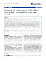
Photomicrograph showing the presence of haemosiderin deposits, foamy histiocytes and giant cells confirming pig-mented villonodular synovitis (haematoxylin and eosin stain)Figure 2
Photomicrograph showing the presence of haemosi-
derin deposits, foamy histiocytes and giant cells con-
firming pigmented villonodular synovitis
(haematoxylin and eosin stain).
Publish with BioMed Central and every
scientist can read your work free of charge
"BioMed Central will be the most significant development for
disseminating the results of biomedical research in our lifetime."
Sir Paul Nurse, Cancer Research UK
Your research papers will be:
available free of charge to the entire biomedical community
peer reviewed and published immediately upon acceptance
cited in PubMed and archived on PubMed Central
yours — you keep the copyright
Submit your manuscript here:
/>BioMedcentral
Journal of Medical Case Reports 2009, 3:121 />Page 3 of 3
(page number not for citation purposes)
Competing interests
The authors declare that they have no competing interests.
Authors' contributions
SS made substantial contributions in acquiring data,
reviewing the literature and preparing the manuscript. BR
performed the knee replacement operation and also con-
tributed in reviewing the literature and drafting the man-
uscript. SSB gave final approval to the draft to be
published. All authors read and approved the final manu-
script.
Consent
Written informed consent was obtained from the patient
for publication of this case report and any accompanying
images. A copy of the written consent is available for
review by the Editor-in-Chief of this journal.
References
1. Pearse OE, Klass B, Bendall SP: Pigmented villonodular synovitis
of the ankle occurring in a patient on anticoagulation ther-
apy. J Surg Orthop Adv 2004, 13(4):217-219.
2. Myers BW, Masi AT: Pigmented villonodular synovitis and ten-
osynovitis: a clinical epidemiologic study of 166 cases and lit-
erature review. Medicine 1980, 19:223-238.
3. Granowitz SP, O'Antorio J, Mankin H: The pathogenesis and long
term end results of pigmented villonodular synovitis. Clin
Orthop Relat Res 1976, 114:335-351.
4. Jaffe HL, Lichtenstein L, Sutro CJ: Pigmented villonodular synovi-
tis, bursitis and tenosynovitis. Arch Pathol 1941, 31:731-765.
5. Leszczynski J, Huckell JR, Percy JS, LeRiche JC, Lentle BC: Pig-
mented villonodular synovitis in multiple joints. Occurrence
in a child with cavernous hemangioma of lip and pulmonary
stenosis. Ann Rheum Dis 1975, 34:269-272.
6. Rao AS, Vigorite VJ: Pigmented villonodular synovitis (giant cell
tumor of tendon sheath and synovial membrane). Review of
81 cases. J Bone Joint Surg Am 1984, 66:76-94.
7. Wendt RG, Wolfe F, McQueen D, Murphy P, Solomon H, Housh-
older M: Polyarticular pigmented villonodular synovitis in
children: evidence for genetic contribution. J Rheumatol 1986,
13(5):921-926.
8. Schumacher HR, Lotke P, Athreya B, Rothfuss S: Pigmented villon-
odular synovitis: light and electron microscopic studies.
Semin Arthritis Rheumatol 1982, 129(1):32-43.
9. Matsui H, Takahashi Y, Matsunaga T, Tanaka-Horie T, Minowa H, Sug-
imoto M, Tsukino R, Mii Y, Giddings J, Yoshioka A: Successful
arthroscopic treatment of pigmented villonodular synovitis
of the knee in a patient with congenital deficiency of plas-
minogen activator inhibitor-1 and recurrent haemarthrosis.
Haemostasis 2001, 31:106-112.