Báo cáo y học: "Undifferentiated giant cell type carcinoma of the gallbladder with sarcomatoid dedifferentiation: a case report and review of the literature" ppsx
Bạn đang xem bản rút gọn của tài liệu. Xem và tải ngay bản đầy đủ của tài liệu tại đây (623.14 KB, 5 trang )
Case report
Open Access
Undifferentiated giant cell type carcinoma of the gallbladder
with sarcomatoid dedifferentiation:
a case report and review of the literature
Andreas Manouras
1
, Michael Genetzakis
1
, Emmanuel E. Lagoudianakis
1
,
Haridimos Markogiannakis
1
*, Artemisia Papadima
2
, George Agrogiannis
3
,
Hariklia Gakiopoulou
3
, Panagiotis Kekis
1
, Konstantinos Filis
1
and
Efstratios Patsouris
3
Addresses:
1
First Department of Propaedeutic Surgery, Hippokrateion Hospital, Athens Medical School, University of Athens, Q. Sofias 114 av.,
11527 Athens, Greece,
2
Department of Anesthesiology, Hippokrateion Hospital, Q. Sofias 114 av., 11527 Athens, Greece and
3
First Department of
Pathology, Athens Medical School, University of Athens, Q. Sofias 114 av., 11527 Athens, Greece
Email: AM - ; MG - ; EEL - ; HM* - ;
AP - ; GA - ; HG - ; PK - ; KF - ;
EP -
* Corresponding author
Published: 18 March 2009 Received: 22 December 2007
Accepted: 22 January 2009
Journal of Medical Case Reports 2009, 3:6496 doi: 10.1186/1752-1947-3-6496
This article is available from: />© 2009 Manouras et al; licensee Cases Network Ltd.
This is an Open Access article distributed under the terms of the Creative Commons Attribution License (
/>which permits unrestricted use, distribution, and reproduction in any medium, provided the original work is properly cited.
Abstract
Introduction: Undifferentiated gallbladder carcinoma is a rare entity. Among unusual types of
undifferentiated gallbladder carcinoma, giant cell type carcinoma is infrequent and, moreover, very few
cases of such neoplasms with osteoclast-like giant cells have been documented. We report a case of
undifferentiated gallbladder carcinoma presenting an unusual immunophenotype that was shown to be of
giant cell type with sarcomatoid dedifferentiation infiltrated by osteoclast-like multinucleated cells.
Case presentation: An 84-year-old Greek man presented with right upper quadrant pain, high
fever, rigors, anorexia and weight loss during the past month. Clinical examination revealed
tenderness in the right upper abdominal quadrant and a palpable gallbladder. Blood tests showed
elevated white blood-cell count and transaminases. Abdominal ultrasound and computed tomography
demonstrated a markedly distended gallbladder, measuring 16 cm x 8 cm, with oedema and
pericholecystic fluid, consistent with gallbladder empyema. After an open cholecystectomy and an
uneventful recovery, the patient was discharged on the 4
th
postoperative day. On cut surface, a 2cm
solid mass was identified, obstructing the lumen in the neck of the gallbladder. Histopathology and
immunohistochemistry offered the diagnosis of an undifferentiated, giant cell type carcinoma of the
gallbladder with sarcomatoid dedifferentiation infiltrated with osteoclast-like giant cells.
Conclusions: Undifferentiated, giant cell type carcinoma of the gallbladder with sarcomatoid
dedifferentiation infiltrated with osteoclast-like giant cells is a very infrequent neoplasm. Controversy
exists over its nature, as related knowledge remains incomplete. Thorough histopathological and
immunohistochemical evaluation is imperative for diagnosis. Due to their rarity, the biological
behaviour and prognosis of these tumours remain unclear.
Page 1 of 5
(page number not for citation purposes)
Introduction
The incidence of gallbladder carcinoma is 1.2 cases per
100 000 per year, increasing with age, particularly after the
fifth decade of life [1,2]. Risk factors include cholelithiasis,
calcified gallbladder wall, adenomatous gallbladder
polyps, obesity, oestrogen, choledochal cysts and chemical
carcinogens [2–4]. Adenoma-carcinoma sequence is
highly suspected for gallbladder cancer’s aetiology [1,2].
Mainly of epithelial cell origin, the majority of gallbladder
carcinomas are adeno-carcinomas of varying degrees of
differentiation. Other, less common histological types
include clear cell, mucinous, squamous and adenosqua-
mous cell, signet ringcell, small cell, spindle and giant cell, as
well as undifferentiated carcinomas with the latter account-
ing for 10.4% to 10.9% of gallbladder carcinomas [1].
Among undifferentiated gallbladder carcinomas, giant cell
type carcinomas may also rarely be encountered; in such
cases, it is assumed that anaplastic giant cell components
may probably represent dedifferentiation of a pre-existing,
well-differentiated adenocarcinoma [2]. Moreover, several
types of gallbladder carcinoma may contain a variable
number of osteoclast-like giant cells differentiating their
biological behaviour as well as prognosis [5,6]. Here, we
present a case of undifferentiated gallbladder carcinoma
exerting an unusual immunophenotype that was shown to
be of giant cell type with sarcomatoid dedifferentiation
infiltrated by osteoclast-like multinucleated cells.
Case presentation
An 84-year-old Greek man with an unremarkable medical
history was admitted to our hospital with right upper
quadrant pain, high fever, rigors, anorexia and weight loss
over the preceding month. Clinical examination revealed
tenderness in the right upper abdominal quadrant and a
palpable gallbladder. Blood tests showed elevated white
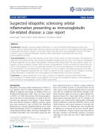
blood cell count and transaminases. Abdominal ultrasound
and computed tomography (CT) demonstrated a markedly
distended gallbladder, measuring 16 cm x 8 cm, with
oedema and pericholecystic fluid consistent with gallblad-
der empyema (Figure 1). Relying on the patient’s clinical
picture and the results of the imaging studies, a diagnosis of
gallbladder empyema was made. Due to this diagnosis and
the big size of the gallbladder, an open cholecystectomy was
decided upon. No other abnormality was identified intra-
operatively during the assessment of the peritoneal cavity,
including lymphadenopathy and liver or peritoneal pathol-
ogy. Based on the pre-operative as well as the intra-operative
findings, open cholecystectomy was performed; no lymph
nodes were excised. After an uneventful recovery, the patient
was discharged on the 4
th
postoperative day.
On cut surface, a 2 cm firm mass obstructing the lumen in
the neck of the gallbladder was identified. The mass was
solid, yellowish-grey and granular with focal areas of
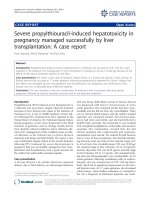
necrosis. Under light microscopy, the tumour demon-
strated invasion of the muscularis propria in some areas,
with foci of necrosis and haemorrhage (Figure 2A). No
extension to the gallbladder serosa was found. Examina-
tion of multiple regions under light microscopy did not
reveal any vascular invasion. Additionally, no evidence of
carcinoma was present in situ.
The neoplastic tissue consisted of diffusely arranged and
focally syncytial large and pleomorphic tumour giant cells,
with a significant infiltrating population of large multi-
nucleated hyperchromatic cells. The giant tumour cells
demonstrated hypochromatic nuclei with prominent
nucleoli and severe mitotic activity (Figure 2B). Hetero-
logous elements, such as osteoid and cartilaginous
differentiated cells, were not identified. In addition, there
was no evidence of any glandular structures. Following
haematoxylin-eosin study and according to the morphol-
ogy of the tumour cells, our differential diagnosis was one
Figure 1.
Abdominal computed tomography scan demonstrated a
distended gallbladder with oedema and pericholecystic fluid.
Figure 2.
(A) Diffuse infiltration by tumour cells. Invasion of muscularis
propria is also present (x20). (B) Notice the pleomorphic
hypochromatic nuclei with prominent nucleoli, and scattered
multinucleated hyperchromatic cells. Haemorrhage and
necrotic areas are also present (x200). (C) Total
immuno-negativity to anti-epithelial membrane antigen (x100).
Page 2 of 5
(page number not for citation purposes)
Journal of Medical Case Reports 2009, 3:6496 />of undifferentiated sarcomatoid carcinoma, sarcoma or
carcinosarcoma of the gallbladder.
Immunohistochemistry was performed using commer-
cially available antibodies against epithelial, neuroendo-
crine and mesenchymal markers. Secondary antibody
conjugated with peroxidise-labelled polymer (Envision,
Dako Carpinteria,CA, USA) 3’ diaminobenzidine was used
as chromogen, and finally the slides were lightly counter-
stained with haematoxylin. The results of immunochem-
istry stains are presented in Tabl e 1. The tumour
demonstrated remarkable immunonegativity to epithelial
markers such as epithelial membrane antigen (EMA),
carcinoembryonic antigen (CEA), AE1/AE3 and only mild
and focal positivity to CAM5.2 (Table 1 and Figures 2C
and 3A). On the other hand, mesenchymal markers
(vimentin, smooth-muscle actin: SMA) were strongly
positive (Table 1, Figures 3B and 3C). The multinucleated
cells showed positivity to CD68 stains (Table 1).
Osteoclast-like giant cells were considered to be of
histiocytic origin because of their PGM1 and KP1
positivity. Markers for neuro-endocrine tumours, lympho-
mas and melanoma were negative. The entire procedure
was repeated by another technician for more reliability,
with exactly the same results. Electron microscopy was not
performed.
Despite the negativity to almost all epithelial markers
based on the monophasic tumour immuno-reactivity
(that is, there was no evidence of two different compo-
nents that would have lead to the diagnosis of a
carcinosarcoma), the absence of heterologous compo-
nents, and the focal positivity to CAM5.2 (which is a
strong marker for the epithelial origin of a tumour), our
final diagnosis was that of a primary, undifferentiated,
giant cell type carcinoma of the gallbladder with sarco-
matoid dedifferentiation infiltrated with osteoclast-like
giant cells.
Following histopathological diagnosis, further thorough
investigation did not reveal any abnormality confirming,
thus, the diagnosis of a primary gallbladder neoplasm. The
patient died 2 months after the operation from dissemi-
nated disease; particularly, head, chest and abdominal CT
scans showed multiple brain, lung and hepatic metastases.
Discussion
The patient in this case report presented with a clinical
picture of a gallbladder empyema. This clinical diagnosis
was furthermore supported by the radiological as well as
the intra-operative findings. The diagnosis of gallbladder
neoplasm was only made following histopathological
evaluation of the resected specimen. The 2 cm solid lesion
obstructed the lumen in the neck of the gallbladder, thus
leading to the clinical presentation and the imaging and
intra-operative findings of gallbladder empyema in our
patient. Incidental finding of gallbladder carcinoma follow-
ing cholecystectomy for symptomatic cholelithiasis, acute
cholecystitis, gallbladder empyema or even asymptomatic
cholelithiasis has also been observed by others [3–5].
Our case proved difficult to classify. The final histopatho-
logical diagnosis resulted from a combination of mor-
phology and immunohistochemistry. The morphological
characteristics were those of a tumour of epithelial origin.
The positive response to CAM5.2, a strong epithelial
marker, was an additional characteristic for a carcinoma.
According to the World Health Organization (WHO) 2000
classification, undifferentiated carcinoma lacks glandular
structures; in the presented case, there was no evidence of
such structures, leading to the diagnosis of an undiffer-
entiated gallbladder carcinoma. The neoplastic tissue
consisted of diffusely arranged giant cells, with scattered
multinucleated cells. The multinucleated cells were mor-
phologically and immunohistochemically compared to
those of primary giant cell tumours as well as to osteoclast-
like giant cells and were shown to resemble the latter more
closely. The unusual feature of the immunophenotype of
Table 1. Results of immunohistochemistry.
Antigen Result
EMA -
AE1/AE3 -
CEA -
CAM 5.2 +
CD68 (PGM1) ++
CD68 (KP1) ++
S100 -
Vimentin +++
SMA ++
F VIII -
CD4 -
CD34 -
UCHL1 +/-
LYS -
HMB45 -
CD15 -
CD30 -
CD23 -
(See Abbreviations for full names of the antigens).
Figure 3.
(A) Focal immunopositivity to anti-cytokeratin CAM5.2
(x400). (B) Intense staining to anti-vimentin (x100). (C)
Positively stained tumour cells against anti-smooth-muscle
actin (x400).
Page 3 of 5
(page number not for citation purposes)
Journal of Medical Case Reports 2009, 3:6496 />the presented case is that, despite the epithelioid
morphology, the tumour cells demonstrated only focal
immuno-positivity to cytokeratins (CAM5.2) and, at the
same time, strong positivity to mesenchymal markers (e.g.
vimentin). This combination, showing a sarcomatoid
dedifferentiation of the tumour, is very rare. A possible
explanation for this unusual feature is that keratin
expression may decrease, while vimentin expression may
increase, as the tumour presents sarcomatoid dedifferen-
tiation. The final diagnosis in our patient was therefore
that of undifferentiated gallbladder carcinoma of giant cell
type with osteoclast-like giant cells and sarcomatoid
dedifferentiation.
Gallbladder carcinomas with osteoclast-like giant cells
should be distinguished from giant cell carcinomas. The
latter are composed of pleomorphic, undifferentiated
giant cells with bizarre nuclei, showing immunohisto-
chemical evidence of epithelial derivation, while display-
ing an identifiable transition between adenocarcinoma
and giant cells [5,6]. However, in our case, there were no
demonstrable foci of adenocarcinoma in the tumour.
Furthermore, the malignant giant cells were immuno-
negative to epithelial staining with only the focal staining
with antiCAM5.2 suggestive of their probable histogenesis.
CAM5.2 is a keratin mixture, containing a range of
cytokeratins between 39 kd and 50 kd, which is a strong
marker for the epithelial origin of a tumour.
Moreover, undifferentiated carcinomas with osteoclast-
like giant cells are rare, mainly pancreatic and periampu-
lary neoplasms that morphologically mimic giant cell
tumours of bones [5,7]. The terminology, histogenesis and
biological behaviour of these tumours remain controver-
sial. Several carcinomas may contain a variable number
of osteoclast-like giant cells [7–12]. In addition, some
anaplastic, spindle and giant cell carcinomas of the
gallbladder and extrahepatic bile ducts show numerous
osteoclast-like giant cells that may simulate giant cell
tumours [5]. It is crucial, however, to separate these two
types of neoplasm because of striking differences in
prognosis. Although prognostic significance of osteo-
clast-like giant cells is yet to be determined, a more
favourable prognosis has been suggested for carcinomas in
breast and pancreas [8,13]. However, further studies are
required referring specifically to gallbladder cancer.
Epithelial, histiocytic or mesenchymal metaplasia has
been suggested to explain the origin of osteoclast-like
giant cells. Immunohistochemical analysis has demon-
strated the giant cells to be of histiocytic origin lacking of
epithelial differentiation. These findings may imply that
osteoclast-like giant cells are a specialised form of
macrophage [6].
To the best of our knowledge, only 4 cases of osteoclastic-
like giant cells infiltrating a gallbladder carcinoma have
been reported in the literature so far. The first were
presented in 1992 by Grosso et al. [12] and Ito et al. [14],
respectively. The neoplasm reported by Grosso et al. was
an adenosquamous carcinoma and that by Ito et al. a giant
cell tumour. Subsequently, two more cases were reported
by Albores-Saavedra et al. [5] and by Akatsu et al. [6],
respectively, in 2006, with the latter being an adenosqua-
mous carcinoma. Like our patient, in the case reported by
Albores-Saavedra et al., there was also focal cytokeratin
positivity to CAM5.2 of the giant cells, and the osteoclast-
like giant cells showed immunoreactivity for CD68.
Therefore, our case represents the 5
th
reported case of a
gallbladder carcinoma with osteoclast-like giant cells, but
the 3
rd
case of undifferentiated giant cell type and the 1
st
case exhibiting sarcomatoid dedifferentiation.
Conclusions
Undifferentiated, giant cell type carcinoma of the gall-
bladder with sarcomat oid dedifferentiation infiltrated
with osteoclast-like giant cells is a very rare neoplasm.
Extraskeletal carcinomas exhibiting osteoclast-like giant
cells have been suggested to represent a distinct clinico-
pathological entity with a more favourable prognosis
[7–9,13]. However, the clinical importance of the phe-
nomenon remains unclear, owing to the rarity of such
cases. Our patient died 2 months after the operation from
disseminated disease. Poor prognosis could not be
associated with the presence of osteoclast-like giant cells,
as in our case, the undifferentiated carcinoma showed
dedifferentiation with sarcomatoid features probably
representing a highly virulent phenotype.
Abbreviations
AE1/AE3, cytokeratin AE1/AE3; CAM5.2, cytokeratin
CAM5.2; CEA, carcinoembryonic antigen; CD68 KP1,
KP1 monoclonal antibody to CD68 antigen; CD68
PGM1, PGM1 monoclonal antibody to CD68 antigen;
CT, computed tomography; EMA, epithelial membrane
antigen; F VIII, factor VIII; HMB45, melanocytic marker -
melanoma associated mRNA sequence gp100; SMA,
smooth muscle actin; S100, S-100 protein; UCHL1,
ubiquitin C-terminal hydrolase 1; WHO, World Health
Organization.
Consent
Written informed consent was obtained from the patient
for publication of this case report and any accompanying
images. A copy of the written consent is available for
review by the Editor-in-Chief of this journal.
Competing interests
The authors declare that they have no competing interests.
Page 4 of 5
(page number not for citation purposes)
Journal of Medical Case Reports 2009, 3:6496 />Authors’ contributions
AM carried out the operation and contributed to acquisi-
tion of consent and critical review of the manuscript. MG
contributed to manuscript conception, research, acquisi-
tion of data, drafting and writing of the manuscript. EEL
contributed to manuscript conception, research, acquisi-
tion of data, drafting and writing of the manuscript. HM
contributed to manuscript conception, research, acquisi-
tion of data, drafting and writing of the manuscript. AP
contributed to the preoperative and postoperative man-
agement of the patient and to critical review of the
manuscript. GA and HG carried out the histopathologic
evaluation and contributed to writing of the manuscript.
PK assisted in the operation and contributed to critical
review of the manuscript. KF contributed to organising
and drafting of the manuscript, and critically revised the
manuscript. EP carried out the histopathologic evaluation,
contributed to organising and drafting of the manuscript,
and critically revised the manuscript. All authors read and
approved the final manuscript.
References
1. Guo KJ, Yamaguchi K, Enjoji M: Undifferentiated carcinoma of
the gallbladder. A clinicopathologic, histochemical, and
immunohistochemical study of 21 patients with a poor
prognosis. Cancer 1988, 61(9):1872-1879.
2. Albores-Saavedra J, Cruz-Ortiz H, Alcantara-Vazques A, Henson DE:
Unusual types of gallbladder carcinoma. A report of 16 cases.
Arch Pathol Lab Med 1981, 105(6):287-293.
3. Bartlett DL, Fong Y, Fortner JG, Brennan MF, Blumgart LH:
Long-term results after resection for gallbladder cancer.
Implications for staging and management. Ann Surg 1996,
224(5):639-646.
4. Kondo S, Nimura Y, Hayakawa N, Kamiya J, Nagino M, Uesaka K:
Extensive surgery for carcinoma of the gallbladder. Br J Surg
2002, 89(2):179-184.
5. Albores-Saavedra J, Grider DJ, Wu J, Henson DE, Goodman ZD:
Giant cell tumor of the extrahepatic biliary tree: a clinico-
pathologic study of 4 cases and comparison with anaplastic
spindle and giant cell carcinoma with osteoclast-like giant
cells. Am J Surg Pathol 2006, 30(4):495-500.
6. Akatsu T, Kameyama K, Kawachi S, Tanabe M, Aiura K, Wakabayashi
G, Ueda M, Shimazu M, Kitajima M: Gallbladder carcinoma with
osteoclast-like giant cells. J Gastroenterol 2006, 41(1):83-87.
7. Stracca-Pansa V, Menegon A, Donisi PM, Bozzola L, Fedeli F, Quarto F,
Nappi O, Pettinato G: Gastric carcinoma with osteoclast-
like giant cells. Report of four cases. Am J Clin Pathol 1995,
103(4):453-459.
8. Gjerdrum LM, Lauridsen MC, Sørensen FB: Breast carcinoma with
osteoclast-like giant cells: morphological and ultrastructural
studies of a case with review of the literature. Breast 2001,
10(3):231-236.
9. Baschinsky DY, Frankel WL, Niemann TH: Gastric carcinoma
with osteoclast-like giant cells. Am J Gastroenterol 1999,
94(6):1678-1681.
10. Odeh M, Misselevich I, Oliven A, Boss JH: Small intestinal
carcinoma with osteoclast-like giant cells. Am J Gastroenterol
1995, 90(7):1177-1179.
11. Ikeda T, Seki S, Maki M, Noguchi N, Kawamura T, Arii S, Igari T, Koike
M, Hirokawa K: Hepatocellular carcinoma with osteoclast-like
giant cells: possibility of osteoclastogenesis by hepatocyte-
derived cells. Pathol Int 2003, 53(7):450-456.
12. Grosso LE, Gonzalez JG: Stromal osteoclast-like giant cells in an
adenosquamous carcinoma of the gallbladder. Hum Pathol
1992, 23(6):703-706.
13. Dworak O, Wittekind C, Koerfgen HP, Gall FP: Osteoclastic giant
cell tumor of the pancreas. An immunohistological study and
review of the literature. Pathol Res Pract 1993, 189(2):228-231;
discussion: 232-234.
14. Ito M, Hsu CT, Naito S, Matsuo T, Onizuka S, Sekine I, Fujii H,
Matsuoka Y: Osteoclast-like giant cell tumour of the gallblad-
der.
Virchows Arch A Pathol Anat Histopathol 1992, 420(4):359-366.
Page 5 of 5
(page number not for citation purposes)
Journal of Medical Case Reports 2009, 3:6496 />Do you have a case to share?
Submit your case report today
• Rapid peer review
• Fast publication
• PubMed indexing
• Inclusion in Cases Database
Any patient, any case, can teach us
something
www.casesnetwork.com