Báo cáo y học: "Laparoscopic-assisted resection of a giant colonic diverticulum: a case report" ppsx
Bạn đang xem bản rút gọn của tài liệu. Xem và tải ngay bản đầy đủ của tài liệu tại đây (1.07 MB, 6 trang )
Case report
Open Access
Laparoscopic-assisted resection of a giant colonic diverticulum:
a case report
Jacqueline E Collin
1
*, Gurprit SS Atwal
2
, William K Dunn
3
and
Austin G Acheson
1
Address:
1
Department of Colorectal Surgery, Queens Medical Centre, Nottingham Universities NHS Trust, Nottingham NG7 2UH, UK,
2
Department of Histopathology, Queens Medical Centre, Nottingham Universities NHS Trust, Nottingham NG7 2UH, UK and
3
Department of
Radiology, Queens Medical Centre, Nottingham Universities NHS Trust, Nottingham NG7 2UH, UK
Email: JEC* - ; GSSA - ; WKD - ;
AGA -
* Corresponding author
Published: 28 May 2009 Received: 8 February 2008
Accepted: 23 January 2009
Journal of Medical Case Reports 2009, 3:7075 doi: 10.1186/1752-1947-3-7075
This article is available from: />© 2009 Collin et al; licensee Cases Network Ltd.
This is an Open Access article distributed under the terms of the Creative Commons Attribution License (
/>which permits unrestricted use, distribution, and reproduction in any medium, provided the original work is properly cited.
Abstract
Introduction: Diverticular disease of the colon is a common benign condition. The majority of
patients with diverticular disease are asymptomatic and are managed non-operatively, however
complications such as perforation, bleeding, fistulation and stricture formation can necessitate
surgical intervention. A giant colonic diverticulum is defined as a diverticulum larger than 4cm in
diameter. Despite the increasing incidence of colonic diverticular disease, giant colonic diverticula
remain a rare clinical entity.
Case presentation: This is the first reported case of laparoscopic-assisted resection of a giant
colonic diverticulum. We discuss the symptoms and signs of this rare complication of diverticular
disease and suggest investigations and management. Reflecting on this case and those reported in the
literature to date, we highlight potential diagnostic difficulties and consider the differential diagnosis of
intra-abdominal gas-filled cysts.
Conclusion: The presence of a giant colonic diverticulum carries substantial risk of complications.
Diagnosis is based on history and examination supported by abdominal X-ray and computed
tomography findings. In view of the chronic course of symptoms and potential for complications,
elective surgical removal is recommended. Colonic resection is the treatment of choice for this
condition and, where possible, should be performed laparoscopically.
Introduction
Diverticular disease of the colon is a common benign
condition that occurs in excess of 60% in those aged over
70 years [1,2]. It is generally a disease of the western world
and the incidence appears to be increasing [3,4]. The
majority of patients with diverticular disease have
involvement of the sigmoid colon. These patients are
frequently asymptomatic, when the condition is known as
Page 1 of 6
(page number not for citation purposes)
diverticulosis, and the diagnosis is made incidentally.
Diverticular disease refers to symptomatic diverticula;
patients commonly present with bloating, abdominal
pain, flatus and rectal bleeding. Inflammation of diverti-
cula, known as diverticulitis, classically causes left-sided
abdominal pain, change in bowel habit with passage of
mucous or fresh blood, and systemic upset.
About 5% of patients who have symptomatic diverticula
experience complications such as perforation, bleeding,
fistulation and stricture formation which can necessitate
surgical intervention.
A giant colonic diverticulum (GCD) is defined as a
diverticulum larger than 4cm in diameter [4]. Some as
large as 40cm have been reported in the literature [5]. The
mean age of presentation of GCD mirrors that of
diverticular disease with the majority presenting after the
sixth decade [1,4].
The presentation of GCD is variable, ranging from the
asymptomatic patient (4%) to a host of non-specific
gastrointestinal (GI) symptoms with only 10% of patients
presenting with an abdominal mass [4]. GCD carries a
substantial risk of complications and elective surgical
removal is recommended [6].
Despite the increasing incidence of colonic diverticular
disease, GCD remains a rare clinical entity [7]. We report a
case of a 53-year-old man who underwent a laparoscopic-
assisted sigmoid colectomy for treatment of a sympto-
matic giant diverticulum. This is the first reported case of
laparoscopic-assisted resection of a GCD.
Case presentation
A 53-year-old white Italian man initially presented to
gastroenterologists with a 5-week history of dyspepsia,
epigastric pain and a palpable mass in the left hypochron-
drium. There was no history of anorexia, dysphagia,
weight loss, change in bowel habit or gastrointestinal
blood loss. His past medical history included early
Alzheimer’s disease and discoid lupus.
Examination revealed a well circumscribed, mobile mass
in the left hypochrondrium extending above the level of
the ribs raising the possibility of an enlarged spleen. There
was no palpable lymphadenopathy.
A blood film showed atypical myelomonocytic cells but a
subsequent bone marrow aspiration was normal. All
other routine blood tests were within normal limits. An
abdominal ultrasound scan demonstrated a normal
spleen and a separate gas-filled cyst in the left
hypochondrium.
Over the next few weeks, the patient developed diarrhoea
and lost 3kg in weight. He reported that the mass appeared
to be fluctuating in size.
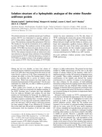
An abdomen computed tomography (CT) scan (Figure 1)
demonstrated a large gas-filled structure measuring 11cm x
12cm, appearing to arise from the sigmoid colon,
displacing the adjacent small and large bowel loops. The
features were consistent with a giant sigmoid diverticulum.
He was referred to colorectal surgeons and a barium
enema was performed to further assess the extent of the
diverticular disease. This confirmed moderate sigmoid
diverticulosis but did not demonstrate direct communica-
tion between the colon and the giant cyst (Figure 2). The
diagnosis of GCD was discussed with the patient and
definitive surgical management was advised. Initially, the
patient was reluctant to have surgery, but over the next 6
months, he experienced two further episodes of acute
abdominal pain necessitating hospital admission. Both
episodes were similar in nature with pain as the
predominant symptom; an abdominal X-ray (AXR) taken
on admission demonstrated the gas-filled structure and in
the absence of raised inflammatory markers, a normal
white cell count and no fever, the diagnosis of enlarging
GCD was made. Both episodes settled quickly with bowel
rest and intravenous fluids. The patient then agreed to
surgical intervention.
A laparoscopic-assisted sigmoid colectomy was performed
6 weeks later. Four 12mm ports were inserted and
Figure 1. Abdominal computed tomography demonstrating
a large gas-filled structure in the left upper abdomen arising
from the sigmoid colon.
Page 2 of 6
(page number not for citation purposes)
Journal of Medical Case Reports 2009, 3:7075 />pneumoperitoneum achieved. Three of the ports were
positioned along the lateral edge of the right rectus
abdominus muscle and triangulated to provide optimum
access to the left colon. The fourth port was in the left iliac
fossa. The large cystic structure was clearly visible in the left
hypochondrium at the apex of a long mobile loop of
sigmoid colon on the anti-mesenteric border (Figure 3).
The remaining sigmoid colon had macroscopic evidence
of mild diverticulosis. The diverticulum was attached to
the lateral abdominal wall adjacent to the spleen by
adhesions. These adhesions were divided laparoscopically
by a combination of scissor diathermy and ultracision. The
sigmoid colon was then fully mobilised from lateral to
medial but no attempt was made to divide the mesenteric
vessels intracorporeally in view of the fact this was benign
disease and the sigmoid was long and tortuous. The
mobilised sigmoid colon was externalised through a 7cm
incision in the left iliac fossa. A wound protector was used
during extraction of the cyst and a decision was made not
to decompress it before removal in order to keep possible
contamination down to a minimum. The sigmoid colon
was resected along with the diverticulum and a hand-sewn
primary anastomosis was performed extracorporeally.
The patient made an excellent postoperative recovery and
was discharged on the fourth postoperative day.
Macroscopic assessment of the segmental colonic resection
confirmed the presence of diverticular disease with an
associated giant cyst measuring 11cm in maximal
diameter. The wall of the cyst measured 0.6 to 1cm in
thickness. Microscopically, it did not contain any elements
of bowel wall and instead was composed of reactive scar
tissue with foreign body type giant cell reaction. The
presence of plant material admixed with inflammatory
debris was thought to be indicative of faecal matter and
suggested a direct communication between the cyst and
bowel lumen. However, this was not identified histologi-
cally. There was no evidence of dysplasia or malignancy. In
accordance with the classification suggested by Steen-
voorde et al. [4], the histological features were consistent
with Type II GCD (Table 1).
Discussion
Diverticular disease of the colon is a significant cause of
morbidity and mortality in the western world and its
frequency increased throughout the whole of the 20
th
century [3,8]. Since it is a disease of the elderly, and with
an ageing population, it can be expected to occupy an
increasing portion of the surgical and gastroenterological
workload [3,8].
GCDs are defined as those that are larger than 4cm in
diameter [4, 5] and with the increasing incidence of
diverticular disease [3,8], it is likely that the incidence of
these giant lesions will increase further. Awareness of the
presenting symptoms, investigations, differential diagno-
sis and management is therefore important.
As in our patient, it is not unusual for these patients to
undergo multiple investigations before making the correct
diagnosis. Plain supine abdominal X-ray is the simplest
and most readily available investigation and should be
used as the first line in suspected cases. If a large air filled
Figure 2. Barium enema: the air filled cavity did not fill with
barium nor did it change in size on insufflation.
Figure 3. Externalised sigmoid colon and anti-mesenteric
giant cyst.
Page 3 of 6
(page number not for citation purposes)
Journal of Medical Case Reports 2009, 3:7075 />structure with or without fluid levels is visualised then an
abdominal CT scan would be indicated. Barium enema
failed to demonstrate a communication between the giant
diverticulum and the colon in approximately one-third of
reported cases [1,4]. It is therefore not surprising that no
communication was identified in our patient. Barium
enema can be useful at providing valuable information
regarding the extent of further diverticula.
The use of abdominal ultrasound has been reported to be
helpful in only 25% of cases [4]. Early colonoscopy is
advised in the setting of persistent or frequent acute
diverticulitis to rule out concurrent pathology [9]. Our
patient was admitted acutely on two occasions a few
months apart, however, the symptoms and signs were not
suggestive of acute diverticulitis but were felt to be in
keeping with enlarging GCD therefore colonoscopy was
not performed.
The role of colonoscopy in diagnosing GCD is limited. The
ostium between the diverticulu m and the colon is
frequently too small to be detected [1,2,4] and even in
cases with wide necked GCD, the ostium is not detected on
sigmoidoscopy [1]. The combination of a large soft,
mobile mass in an elderly patient and a lucent cystic
structure related to the sigmoid colon on AXR should
suggest the diagnosis of a GCD [6].
Other causes for intra-abdominal gas-filled cysts, radi-
ologically mimicking GCD [2], along with their principal
distinguishing features, are summarised in Table 2.
Steenvoorde et al. suggested a histological classification
of GCD based on three subtypes (Table 1). The distinction
between type I and II has not always been made with both
categories being discussed as one entity in many papers
[5]. Theories behind the formation of GCD type I and II
are speculative and not mutually exclusive. The suggested
aetiology of type I is based on the premise that the
communication between the GCD and the colon is small
enough to preclude the escape of air from the diverticulum
[1]. The two most widely accepted theories are, a
unidirectional ball-valve mechanism causing gas entrap-
ment and infection with gas producing organisms leading
to progressive diverticula enlargement [5]. However, such
theories do not convincingly explain the existence of type I
GCD with wide necks.
Type II is postulated to form following a subserosal
perforation resulting in a walled off abscess cavity that
gradually enlarges to giant size [7]. Type III contains all
layers of bowel wall and structurally resembles a duplica-
tion cyst [7] but is in continuity with the gut lumen and
occurs in adults. Approximately 20% of GCD show no
evidence of a communicating ostium between the colon
and the diverticulum and it is thought that this tract may
be lost due to inflammatory changes [5].
Surgical management of a GCD involves either removing
the diverticulum in isolation or colectomy. Diverticulect-
omy is not recommended as the mouth of the diverticulum
may be wide and the surrounding inflammation could
increase the potential for breakdown of the colonic closure
[2]. Giant diverticula appear mostly (81%) in the sigmoid
colon [5] with 50% of patients having concurrent sigmoid
diverticula [4], thus sigmoid colectomy with primary
end-end anastomosis [7] is the preferred operation.
Resection is frequently difficult due to the inflammatory
diverticulum and it is often densely adherent to surround-
ing structures [2]. In complicated or emergency cases, the
safest surgical solution may be a Hartmann’s procedure [7].
The advent of laparoscopic colorectal surgery has had a
significant impact on the postoperative recovery period for
patients undergoing surgical resections for both benign
and malignant colorectal disease. The most important
advantages to the patient of laparoscopic surgery are
reduction in pain, more rapid recovery of bowel function,
better cosmetic results and a shorter hospital stay [5,6,10].
Our patient was fit for discharge on day four and this
undoubtedly was due to the minimally invasive surgery
performed. Based on our experience in this patient along
with the recommendations of the Cochrane review group
[10], surgical remo val a GCD should be min imally
invasive using laparoscopic techniques.
Conclusion
Giant colonic diverticulum is a rare entity that is associated
with a significant complication rate. The presentation of
GCD is variable ranging from the asymptomatic patient
(4%) to a host of gastrointestinal symptoms including
abdominal pain (68%), constipation (18%), rectal bleed-
ing (13%), vomiting (12%), abdominal distension (11%),
diarrhoea (11%) and abdominal mass (10%) [4]. Accurate
Table 1. Histological classification of giant colonic diverticulum; from Steevoorde et al. [4]
Type Name Aetiology Histology
I Pseudo-diverticulum Unidirectional ball-valve mechanism
Gas producing organism
Remnants muscularis mucosa/muscularis propria
II Inflammatory Local perforation of mucosa with abscess cavity Reactive scar tissue, no bowel tissue
III True diverticulum Congenital All three layers of bowel tissue, communicating with gut lumen
Page 4 of 6
(page number not for citation purposes)
Journal of Medical Case Reports 2009, 3:7075 />diagnosis, although difficult, can be achieved using a
combination of clinical examination, plain AXR and CT
scanning.
The presence of a GCD carries substantial risk of
complications (12% to 19%) including inflammation,
perforation, abscess formation, fistula formation, urinary
obstruction [7], volvulus, small bowel obstruction and
rarely, the development of adenocarcinoma [1,4].
In view of the chronic course of symptoms and potential
for complications, elective surgical removal is recom-
mended [6]. Colonic resection is the treatment of choice
for this condition and, where possible, should be
performed laparoscopically.
Abbreviations
AXR, abdominal X-ray; CT, computerised tomography;
GCD, giant colonic diverticulum; GI, gastrointestinal; IBD,
inflammatory bowel disease; PR, per rectum; RUQ, right
upper quadrant; Tech
99
, technetium-99; USS, ultrasound
scan.
Consent
Written informed consent was obtained from the patient
for publication of this case report and any accompanying
images. A copy of the written consent is available for
review by the Editor-in-Chief of this journal.
Competing interests
The authors declare that they have no competing interests.
Table 2. Differential diagnosis of intra-abdominal gas-filled cysts
Condition Age at
presentation
(years)
Diagnostic
investigation
Distinguishing features
GCD >60 AXR, CT >4cm in size, air filled cyst
Usually arises from the sigmoid colon
Anti-mesenteric border [2]
Associated diverticular disease
60% palpable abdominal mass [4–6]
Pneumatosis cystoides 30–50 [11] CT Usually asymptomatic
Symptoms: abdominal distension, discomfort, mucoid stools
15% primary/idiopathic
85% secondary: IBD, diverticulosis, pulmonary disease
Numerous small pockets within bowel wall
Affects small and large bowel [11]
Meckels diverticulum <30 Tech
99
, CT 2% population, 95% asymptomatic
<2cm in length
PR bleeding most common presenting symptom in children
Other symptoms: abdominal obstruction, inflammation, intussusception, ulceration
and perforation
Contain all layers of bowel wall
Anti-mesenteric border, within 100cm of ileocaecal valve
Volvulus
(caecal/sigmoid)
>70 AXR,
Sigmoidoscopy
Associated bowel obstruction
Redundant sigmoid colon, past history of chronic constipation
Haustra visible on distended loop on AXR [12]
Duplication cysts <2 CT, USS, AXR Anywhere along GI tract, most common in ileum
Can be single/multiple
50% have associated anomalies
Wide range of symptoms pending location
Mesenteric side, elongated in shape
90% Non-communicating with gut lumen
All bowel layers [12]
Emphysematous cystitis >40 AXR, CT, USS Due to bacterial fermentation of urinary glucose
Gas production in bladder lumen and wall
Assoc with diabetes, neurogenic bladder, bladder outlet obstruction, recurrent
urinary tract infections
Symptoms include dysuria, frequency, pneumaturia
Distended tympanic mass arising from pelvis
Most commonly due to Escherichia coli
Emphysematous
cholecystitis [12]
>40 AXR, CT RUQ pain, vomiting, pyrexia +/− RUQ mass
Increased risk with diabetes and gallstones
Infection usually due to Clostridium perfringes
More risk of gangrene and perforation than with acute cholecystitis
Intra-abdominal abscess – CT Source of intra-abdominal sepsis
Swinging pyrexia
Palpable mass
Page 5 of 6
(page number not for citation purposes)
Journal of Medical Case Reports 2009, 3:7075 />Authors’ contributions
JEC was involved clinically with the case, researched the
article, and drafted and revised the manuscript coordinat-
ing the authors’ contributions. GSSA confirmed the
histological diagnosis and histological classification and
contributed to the overall report, reviewing and revising
the manuscript. WKD confirmed the radiological diag-
nosis and assisted with the section on radiological
differential diagnosis of gas-filled structures. AA worked
with JEC in establishing both the concept and design of
the report. AA critically appraised the article, guiding its
progress from draft to final version. All authors read and
approved the final manuscript.
References
1. LeviDM,LeviJU,BergauDK,WengerJ:Giant colonic
diverticulum: an unusual manifestation of a common disease.
Am J Gastroenterol 1993, 88:139-142.
2. Oliveira NC, Welch JP: Giant diverticula of the colon: a clinical
assessment. Am J Gastroenterol 1997, 92:1092-1096.
3. Kang JY, Hoare J, Tinto A, Subramanian S, Ellis C, Majeed A,
Melville D, Maxwell JD: Diverticular disease of the colon on
the rise: a study of hospital admissions in England between
1989/1990 and 1999/2000. Aliment Pharmacol Ther 2003,
17:1189-1195.
4. Steenvoorde P, Vogelaar FJ, Oskam J, Tollenaar RAEM: Giant
colonic diverticula. Review of diagnostic and therapeutic
options. Dig Surg 2004, 21:1-6.
5. Choong CK, Frizelle FA: Giant colonic diverticulum report of
four cases and review of the literature. Dis Colon Rectum 1998,
41:1178-1185.
6. Fox AT, Singh G: The abdominal gas filled cyst. CME Gastroenterol
Hepatol Nutr 2000, 3:66-67.
7. Chaiyasate K, Yavuzer R, Mittal V: Images in surgery: giant
sigmoid diverticulum. Surgery 2006, 139:276-277.
8. Kang JY, Melville D, Maxwell JD: Epidemiology and management
of diverticular disease of the colon. Drugs Aging 2004,
21:211-228.
9. Lahat A, Yanai H, Sakhnini E, Menachem Y, Bar-Meir S: Role of
colonoscopy in patients with persistent acute diverticulitis.
World J Gastroenterol 2008, 14:2763-2766.
10. Schwenk W, Haase O, Neudecker J, Muller JM: Short term benefits
for laparoscopic colorectal resection. Cochrane Database Syst Rev
2005, 20:CD003145.
11. Galanduik S, Fazio VW: Pneumatosis cystoides intestinalis: a
review of the literature. Dis Colon Rectum 1986, 29:358-363.
12. Foster DR, Ross B: Giant sigmoid diverticulum: clinical and
radiological features. Gut 1977, 18:1051-1053.
Page 6 of 6
(page number not for citation purposes)
Journal of Medical Case Reports 2009, 3:7075 />Do you have a case to share?
Submit your case report today
• Rapid peer review
• Fast publication
• PubMed indexing
• Inclusion in Cases Database
Any patient, any case, can teach us
something
www.casesnetwork.com