Báo cáo y học: " Recurring staphylococcal scalded skin syndrome in a very low birth weight infant: a case report" pptx
Bạn đang xem bản rút gọn của tài liệu. Xem và tải ngay bản đầy đủ của tài liệu tại đây (371.58 KB, 3 trang )
Case report
Open Access
Recurring staphylococcal scalded skin syndrome in a very low birth
weight infant: a case report
Carola EPM Duijsters
1
*, Feico JJ Halbertsma
1
, René F Kornelisse
2
,
Niek LA Arents
3
and Peter Andriessen
1
Addresses:
1
Department of Pediatrics, Division of Neonatology, Máxima Medical Center Veldhoven, The Netherlands
2
Department of Pediatrics, Division of Neonatology, Erasmus MC-Sophia Children’s Hospital, Rotterdam, The Netherlands
3
Laboratories of Pathologic Anatomy and Medical Microbiology Veldhoven, The Netherlands
Email: CEPMD* - ; FJJH - ; RFK - ; NLAA - ;
PA -
* Corresponding author
Received: 18 July 2008 Accepted: 12 January 2009 Published: 12 August 2009
Journal of Medical Case Reports 2009, 3:7313 doi: 10.4076/1752-1947-3-7313
This article is available from: />© 2009 Duijsters et al.; licensee Cases Network Ltd.
This is an Open Access article distributed under the terms of the Creative Commons Attribution License (
/>which permits unrestricted use, distribution, and reproduction in any medium, provided the original work is properly cited.
Abstract
Introduction: Staphylococcal scalded skin syndrome is an extensive desquamative erythematous
condition caused by exfoliative toxins of Staphylococcus aureus. This disease usually affects neonates
and generally responds rapidly to antibiotic therapy.
Case presentation: We describe the case of a premature baby boy, weighing 1030 g, born after
26 6/7 weeks gestation, who developed two episodes of Staphylococcal scalded skin syndrome on
days 19 and 48 of life. Cultures obtained during the first period did not reveal Staphylococcus aureus,
but diagnosis was based on typical clinical grounds.
Although the initial diagnosis was irritation by the fixation material of a nasal continuous positive
airway pressure tube, the infant showed rapidly progressing skin blistering and exfoliation,
characteristic of Staphylococcal scalded skin syndrome. After administration of antibiotic treatment,
complete recovery was seen. In the second period, diagnosis of Staphylococcal scalded skin syndrome
was made clinically and confirmed by results of microbiologic investigations. Staphylococcus aureus was
cultured from the nose, skin lesions and the pharynx. The strain appeared to produce exfoliative
toxin A. The clinical response to similar antibiotic treatment was identical to the first period of
Staphylococcal scalded skin syndrome.
Conclusion: This case report discusses an unusual presentation of recurring Staphylococcal scalded
skin syndrome in a baby with a very low birth weight.
Introduction
Staphylococcal scalded skin syndrome (SSSS) is a rapidly
expanding exfoliative disease of the skin characterized by
blistering and epidermal peeling. The disease is induced by
exfoliative toxins (ET) of Staphylococcus aureus and typically
occurs in newborn babies with an onset between the first
3 and 16 days of life [1]. In this case report we discuss an
unusual presentation of recurring SSSS in a baby with a
Page 1 of 3
(page number not for citation purposes)
very low birth weight (VLBW). Furthermore, we emphasize
the importance of early diagnosis of SSSS because of
the risk of nosocomial spread to other patients in the
Neonatal Intensive Care Unit (NICU), the morbidity and
the increased risk of mortality in children [2].
Case presentation
A baby boy weighing 1030 g was born at 26 6/7 weeks
gestation by vaginal delivery. Pregnancy was uncompli-
cated until 2 days before delivery when spontaneous
rupture of membranes occurred. The mother received toco-
lytic treatment but labour continued and the baby was de-
livered. Apgar scores were 2, 6 and 9 at 1, 5 and 10 minutes
after birth, respectively.
The infant was transported to the NICU with nasal
conti nuous positive airway pressure (CPAP). Because
rupture of membranes existed for more than 48 hours,
antibiotics (amoxicillin-clavulanic acid and gentamicin)
were started. Blood culture remained negative, but group B
streptococcus was cultured from the infant’s ear and the
placenta. We found no clues of infection clinically or in
laboratory tests. Amoxicillin-clavulanic acid was contin-
ued for 7 days and gentamicin for 3 days.
On day 19 of life, a 5 mm red skin lesion with epidermal
peeling was noticed on the left nostril were the nasal CPAP
tube was fixed. Within 6 hours the lesions expanded and
new lesions of blisters and exfoliation appeared on
shoulders, trunk and the peri-umbilical region. This left
an erythematous and moist surface (Fig. 1). The infant was
sensitive to manipulation and extremely irritable. The
clinical appearance was interpreted as SSSS and blood
culture, culture of a blister, and swabs of the nose and
perineum were obtained.We started a combination therapy
of intravenous flucloxacillin (75 mg/kg in 3 doses) for 10
days, local administration of fusidic acid and adequate pain
treatment with intravenous acetaminophen and morphine
following premature infant pain profile scores. The patient
was isolated. One day after initiation of antibiotic therapy,
the lesions ceased to increase or expand. Complete recovery
of the affected skin was seen within 11 days. Blood culture
remained negative, as were swabs of the nose and
perineum. Culture of the skin lesion revealed coagulase-
negative staphylococci. Skinbiopsy was not performed.The
patient was discharged to the pediatric ward approximately
one month after birth.
On day 48 of life, suspicion of SSSS arose again and the
patient was transferred back to the NICU. The infant
developed discrete exfoliation of the nose and the distal
part of digits I and III. Within 24 hours, the scrotum, all
fingers and the inguinal and peri-umbilical regions were
affected. Again we noticed rapidly expanding skin lesions
with blistering, epidermal peeling and erythroderma. The
boy had no clinical signs of infection, but became agitated
when touched. After obtaining cultures from the skin
(digits I and III), the nose, the pharynx and the blood,
intravenous flucloxacillin was administered for 15 days.
Suppor tive treatment consisted of adequate analgesic
treatment, aseptic care and isolation of the patient. As in
the first episode of SSSS, no further increase in the lesions
was noticed after 36 hours and complete resolution
appeared after 1 week. S. aureus was cultured from the
nose, the fingers and the pharynx. Blood culture was again
sterile. The material was sent to The National Institute for
Public Health and the Environment for polymerase chain
reaction analysis of the exfoliative toxin genes of the strain.
The S. aureus strain was shown to possess the eta gene for
Exfoliative Toxin A (ET-A). This confirmed our clinical
diagnosis of SSSS. The course of further treatment of the
patient was uneventful and discharge to home took
place on day 74. Follow-up in our out-patient clinic till
6 months after discharge was completely satisfactory
without any relapse of symptoms of SSSS.
Discussion
SSSS is a clinical manifestation of infection caused by
exfoliative toxin producing Staphylococci, usually phage II
S. aureus strains. These toxins, especially ET-A and ET-B,
spread hematogenously and cause erythema, blistering
and superficial scalding of the skin by targeting the protein
desmoglein I in the zona granulosa of the epidermis [3].
Of the S. aureus strains 5% to 6% produce ET, with over
80% of the ET as ET-A. In this case report, investigations
for exfoliative toxins were positive for ET-A. This result
confirmed our clinical diagnosis of SSSS.
The differential diagnosis of skin exfoliation in neonates
includes SSSS, bullous impetigo, chemical burns, drug or
viral-induced toxic epidermal necrolysis, epidermolysis
bullosa, bullous mastocytosis and neonatal pemphigus
[4-6].
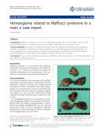
Figure 1. Red skin lesions with epidermal peeling on the face
and body of a baby boy with a very low birth weight.
Page 2 of 3
(page number not for citation purposes)
Journal of Medical Case Reports 2009, 3:7313 />Diagnosis of SSSS is mainly based on clinical criteria and
only confirmed by exotoxin producing S. aureus. Diagnosis
based on blood or tissue cultures often occurs later than
clinical diagnosis. Therefore, fast recognition of this
disease is required to start systemic antibiotic treatment
with b-lactamase-resistant penicillin at an early stage.
Although this treatment is easy and effective within 24 to
36 hours, mortality is still 3% to 11% in children [2] and
over 50% in adults [3]. Besides antibiotic therapy and
supportive and aseptic skin care, adequate analgesic treat-
ment (morphine), minimal handling and appropriate
management of fluid and electrolyte balance are necessary
[5,7]. To prevent outbreaks of SSSS involving a large number
of newborn babies in neonatal wards, it is important to
isolate the infected infant in an incubator [8].
Epidemiological studies showed a prevalence of 3% to 6%
of toxin producing S. aureus carriage, with a prevalence of
3% in antenatal women [8]. Hargiss et al. [9] reported that
up to 60% of neonates discharged from hospital may be
nasal carriers of S. aureus. Although these data may suggest
a high incidence of SSSS, Mockenhaupt et al. [2] calculated
an overall incidence between 0.09 and 0.13 cases per
million habitants per 5 year with 95% confidence intervals
of 0 to 4. SSSS predominantly occurs in neonates, with
onset between 3 and 16 days and is less common in
preterm infants [1]. Factors that may be responsible for the
higher incidence in neonates in comparison to adults
include renal immaturity, resulting in diminished clear-
ance of toxins and a lack of specific antibodies against
staphylococcal toxins [7].
Less than 10 cases of SSSS in infants with VLBW have been
reported in the literature [4-6,10-12]. Recurrence of SSSS
appears to be even more uncommon, especially among
preterm neonates. Dobson et al [13] describe an adult
patient who developed SSSS 8 days following the cessation
of antibiotics for a chest infection and pressure sores.
Diagnosis was made on a clinical basis and the patient was
treated with intravenous cefuroxime. Recrudescence of the
disease appeared after switching to oral cefaclor [13].
Rieger-Fackeldey et al. [14] report SSSS in an extremely low
birth weight infant with a relapse 4 weeks after the primary
infection. Another case report mentions SSSS in two male
siblings aged 5 and 10 years old. This condition recurred in
the same children within a period of about 12 months
[15]. Our case report in which we describe a preterm infant
with a birth weight of 1030 g with two episodes of SSSS on
days 19 and 48 of life seems to be unique in the literature.
Conclusion
We describe an unusual case of recurring SSSS in a VLBW
infant and underline the necessity of early diagnosis and
treatment of this disease.
Consent
Written informed consent was obtained from the patient's
parents for publication of this case report and any
accompanying images. A copy of the written consent is
available for review by the Editor-in-Chief of this journal.
Competing interests
The authors declare that they have no competing interests.
Authors’ contributions
All authors contributed to acquisition of case details and
their analysis and interpretation. CD wrote the first draft of
the manuscript. FH, RK, NA and PA revised the manuscript.
All authors have given final approval of this manuscript to
be published.
References
1. Dancer SJ, Simmons NA, Poston SM, Noble WC: Outbreak of
staphylococcal scalded s kin syndrome among neonates.
J Infect 1988, 16:87-103.
2. Mockenhaupt M, Idzko M, Grosber M, Schopf E, Norgauer J:
Epidemiology of staphylococcal scalded skin syndrome in
Germany. J Invest Dermatol 2005, 124:700-703.
3. Cribier B, Piemont Y, Grosshans E: Staphylococcal scalded skin
syndrome in adults. A clinical review illustrated with a new
case. J Am Acad Dermatol 1994, 30:319-324.
4. Coleman JC, Dobson NR: Diagnostic dilemma: extremely low
birth weight baby with staphylococcal scalded-skin syndrome
or toxic epidermal necrolysis. J Perinatol 2006, 26:714-716.
5. Hutten M, Heimann K, Baron JM, Wenzl TG, Merk HF, Ott H:
Staphylococcal scalded skin syndrome as a harbinger of late-
onset staphylococcal septicaemia in a premature infant of
very low birth weight. Acta Derm Venereol 2008, 88:416-417.
6. Makhoul IR, Kassis I, Hashman N, Sujov P: Staphylococcal scalded-
skin syndrome in a very low birth weight premature infant.
Pediatrics 2001, 108:E16.
7. Haveman LM, Fleer A, de Vries LS, Gerards LJ : Congenital
staphylococcal scalded skin syndrome in a premature infant.
Acta Paediatr 2004, 93:1661-1662.
8. Ladhani S, Evans RW: Staphylococcal scalded skin syndrome.
Arch Dis Child 1998, 78:85-88.
9. Hargiss C, Larson E: The epidemiology of Staphylococcus
aureus in a newborn nursery from 1970 through 1976.
Pediatrics 1978, 61:348-353.
10. Haveman LM, Fleer A, Gerards LJ: Staphylococcal scalded skin
syndrome in two very low birth weight infants. J Perinat Med
2003, 31:515-519.
11. Saiman L, Jakob K, Holmes KW, Whittier S, Garzon MC, Rago JV
et al.: Molecular epidemiology of staphyloco ccal scalded
skin syndrome in premature infants. Pediatr Infect Dis J 1998,
17:329-334.
12. Kapoor V, Travadi J, Braye S: Staphylococcal scalded skin
syndrome in an extremely premature neonate: a case report
with a brief review of literature. J Paediatr Child Health 2008,
44:374-376.
13. Dobs on CM, King CM: Adult staphylococcal s calded skin
syndrome: histological pitfalls and new diagnostic perspec-
tives. Br J Dermatol 2003, 148:1068-1069.
14. Rieger-Fackeldey E, Plano LR, Kramer A, Schulze A: Staphylococcal
scalded skin syndrome related to an exfoliative toxin A- and
B-producing strain in preterm infants. Eur J Pediatr 2002,
161:649-652.
15. Machang’u RS, Mgode G, Gisakanyi N: Recurrent staphylococcal
scalded skin syndrome in children: report of two cases. East Afr
Med J 1997, 74:603-604.
Page 3 of 3
(page number not for citation purposes)
Journal of Medical Case Reports 2009, 3:7313 />