Báo cáo y học: "Appearance of a double bubble in achalasia cardia: a case report" docx
Bạn đang xem bản rút gọn của tài liệu. Xem và tải ngay bản đầy đủ của tài liệu tại đây (5.82 MB, 5 trang )
BioMed Central
Page 1 of 5
(page number not for citation purposes)
Journal of Medical Case Reports
Open Access
Case report
Appearance of a double bubble in achalasia cardia: a case report
Shaheen E Lakhan*, S Jeevan Kumar and P Ratnakar Kini
Address: Global Neuroscience Initiative Foundation, Los Angeles, CA, USA
Email: Shaheen E Lakhan* - ; S Jeevan Kumar - ; P Ratnakar Kini -
* Corresponding author
Abstract
Introduction: Achalasia cardia is characterized by failure of the lower esophageal sphincter to
relax in response to swallowing and by an absence of peristalsis in the esophageal body. Absence
of a gastric air bubble is a well known radiological finding. Pneumatic balloon dilatation results in
reappearance of the gastric bubble.
Case presentation: We report the case of a 43-year-old Indian man with achalasia cardia whose
chest X-ray at the time of presentation showed an air bubble in the gastric region causing a
diagnostic quandary. Successful dilatation of the lower esophageal sphincter resulted in the
appearance of another air bubble in the gastric region. Proper analysis showed that the first bubble
was actually a colonic air bubble of the splenic flexure and the appearance of the second bubble
was the anticipated gastric air bubble.
Conclusion: In patients presenting with achalasia cardia, a colonic air bubble may be seen in the
gastric region causing diagnostic difficulty. In these patients, a gastric air bubble may appear after
pneumatic dilatation. At the end of the procedure, there will be two air bubbles ("double bubble"):
a colonic and a gastric air bubble. To our knowledge, this finding has not been reported in the
literature thus far.
Introduction
Achalasia cardia is characterized by failure of the lower
esophageal sphincter to relax in response to swallowing
and by an absence of peristalsis in the esophageal body.
Absence of a gastric air bubble is a well known radiologi-
cal finding in patients with achalasia cardia.
Case presentation
A 43-year-old Indian man was referred to our gastroenter-
ology department with complaints of dysphagia for both
solids and liquids. The symptom was non-progressive. He
also had recurrent vomiting and he regurgitated undi-
gested food. He had considerable weight loss. The physi-
cal examination was unremarkable. Ultrasound scan of
the abdomen showed normal findings. Upper gastrointes-
tinal endoscopy showed a dilated esophageal body and
no peristalsis was seen. The lower esophageal sphincter
was tightly closed. With gentle pressure, the endoscopist
was able to negotiate the endoscope into the stomach.
Retroflexion of the endoscope revealed no mass lesion in
the esophagogastric junction or in the cardia. All of these
features pointed towards the diagnosis of primary achala-
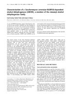
sia cardia. Chest X-ray of the patient showed an air bubble
below the left hemi-diaphragm in the gastric region which
is not expected in a case of achalasia cardia (Figures 1 and
2). Barium swallow in this patient showed dilated esopha-
gus, "bird beak" appearance of the distal esophagus and
an esophageal air fluid level.
Published: 13 December 2008
Journal of Medical Case Reports 2008, 2:383 doi:10.1186/1752-1947-2-383
Received: 31 January 2008
Accepted: 13 December 2008
This article is available from: />© 2008 Lakhan et al; licensee BioMed Central Ltd.
This is an Open Access article distributed under the terms of the Creative Commons Attribution License ( />),
which permits unrestricted use, distribution, and reproduction in any medium, provided the original work is properly cited.
Journal of Medical Case Reports 2008, 2:383 />Page 2 of 5
(page number not for citation purposes)
The presence of an air bubble below the left hemi-dia-
phragm in the gastric region as seen in the chest X-ray
posed a diagnostic challenge. But since the clinical his-
tory, examination, upper gastrointestinal endoscopy and
barium swallow X-ray were suggestive of achalasia cardia,
a final diagnosis of achalasia cardia was made and pneu-
matic balloon dilatation of the lower esophageal sphinc-
ter was planned.
Under sedation, pneumatic balloon dilatation of the
lower esophageal dilatation was carried out. The proce-
dure was performed under endoscopic vision. The bal-
loon was placed across the lower esophageal sphincter
and inflated. The balloon was kept in the inflated position
for 2 minutes. With the help of the retroflexed endoscope,
active bleeding was seen across the chest junction which
indicated a successful dilatation of the lower esophageal
sphincter. There were no procedure-related complica-
tions.
A chest X-ray after the procedure showed two air bubbles
under the left hemi-diaphragm in the gastric region. A sec-
Chest X-ray showing air bubble below the left hemi-diaphragm in the gastric regionFigure 1
Chest X-ray showing air bubble below the left hemi-diaphragm in the gastric region.
Journal of Medical Case Reports 2008, 2:383 />Page 3 of 5
(page number not for citation purposes)
ond air bubble had appeared adjacent to the previous one
which was present before dilatation. Since a successful dil-
atation of the lower esophageal sphincter results in the
appearance of the gastric bubble, the second air bubble that
appeared was the gastric air bubble. Careful examination of
the first air bubble present before the procedure, showed
haustral markings. This confirmed that the air bubble was
a colonic air bubble. The "double bubble" is thus a colonic
air bubble and a gastric air bubble (Figure 3).
The colonic air bubble was seen before the procedure
mimicking the gastric air bubble which caused diagnostic
confusion. After successful pneumatic dilatation, the gas-
tric air bubble appeared below the left hemi-diaphragm,
which is an anticipated event. This appearance of a double
bubble in a case of achalasia cardia not only causes diag-
nostic problems, but is also very unusual and has not been
reported in the literature before.
Discussion
Achalasia is a primary esophageal motility disorder
involving the body of the esophagus and lower esopha-
geal sphincter affecting equally both genders and all ages
[1]. Although endoscopy is considered to have a poor sen-
sitivity and specificity in the diagnosis of achalasia, it has
an important role in ruling out secondary causes of acha-
lasia (i.e. pseudoachalasia). A chest X-ray can give impor-
tant information. It may show the absence of a gastric air
bubble. Barium swallow will show dilated esophagus,
"bird beak" appearance of the distal esophagus and an
esophageal air fluid level. In up to 20% of achalasia
patients, however, these classic X-ray findings are not
present. Manometry is the gold standard for diagnosing
achalasia cardia [1]. In patients with typical radiographic
findings of achalasia, the barium study can be used to
guide treatment without a need for manometry. If radio-
graphic findings are equivocal, however, manometry may
Chest X-ray; red arrow shows the air bubble below the left hemi-diaphragm before pneumatic balloon dilatationFigure 2
Chest X-ray; red arrow shows the air bubble below the left hemi-diaphragm before pneumatic balloon dilatation. This is an
atypical finding.
Journal of Medical Case Reports 2008, 2:383 />Page 4 of 5
(page number not for citation purposes)
be required for a more certain diagnosis [2]. But manom-
etry is not available in all medical centers. In centers
where manometry is not available, clinical history, endos-
copy, chest X-ray and barium swallow are all taken
together to diagnose achalasia cardia. With respect to
treatment, Heller's myotomy and pneumatic balloon dil-
atations of the lower esophageal sphincter are considered
definitive treatments for achalasia [3].
Since achalasia cardia is associated with failure of the
lower esophageal sphincter to relax, not enough air passes
across into the stomach. This is manifested as an absent
gastric bubble in the abdominal X-rays. Though this is not
a sensitive method, absence of a gastric air bubble in the
chest X-ray is one of the significant findings for diagnos-
ing achalasia [4]. After successful dilatation of the lower
esophageal sphincter, the gastric air bubble reappears in
the chest X-ray.
Sometimes achalasia presents with atypical presentations
and atypical findings. There are case reports of achalasia
presenting as acute airway obstruction and recurrent
pneumonitis [5,6]. In patients with atypical presentation
and findings, the diagnosis is often delayed. In our
patient, there was an atypical finding in the form of the
presence of an air bubble below the left hemi-diaphragm
in the gastric region in the chest X-ray film.
Based on the clinical history, examination, upper gas-
trointestinal endoscopy and barium swallow X-ray find-
Chest X-ray showing the double bubble after successful dilatation of the lower esophageal sphincterFigure 3
Chest X-ray showing the double bubble after successful dilatation of the lower esophageal sphincter. Red arrows indicate the
presence of air in the colon. Note the haustrations. Black arrow indicates the presence of air in the stomach which appeared
after dilatation.
Publish with BioMed Central and every
scientist can read your work free of charge
"BioMed Central will be the most significant development for
disseminating the results of biomedical research in our lifetime."
Sir Paul Nurse, Cancer Research UK
Your research papers will be:
available free of charge to the entire biomedical community
peer reviewed and published immediately upon acceptance
cited in PubMed and archived on PubMed Central
yours — you keep the copyright
Submit your manuscript here:
/>BioMedcentral
Journal of Medical Case Reports 2008, 2:383 />Page 5 of 5
(page number not for citation purposes)
ings, a provisional diagnosis of achalasia cardia was made.
Pneumatic balloon dilatation was done to relieve the
symptoms. The chest X-ray taken after the successful pro-
cedure showed the appearance a second air bubble in the
gastric region adjacent to the previous one. This phenom-
enon is an anticipated one. Careful examination of the
first air bubble, which was seen even before the dilatation
was done, showed haustral markings. Haustral markings
are seen in the colon. This led us to the conclusion that the
air bubble which was present before dilatation was indeed
a colonic air bubble in the splenic flexure. Therefore, the
second air bubble, which appeared after successful dilata-
tion of the lower esophageal sphincter, was the gastric air
bubble.
So in our patient, at the end of the dilatation, there were
two air bubbles – a double bubble. A thorough Medline
search was performed. To our knowledge, this finding has
not been reported in the literature thus far. The appear-
ance of a double bubble in patients with achalasia cardia
is an interesting finding following a successful dilatation
of the lower esophageal sphincter. This double bubble
sign may pose a diagnostic challenge in the patients in
whom it is present. Knowledge of this unusual sign may
be helpful in these circumstances.
Conclusion
A colonic air bubble in the splenic flexure may mimic a
gastric air bubble in chest X-ray films. This may cause con-
fusion in the diagnosis of achalasia cardia where the gas-
tric bubble is generally absent. Successful dilatation of the
esophageal sphincter in patients with achalasia cardia
results in reappearance of the gastric air bubble. In
patients whose chest X-ray shows a colonic air bubble in
the gastric region at the time of presentation, the chest X-
ray will show a double bubble after successful dilatation
of the lower esophageal sphincter. The double bubble rep-
resents the colonic air bubble and the gastric air bubble.
Consent
Written informed consent was obtained from the patient
for publication of this case report and any accompanying
images. A copy of the written consent is available for
review by the Editor-in-Chief of this journal.
Competing interests
The authors declare that they have no competing interests.
Authors' contributions
SL, SJK, and PRK secured the case, conducted the literature
review, and participated in the preparation of the manu-
script. All authors read and approved the final manu-
script.
References
1. Vaezi MF, Richter JE: Diagnosis and management of achalasia.
American College of Gastroenterology Practice Parameter
Committee. Am J Gastroenterol 1999, 94(12):3406-3412.
2. Amaravadi R, Levine MS, Rubesin SE, Laufer I, Redfern RO, Katzka
DA: Achalasia with complete relaxation of lower esophageal
sphincter: radiographic-manometric correlation. Radiology
2005, 235(3):886-891.
3. Pohl D, Tutuian R: Achalasia: an overview of diagnosis and
treatment. J Gastrointestin Liver Dis 2007, 16(3):297-303.
4. Orlando RC, Call DL, Bream CA: Achalasia and absent gastric
air bubble. Ann Intern Med 1978, 88(1):60-61.
5. Khan AA, Shah SW, Alam A, Butt AK, Shafqat E, Malik K, Amin J: Ach-
alasia esophagus; presenting as acute air way obstruction. J
Pak Med Assoc 2007, 57(8):423-425.
6. Sharma GL, Kumar A, Mukund A, Kedia A: Atypical presentation
of achalasia cardia: a case report. Indian J Radiol Imaging 2005,
15:175-176.