Báo cáo y học: " Extramammary Paget''''s disease of the penis: a case report and review of the literature" pot
Bạn đang xem bản rút gọn của tài liệu. Xem và tải ngay bản đầy đủ của tài liệu tại đây (1.97 MB, 4 trang )
BioMed Central
Page 1 of 4
(page number not for citation purposes)
Journal of Medical Case Reports
Open Access
Case report
Extramammary Paget's disease of the penis: a case report and
review of the literature
Kingsley C Ekwueme
1
, Hani D Zakhour
2
and Nigel J Parr*
1
Address:
1
Regional Cancer Centre, Department of Urology, Wirral University Teaching Hospital, Arrowe Park Road, Upton, Wirral, CH49 5PE, UK
and
2
Department of Histopathology and Clinical Cytology, Wirral University Teaching Hospital, Arrowe Park Road, Upton, Wirral, CH49 5PE, UK
Email: Kingsley C Ekwueme - ; Hani D Zakhour - ;
Nigel J Parr* -
* Corresponding author
Abstract
Introduction: Extramammary Paget's disease is a rare cutaneous, slow growing, intraepithelial
adenocarcinoma developing in the apocrine gland-bearing areas. Isolated Paget's disease of the
penis is extremely rare.
Case presentation: We describe the case of an 87-year-old Caucasian male who presented with
a non-healing erythematous plaque on the shaft of the penis previously misdiagnosed as Bowen's
disease. The diagnosis was made histologically on the excised specimen and was supported by
immunohistochemical staining.
Conclusion: Extramammary Paget's disease is a rare disease which can mimic various types of
dermatosis. A high index of suspicion is required, combined with biopsy and immunohistochemical
staining in order to make the correct diagnosis. Long-term follow-up is mandatory in these patients
in order to identify and treat any subsequent recurrence or concurrent malignancy.
Introduction
Extamammary Paget's disease (EMPD) is a rare cutaneous,
intraepithelial adenocarcinoma involving primarily the
epidermis but occasionally extending into the underlying
dermis. It has predilection for apocrine gland-bearing
areas: mostly the perineum, vulva, axilla, scrotum and
penis. Isolated Paget's disease of the penis is rare and only
a few cases have been reported in the literature [1].
We describe a case of EMPD confined to the shaft of the
penis and initially misdiagnosed on punch biopsy. We
also review the literature and highlight the need for a high
index of suspicion in the diagnosis of this rare neoplasm.
Case presentation
An 87-year-old Caucasian male was referred to our centre
by a dermatologist, having undergone punch biopsy of a
penile lesion with the initial histology reported as show-
ing Bowen's disease. The patient gave a 6-month history of
an enlarging lesion on the shaft of his penis prior to pres-
entation to the dermatologist, which had been treated
with topical agents and antibiotics. Nevertheless, the der-
matologist was clinically suspicious of an invasive lesion
prompting referral for wide excision. The patient had had
a similar lesion at the same location 10 years earlier which
was excised by his general practitioner but no histology
report could be traced. He had no other lumps anywhere
in the rest of the body and no family history of similar dis-
Published: 6 January 2009
Journal of Medical Case Reports 2009, 3:4 doi:10.1186/1752-1947-3-4
Received: 5 February 2008
Accepted: 6 January 2009
This article is available from: />© 2009 Ekwueme et al; licensee BioMed Central Ltd.
This is an Open Access article distributed under the terms of the Creative Commons Attribution License ( />),
which permits unrestricted use, distribution, and reproduction in any medium, provided the original work is properly cited.
Journal of Medical Case Reports 2009, 3:4 />Page 2 of 4
(page number not for citation purposes)
ease. His co-morbidities included ischaemic heart disease,
Alzheimer's disease and venous ulcers.
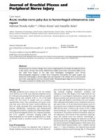
Examination revealed a 2.5 cm erythematous, fleshy, exo-
phytic plaque at the base of the shaft of the penis (Figure
1). There was a satellite lesion proximal to this. The
patient had no palpable inguinal lymphadenopathy. A
clinical suspicion of an invasive squamous cell carcinoma
was made and the patient underwent a wide local excision
of the penile and satellite lesions. Frozen-section exami-
nation was not performed. The scrotal skin was advanced
and primary closure performed. The foreskin was
retracted in order to achieve a tension-free closure.
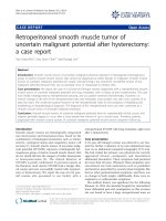
The specimen measured 30 × 50 × 50 mm. Light micros-
copy showed intraepithelial proliferation of neoplastic;
large, pale cells, located predominantly in the basal and
parabasal layers of the epithelium (Figure 2), with mar-
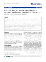
gins apparently clear. Immunohistochemical stains
showed specific staining characteristics with strong posi-
tivity for epithelial membrane antigen (EMA), the cytok-
eratin (CK) CK7, CAM 5.6 and HER2 protein over
expression. CK20 staining was negative. These immuno-
histochemical appearances supported the histological
diagnosis of EMPD (Figure 3). Immunohistochemical
staining also revealed that there were occasional cells in
proximity to the margins.
This patient's histology was discussed at our weekly multi-
disciplinary cancer meeting and the consensus was not to
screen for an underlying non-cutaneous malignancy in
view of the patient's age and co-morbidities. Furthermore,
a decision was made not to attempt wider excision. At 6-
months follow-up, our patient had no local recurrence or
palpable inguinal lymph nodes.
Discussion
EMPD localised to the penis is extremely rare and only few
cases have been reported. The first description of EMPD
was by Crocker in 1889 when he reported a case affecting
the penis and scrotum. EMPD is commoner in females
and the elderly population, with a predilection for apo-
crine gland-bearing areas, most especially the vulva, peri-
anal areas, axilla and penoscrotal region. Other sites
reported include the groin, external auditory canal, chest
and eyelids.
Clinically, presentation is often non-specific and can
mimic any form of dermatosis. Differential diagnoses
include Bowen's disease, tinea cruris, contact dermatitis,
lichen simplex, lichen planus, psoriasis and seborrheic
dermatitis. This can result in delayed presentation as was
the case with our patient. In order to make the correct
diagnosis, a high index of suspicion is required. The diag-
nosis is, however, made on histological grounds and sup-
ported by immunohistochemical analysis. Positive
staining for CK7, a low molecular weight CK, in conjunc-
tion with immunonegativity for high molecular weight
CKs, have consistently been proven to be the most useful
diagnostic markers [2]. This observation was confirmed in
our case.
A recent classification based on the origin of the Paget's
cells has been proposed by Wilkinson and Brown [3].
They classified vulval Paget's disease (PD) into two broad
groups – primary (of cutaneous origin) and secondary (of
non-cutaneous origin). For primary PD, Type 1 is primary
intraepithelial PD, Type 2 is primary intraepithelial PD
with invasion and Type 3 is primary intraepithelial PD as
a manifestation of underlying adenocarcinoma of skin
Photograph of the penile lesionFigure 1
Photograph of the penile lesion.
H&E stain of the resected specimen showing a clear marginFigure 2
H&E stain of the resected specimen showing a clear
margin.
Journal of Medical Case Reports 2009, 3:4 />Page 3 of 4
(page number not for citation purposes)
appendage origin. Secondary PD originates from an
underlying non-cutaneous neoplasm. This proposed clas-
sification could help decide on the extent of surgery, pre-
vent unnecessary surgery and influence the outcome.
The true nature of EMPD and its relationship to underly-
ing malignancy remains uncertain. Published reports sug-
gest that up to 42% of patients have associated underlying
secondary or non-cutaneous malignancy [4]. However,
there is a low incidence of internal malignancy with peno-
scrotal EMPD [5]. The location of the internal malignancy
appears to relate to the location of EMPD. Thus, penoscro-
tal and perianal locations are associated with adenocarci-
noma of the genitourinary and digestive tracts,
respectively [6]. Siesling et al. found an increased risk of
developing a second primary cancer in their series [7]. Fol-
lowing diagnosis of EMPD, a thorough search for an
underlying non-cutaneous malignancy is recommended
[6,8]. However, the decision and extent of the search
should be tailored to the patient. Chiu et al. [9] recom-
mend screening for only those with perianal or invasive
disease and young patients.
The treatment of choice is surgery with wide local excision
and immediate reconstruction. Recurrence rates can be up
to 60% [9]. Results of frozen section-guided wide, local
excision suggest a reduction in the recurrence rate to
between 16% and 25% [9,10]. However, the time con-
straints during surgery mean that assessment of the total
margin status by frozen section is difficult and morbidity
is likely to increase with prolonged anaesthetic times in
frail, elderly patients. In their review, Zhu et al. [10] found
a 13% false negative frozen-section analysis. It is unlikely
that rates can be reduced further, as positive margins in
some cases are only diagnosed by immunohistochemis-
try. Other treatment modalities which have been used
with mixed results include Mohs micrographic surgery,
radiotherapy, Nd:YAG and carbon dioxide laser, topical
Fluorouracil and 5% imiquimod cream.
Immunohistochemical stain of a resected specimen showing occasional cells near the marginFigure 3
Immunohistochemical stain of a resected specimen showing occasional cells near the margin.
Publish with BioMed Central and every
scientist can read your work free of charge
"BioMed Central will be the most significant development for
disseminating the results of biomedical research in our lifetime."
Sir Paul Nurse, Cancer Research UK
Your research papers will be:
available free of charge to the entire biomedical community
peer reviewed and published immediately upon acceptance
cited in PubMed and archived on PubMed Central
yours — you keep the copyright
Submit your manuscript here:
/>BioMedcentral
Journal of Medical Case Reports 2009, 3:4 />Page 4 of 4
(page number not for citation purposes)
The prognosis is good when the disease is confined to the
epidermis. However, in the presence of dermal invasion,
the prognosis is poor [10].
Conclusion
PD of the penis is extremely rare. A high index of suspi-
cion, combined with histological examination supported
by immunohistochemical staining of biopsy specimen is
essential to accurate diagnosis. The treatment of choice is
surgery. Frozen section-guided excision reduces the recur-
rence rate. Long-term follow-up is mandatory in these
patients in order to identify and treat any subsequent
recurrence or concurrent malignancy.
Abbreviations
EMPD: extamammary Paget's disease; EMA: epithelial
membrane antigen; CK: cytokeratin; PD: Paget's disease
Consent
Written informed consent was obtained from the patient
for publication of this case report and accompanying
images. A copy of the written consent is available for
review by the Editor-in-Chief of this journal.
Competing interests
The authors declare that they have no competing interests.
Authors' contributions
KCE summarized the case and wrote the manuscript. HDZ
performed the histological examination of the lesion,
reviewed the histology from the referring hospital and
provided the histology micrographs, whilst NJP is the
Principal Surgeon and provided the overall supervision in
the writing of this article. All authors read and approved
the final manuscript.
References
1. Yang WJ, Kim DS, Im YJ, Cho KS, Rha KH, Cho NH, Choi YD:
Extramammary Paget's disease of penis and scrotum. Urology
2005, 65(5):972-975.
2. Liegl B, Leibl S, Gogg-Kamerer M, Tessaro B, Horn LC, Moinfar F:
Mammary and extramammary Paget's disease: an immuno-
histochemical study of 83 cases. Histopathology 2007,
50(4):439-447.
3. Wilkinson EJ, Brown HM: Vulvar Paget disease of urothelial ori-
gin: a report of three cases and a proposed classification of
vulvar Paget disease. Hum Pathol 2002, 33(5):549-554.
4. Lai YL, Yang WG, Tsay PK, Swei H, Chuang SS, Wen CJ: Penoscro-
tal extramammary Paget's disease: a review of 33 cases in a
20-year experience. Plast Reconstr Surg 2003, 112(4):1017-1023.
5. Park S, Grossfeld GD, McAninch JW, Santucci R: Extramammary
Paget's disease of the penis and scrotum: excision, recon-
struction and evaluation of occult malignancy. J Urol 2001,
166(6):2112-2117.
6. Chanda JJ: Extramammary Paget's disease: prognosis and
relationship to internal malignancy. J Am Acad Dermatol 1985,
13(6):1009-1014.
7. Siesling S, Elferink MA, van Dijck JA, Pierie JP, Blokx WA: Epidemi-
ology and treatment of extramammary Paget disease in the
Netherlands. Eur J Surg Oncol 2007, 33(8):951-955.
8. Khoo JJ, Choon SE: Extramammary Paget's disease: a report of
2 cases and a review of the literature. Malays J Pathol 2003,
25(1):73-78.
9. Chiu TW, Wong PS, Ahmed K, Lam SC, Ying SY, Burd A:
Extramammary Paget's disease in Chinese males: a 21-year
experience. World J Surg 2007, 31(10):1941-1946.
10. Zhu Y, Ye DW, Chen ZW, Zhang SL, Qin XJ: Frozen section-
guided wide local excision in the treatment of penoscrotal
extramammary Paget's disease. BJU Int 2007,
100(6):1282-1287.