Báo cáo y học: " Nodular melanoma presenting with rapid progression and widespread metastases: a case report" pot
Bạn đang xem bản rút gọn của tài liệu. Xem và tải ngay bản đầy đủ của tài liệu tại đây (1.57 MB, 4 trang )
BioMed Central
Page 1 of 4
(page number not for citation purposes)
Journal of Medical Case Reports
Open Access
Case report
Nodular melanoma presenting with rapid progression and
widespread metastases: a case report
Mehmet Ali Erkurt*
1
, Ismet Aydogdu
1
, Irfan Kuku
1
, Emin Kaya
1
and
Yalcin Basaran
2
Address:
1
Inonu University Faculty of Medicine, Department of Hematology, Turgut Ozal Medical Center, TR-44069 Malatya, Turkey and
2
Gulhane Military Medical Academy, Department of Internal Medicine, Ankara, Turkey
Email: Mehmet Ali Erkurt* - ; Ismet Aydogdu - ; Irfan Kuku - ;
Emin Kaya - ; Yalcin Basaran -
* Corresponding author
Abstract
Introduction: Melanoma is responsible for 1% to 2% of all cancer deaths around the world.
Nodular melanoma often carries a poor prognosis because of no prodromal radial growth phase,
early distant metastasis and significant tumor volume.
Case presentation: We present a case of progressive melanoma. A 51-year-old man was
admitted to our hospital with dyspnea and skin lesions. These were multiple, dark colored, firm,
and nodular and varied in size. He was diagnosed with melanoma. Temozolomide was administered,
but he died of respiratory failure within a week after diagnosis.
Conclusion: Nodular melanoma tends to spread rapidly and eventually metastasize to vital organs.
It may be fatal within months of recognition.
Introduction
Melanoma is a neoplasm derived from melanocytes of the
skin and other sites. It accounts for 1% to 3% of all malig-
nancies and 1% to 2% of all cancer deaths worldwide.
Recently, melanoma has become a major health problem
in many countries. The worldwide incidence rate is
increasing much more rapidly than for any other malig-
nancies [1]. The mortality and morbidity rate from
melanoma has risen about 2% annually since 1960 [2].
Projections were for 59,940 cases of melanoma and
48,290 cases of in situ melanoma to be newly diagnosed
in the USA in 2007. Of these, 8110 cases were expected to
be fatal [3].
The lifetime risk of melanoma is 1:70 in the population of
the USA and will probably be 1:50 in 2010 [1]. According
to the American Joint Committee on Cancer, melanomas
are classified as superficial spreading melanoma, nodular
melanoma, lentigo melanoma, acral lentiginous
melanoma and unclassified melanoma. Nodular
melanoma, comprising 10% to 15% of cutaneous
melanomas, is the second most common variety of
melanocytic neoplasms and occurs less commonly than
superficial spreading melanoma. The median age of onset
is 49 years. The duration of lesions before diagnosis is rel-
atively short, ranging from a few months to 2 years [4].
Nodular melanoma often presents as an expanding darkly
pigmented cutaneous nodular lesion, usually found on
the sun-exposed areas of the skin, with far fewer such
Published: 6 February 2009
Journal of Medical Case Reports 2009, 3:50 doi:10.1186/1752-1947-3-50
Received: 29 August 2008
Accepted: 6 February 2009
This article is available from: />© 2009 Erkurt et al; licensee BioMed Central Ltd.
This is an Open Access article distributed under the terms of the Creative Commons Attribution License ( />),
which permits unrestricted use, distribution, and reproduction in any medium, provided the original work is properly cited.
Journal of Medical Case Reports 2009, 3:50 />Page 2 of 4
(page number not for citation purposes)
lesions occurring in covered areas. The most common
sites are the trunk in men and the legs in women. Major
risk factors for nodular melanoma include the presence of
multiple dysplastic nevi, positive family history, light
colored skin with an inability to tan, and excessive sun
exposure. Nodular melanoma is known to present with
greater thickness than the other subtypes of melanoma,
therefore, it often carries a poorer prognosis [5]. Even in
its early stages, it has the potential to metastasize to the
vital organs [6]. Herein, we present a patient with
melanoma which was associated with highly invasive and
aggressive behavior.
Case presentation
A 51-year-old man was admitted to our hospital with mul-
tiple enlarging masses in the inguinal and thoracal areas
of2 months' duration, which progressively enlarged and
spread around the whole body. In the 2 weeks before pres-
entation, the lesions had became darkly pigmented, sug-
gesting melanoma. At the time of presentation, he
complained of dyspnea, cough, fever and night sweats. He
had a smoking history of 20 cigarettes a day for the last 30
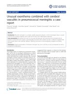
years. Dermatological examination revealed multiple,
dark colored, firm, nodular lesions varying in size (Figure
1A, B). Rhonchi over the whole lung area were noted on
auscultation. The results of routine laboratory studies of
blood and urine were normal except for raised ESR (98
mm/hour), LDH (1311 U/ml) and uric acid (12.8 mg/dl).
Peripheral blood smear and bone marrow examinations
were normal. Chest X-ray showed the presence of hetero-
geneous opacities in the right lung field suggestive of mul-
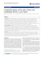
tiple pulmonary metastases. Contrast-enhanced
computed tomography of the abdomen demonstrated a
low density soft tissue lesion 7 × 4.5 cm in diameter with
irregular margins, located within the right atrium, which
was also consistent with the presence of metastases (Fig-
ure 2A). Contrast-enhanced computed tomography of the
thorax showed conglomerated mediastinal lymph nodes
and numerous nodular pulmonary lesions, findings that
suggested the presence of metastases. Pleural and pericar-
dial effusions were also present as well as numerous sub-
cutaneous lesions in the chest wall and brain metastases
confirmed by computed tomography of the brain (Figure
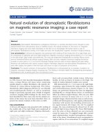
2B). On fine needle aspiration, the nodular lesions were
diagnosed as melanoma (Figure 3). Although the classifi-
cation was not based on histological examination, the
appearance of the masses and the clinical findings of the
patient were consistent with the diagnosis of nodular
melanoma. Temozolomide was administered for 5 con-
secutive days at a daily dose of 150 mg/m
2
/day but the
patient died of respiratory failure within a week after diag-
nosis.
Discussion
Nodular melanoma is more common in men than
women. The trunk is a common site and a discrete nodule
with dark black/brown pigmentation is typical. Ulcera-
tion and bleeding are common complications. Nodular
melanoma has a peak incidence around 50 years of age
[5]. It arises in normal skin or in a precursor lesion, but
without an intervening radial growth phase. A widely
accepted histopathological definition of nodular
melanoma is a melanoma that lacks an in-situ component
beyond three rete ridges of the invasive vertical growth
phase; thus, even in its early stages, nodular melanoma
has the potential to metastasize [6,7]. Acral lentiginous
melanoma is most frequent in the 60 to 70 year age group.
It was so named because of its predilection for acral (dis-
tal) areas of the body, particularly the palms, soles and
subungual areas, and its distinct radial or "lentiginous"
growth phase. Its diagnosis is described as being based on
its histological, intradermal features showing a diffuse
proliferation of large atypical melanocytes along the epi-
dermal-dermal junction which is dispersed in a lentigi-
(A) Clinical image shows a blue red nodule on the skin over the trunkFigure 1
(A) Clinical image shows a blue red nodule on the
skin over the trunk. (B) Clinical image shows multiple,
dark colored, firm, nodular skin lesions varying in size.
A
B
Journal of Medical Case Reports 2009, 3:50 />Page 3 of 4
(page number not for citation purposes)
nous pattern with marked acanthosis and elongation of
the rete ridges. Acral lentiginous melanoma is the only
sub-type of melanoma that occurs at the same rate in all
races, predominantly on an area that seldom receives
much sun exposure. It has been suggested that the etiol-
ogy is different from that of nodular melanoma or that
sun exposure is a lesser risk factor than melanoma else-
where. Also, various histopathologic features including
nodular and acral lentiginous subtypes, vertical growth
phase, high mitotic activity and the presence of micro-
scopic satellites are associated with poor prognosis [8].
Metastatic melanoma usually involves draining lymph
nodes and occasionally adjacent skin first, but eventually
metastasizes to distant visceral sites. The skin and subcu-
taneous lymph nodes (59%) are most commonly
involved followed by lung (36%), brain (20%), liver
(20%), bone (17%) and others (12%) [6,7]. In our
patient, metastatic lesions were seen in the lungs, pleura,
heart and brain at the time of diagnosis. Although the
diagnosis was not confirmed histologically, widespread
metastases that developed in the patient within a short
period of time strongly suggested melanoma. In the liter-
ature [6,7], melanoma is reported to develop metastases
in every organ. Similarly, the patient developed rapidly
progressive vital organ metastases.
The Breslow thickness is the most important prognostic
variable. Tumors of greater. Breslow thickness are more
likely to invade lymphatic or blood vessels allowing a
route of passage for distant spread (Table 1). The number
and localization of metastases are useful for assessing dis-
ease stage and response to therapy. Patients with cutane-
ous, nodal, or gastrointestinal metastases have a median
survival time of 12.5 months; those with pulmonary
metastases have a median survival time of 8.3 months;
and in those with liver, brain, or bone metastases, the
median survival is 4.4 months. The median survival time
is 7 months in patients with a single metastatic site, 4
(A) Computed tomography image shows a lobulated, irregu-larly shaped mass 7 × 4.5 cm in diameter with central hypodensity in the right atriumFigure 2
(A) Computed tomography image shows a lobulated,
irregularly shaped mass 7 × 4.5 cm in diameter with
central hypodensity in the right atrium. (B) Computed
tomography image shows a brain metastasis.
A
B
A cluster of melanin pigment containing melanoma cells is observed in the fine needle aspiration cytology (hematoxylin and eosin staining, 100×)Figure 3
A cluster of melanin pigment containing melanoma
cells is observed in the fine needle aspiration cytology
(hematoxylin and eosin staining, 100×).
Table 1: Prognosis according to Breslow thickness in melanoma
Breslow thickness 5-year survival
In situ 90–100%
Stage I < 1 mm 80–90%
Stage II 1–2 mm 70–80%
Stage III 2.1–4 mm 60–70%
Stage IV > 4 mm 50%
Publish with BioMed Central and every
scientist can read your work free of charge
"BioMed Central will be the most significant development for
disseminating the results of biomedical research in our lifetime."
Sir Paul Nurse, Cancer Research UK
Your research papers will be:
available free of charge to the entire biomedical community
peer reviewed and published immediately upon acceptance
cited in PubMed and archived on PubMed Central
yours — you keep the copyright
Submit your manuscript here:
/>BioMedcentral
Journal of Medical Case Reports 2009, 3:50 />Page 4 of 4
(page number not for citation purposes)
months with two organ sites and 2 months with three or
more metastatic sites. Similarly, the 12-month survival
rate is 36% with a single metastatic site, 13% with two
organ sites, and 0% with three or more metastatic sites [7].
The patient's Breslow thickness was greater than 4 mm. In
our patient, the number of metastases was more than
three and he died 2 months after the onset of his illness,
as expected.
Despite research, no consensus has been reached as to the
optimal management, There is level I evidence for the
treatment of stage III and stage IV patients. Traditionally,
management of melanoma metastatic to distant sites
involves either a single-drug or multi-drug chemothera-
peutic regimen. However, complete response rates have
been poor (< 6%) with a minimal increase in the median
survival [9,10]. Chemotherapy was administered immedi-
ately after the diagnosis of melanoma was suggested, but
he died of respiratory failure because of the rapidly pro-
gressive course of disease with widespread pulmonary,
brain, heart and cutaneous metastases.
Conclusion
Tumor thickness, level of invasion, and number of
involved nodes are the most powerful prognostic indica-
tors in nodular melanoma. As described in our patient,
nodular melanoma has a strong tendency for widespread
dissemination and metastasis to vital organs. Despite
increased therapeutic options for the treatment of
advanced melanoma, the results are disappointing in
patients with widespread metastases.
Abbreviations
ESR: erythrocyte sedimentation rate; LDH: lactate dehy-
drogenase
Consent
Written informed consent was obtained from the patient
for publication of this case report and any accompanying
images. A copy of the written consent is available for
review by the Editor-in-Chief of this journal.
Competing interests
The authors declare that they have no competing interests.
Authors' contributions
This report reflects the opinion of the authors and does
not represent the official position of any institution or
sponsor. MAE was responsible for reviewing previous
research, journal hand searching, and drafting the report.
IK and EK were responsible for provision of published
trial bibliographies, and preparing photographs. YB con-
tributed to the final draft of the manuscript and analysis
of relevant data. IA was responsible for project coordina-
tion. All authors read and approved the final manuscript.
References
1. Burton RC, Coates MS, Hersey P, Roberts G, Chetty MP, Chen S,
Hayes MH, Howe CG, Armstrong BK: An analysis of a melanoma
epidemic. Int J Cancer 1993, 55:765-770.
2. Rigel DS, Carucci JA: Malignant melanoma: prevention, early
detection, and treatment in the 21st century. CA Cancer J Clin
2000, 50:215-236.
3. Jemal A, Siegel R, Ward E, Murray T, Xu J, Thun MJ: Cancer statis-
tics. CA Cancer J Clin 2007, 57:43-66.
4. Balch CM, Buzaid AC, Soong SJ, Atkins MB, Cascinelli N, Coit DG:
Final version of the American Joint Committee on Cancer
staging system for cutaneous melanoma. J Clin Oncol 2001,
19:3635-3648.
5. Bergenmar M, Ringborg U, Mansson Brahme E, Brandberg Y: Nodu-
lar histogenetic type the most significant factor for thick
melanoma: implications for prevention. Melanoma Res 1998,
8:403-411.
6. Balch CM, Soong SJ, Murad TM, Smith JW, Maddox WA, Durant JR:
A multifactorial analysis of melanoma. Prognostic factors in
200 melanoma patients with distant metastases (stage III). J
Clin Oncol 1983, 1:126-134.
7. Barth A, Wanek LA, Morton DL: Prognostic factors in 1521
melanoma patients with distant metastases. J Am Coll Surg
1995, 181:193-201.
8. Bristow IR, Acland K: Acral lentiginous melanoma of the foot
and ankle: A case series and review of the literature. J Foot
Ankle Res 2008, 1:11.
9. Legha SS, Ring S, Papadoupoulos N, Plager C, Chawla S, Benjamin R:
A prospective evaluation of a triple-drug regimen containing
cisplatin, vinblastine, and dacarbazine (CVD) for metastatic
melanoma. Cancer 1989, 64:2024-2029.
10. McClay EF, Mastrangelo MJ, Berd D, Bellet RE: Effective combina-
tion chemo/hormonal therapy for malignant melanoma:
experience with three consecutive trials. Int J Cancer 1992,
50:553-556.