báo cáo khoa học: " Identification of the occurrence and pattern of masseter muscle activities during sleep using EMG and accelerometer systems" docx
Bạn đang xem bản rút gọn của tài liệu. Xem và tải ngay bản đầy đủ của tài liệu tại đây (668.96 KB, 10 trang )
BioMed Central
Page 1 of 10
(page number not for citation purposes)
Head & Face Medicine
Open Access
Methodology
Identification of the occurrence and pattern of masseter muscle
activities during sleep using EMG and accelerometer systems
Hidehiro Yoshimi
1
, Kenichi Sasaguri
1
, Katsushi Tamaki
2
and Sadao Sato*
1
Address:
1
Department of Craniofacial Growth and Development Dentistry, Research Institute of Occlusion Medicine, Research Center of Brain and
Oral Science, Kanagawa, Japan and
2
Oral and Maxillofacial Rehabilitation, Kanagawa Dental College, 82 Inaoka-Cho, Yokosuka, Kanagawa, Japan
Email: Hidehiro Yoshimi - ; Kenichi Sasaguri - ; Katsushi Tamaki - ;
Sadao Sato* -
* Corresponding author
Abstract
Background: Sleep bruxism has been described as a combination of different orofacial motor
activities that include grinding, clenching and tapping, although accurate distribution of the activities
still remains to be clarified.
Methods: We developed a new system for analyzing sleep bruxism to examine the muscle
activities and mandibular movement patterns during sleep bruxism. The system consisted of a 2-
axis accelerometer, electroencephalography and electromyography. Nineteen healthy volunteers
were recruited and screened to evaluate sleep bruxism in the sleep laboratory.
Results: The new system could easily distinguish the different patterns of bruxism movement of
the mandible and the body movement. Results showed that grinding (59.5%) was most common,
followed by clenching (35.6%) based on relative activity to maximum voluntary contraction
(%MVC), whereas tapping was only (4.9%).
Conclusion: It was concluded that the tapping, clenching, and grinding movement of the mandible
could be effectively differentiated by the new system and sleep bruxism was predominantly
perceived as clenching and grinding, which varied between individuals.
Background
Quality of sleep is strongly associated with somatic health
and activity of the body. During sleep, many physiological
events occur, such as sleep talking, sighing, swallowing,
and bruxing along with decreased skeletal muscle activity,
heart rate, body temperature and blood pressure [1]. Brux-
ism sometimes interferes with sleep quality. Sleep brux-
ism is reported to be a common phenomenon in humans
and many studies have shown that bruxism can harm the
dentition, its supporting structures and the temporoman-
dibular joint (TMJ) [2-8]. Many bruxers are not aware of
their behavior, and not all bruxers make noise that bed
partners might notice. The definition of "bruxer" is based
upon patient reports of a history of tooth-grinding occur-
ring more than three times a week for at least six months,
as attested by their sleep partners [6,7]. In addition, brux-
ers exhibited tooth wear, with orofacial jaw muscle
fatigue, tenderness or pain or masseter muscle hypertro-
phy. Recently, we studied the prevalence of bruxism in the
general adult population using a custom-made color-
stained plastic sheet, the BruxChecker, on the maxillary
dentition overnight and found that occlusal contacts
where the color was ground off were seen in the majority
of subjects, indicating sleep bruxism [9].
Published: 11 February 2009
Head & Face Medicine 2009, 5:7 doi:10.1186/1746-160X-5-7
Received: 27 June 2008
Accepted: 11 February 2009
This article is available from: />© 2009 Yoshimi et al; licensee BioMed Central Ltd.
This is an Open Access article distributed under the terms of the Creative Commons Attribution License ( />),
which permits unrestricted use, distribution, and reproduction in any medium, provided the original work is properly cited.
Head & Face Medicine 2009, 5:7 />Page 2 of 10
(page number not for citation purposes)
There is no scientific evidence that bruxism is a type of dis-
ease or abnormal function, however certain conditions
which are caused by bruxism seem to be non-physiologi-
cal phenomena. Rhythmic masticatory muscle activity
does not disrupt nocturnal sleep, further suggesting that
this motor activity is a natural activity occurring during
sleep [8]. Lavigne et al. reported that the patients who
have temporomandibular disorder (TMD) are sometimes
conscious of the existence of sleep bruxism and they pre-
sented evidence to support the positive correlation coeffi-
cient between clinical symptoms of TMD and sleep
bruxism. The influences of bruxism activity on TMD are
not fully established [1].
The diagnosis and treatment planning of bruxism is
becoming more relevant in dentistry, due to many degen-
erative oral diseases that seem to be related to excessive
biomechanical load exerted by the strong masticatory
muscle activities during bruxism. In clinical dentistry,
practitioners must be aware of the criteria by which to dis-
tinguish patients who brux from those who do not. In this
context, it is necessary to define the bruxism described as
the physiological limit of muscle activity during sleep, in
order to distinguish it from the non-physiological range
of bruxism activity.
Previous sleep researches have shown the presence of var-
ious types of sleep bruxism. Phasic/rhythmic (more than
3 bursts), tonic (more than 2 seconds over burst), mixed
(rhythmic+tonic) types [1,10], or steady-state and rhyth-
mic clenching, grinding, and tapping [11]. Various brux-
ism detecting methods have been proposed.
Polysomnography [12-17] and portable EMG [18-21]
were used for measuring sleep bruxism. In addition to
these, stent [22], splint [23], and splints that involves a
piezo-electric element [11,24], were introduced as a brux-
ism-observing technique. Stent or splint techniques may
increase or decrease activity. In these methods, the devices
may influence bruxism activity due to alteration of vertical
dimension, therefore it is not clear whether the data from
these systems is specific or not. The ambulatory EMG
(portable EMG) is adaptable to daily life, but the system
is still not satisfactory due to the presence of considerable
noise from the environment. Sleep laboratory systems,
which include electromyography (EMG), electrokinesiog-
raphy (EKG), electroencephalograpy (EEG), and audio
system are precise, but the mental and physical stress from
the laboratory environment should not be neglected. In
this context, the actual status of bruxism activity during
sleep is not exactly known, and there is no consensus con-
cerning the amount and type of bruxism activity needed
to define a certain type of event.
It is difficult to distinguish between the different activities
by the electromyography (EMG) system alone. In this
study, a newly developed method that may be useful to
assess bruxism, which involves measuring mandibular
movement during sleep, was applied to define different
types of bruxism activities.
The purposes of this study were to investigate whether it is
possible to differentiate the pattern of sleep bruxism using
a newly developed simple device and to determine the
distribution of the different types of bruxism activity.
Attempt to establish the physiological range of bruxism
activity was also considered.
Materials and methods
In this study, 19 volunteers (healthy and young post grad-
uated student and dental college students. 16 males and 3
females, aged 28.5 ± 5.8 years) consented to have their
sleep bruxism activity analyzed. We recruited them unin-
tentionally and they were not bruxer. The experimental
design, procedures and tasks were carefully explained to
the volunteers prior to starting the experiment. Each vol-
unteer slept for the entire night with a bruxism-monitor-
ing system in the sleep laboratory of Kanagawa Dental
College. Experimental procedures were approved by the
Human Ethics Committee of Kanagawa Dental College.
We obtained informed written consent from all subjects,
and we advised them of their right to discontinue the
experiment at any time.
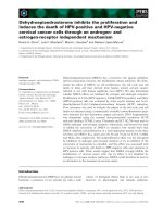
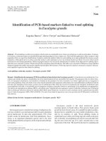
Self-adhesive surface electrodes were placed over the mas-
seter-muscle on a vertical line between the zygomatic arch
and the inferior border of the mandible (Fig. 1). Acceler-
ometers were fastened on the forehead as a reference and
on the middle point of the chin concavity of the mandible
with vinyl polysiloxane and adhesive material. The mus-
cle activity of maximum voluntary contraction in the vol-
unteers was measured 30 minutes before they went to
sleep in order to compare it with actual bruxism activity.
To establish a relative level of contraction before the sleep
bruxism recording, each subject performed at least 3 times
intercuspal-position clenches that were less than 5 sec in
duration at a 100% maximum voluntary contraction
(MVC) effort. The initial MVC data for each subject were
used to normalize all subsequent data so that all EMG sig-
nal could be reported as a percentage of the maximum
(100%) signal.
The new monitoring system of sleep bruxism consisted of
a 2-axis accelerometer (ACC, ADXL202E, Analog Devices
Co. Ltd, Norwood, MA, USA), an electroencephalogram
to measure sleep stage (EEG, Poly Mate AP1124, TEAC
Co. Ltd., Tokyo, Japan) and EMG (EMG, SN 700, Techno
Science Co. Ltd., Tokyo, Japan). An infrared video camera
(Infrared LED CCD camera, KM-033, Koike Musen Denki
Co. Ltd., Tokyo, Japan) recording system which had a
time-lapse video cassette recorder (TLV-3060, Daiwa Co.
Head & Face Medicine 2009, 5:7 />Page 3 of 10
(page number not for citation purposes)
Ltd., Tokyo, Japan) was used for monitoring sleep condi-
tion. Laser Doppler flowmetry (CDF-2000, Cyber Med,
OAS Co. Ltd., Tokyo, Japan) was used to monitor blood-
flow changes. We checked the reactive validation of brux-
ism-analyzing software (G1 System Co. Ltd., Tokyo,
Japan) for body movement through infrared video camera
and EMG data. Various kinds of noises were eliminated
from raw data and identified the existence of mandibular
reaction in the low muscle activity layer.
In this study, the criteria for bruxism activity were as fol-
lows: EMG threshold level was over 5% of activity, mini-
mum time length of bruxism episode was 250 msec of
muscle burst in the case of tapping and over 500 msec of
burst in the cases of clenching and grinding, and mini-
mum inter-episode time was more than 3 sec. Before
measuring jaw movements, coefficient calibration
through calibration voltage and scale value (physical set
value) was calculated. Both calibration voltage and scale
value to terminus point 2 and origin point 1 were estab-
lished. We formulate first degree equations; procure incli-
nations and equations with canceling offset voltage (DC
component). We obtained coefficient calibration data in
this way.
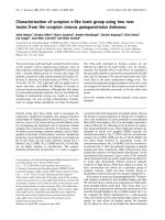
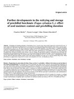
Figure 2 shows a block diagram of the data recording and
analyzing sequence is presented. Briefly, the original raw
data from EMG and ACC had noise elimination using a
50-Hz notch filter and a 60-Hz high-pass filter, followed
by smoothing and absolute-value integration. Step-by-
step categorization of the assembled data provided differ-
ent bruxism patterns. First, tapping activity was catego-
rized in order to eliminate it from the raw data since
tapping was most clearly recognizable and distinctive
from other activities. Tapping movement was character-
ized by rhythmic, sharp and short integral EMG activity as
well as Y-axis movements. The correlation coefficient of
standard tapping wave shape was used to eliminate data
that did not coincide with numerical values. In addition
to these processes, the amplitude of vibration was calcu-
lated according to the following equation to exclude huge
data.
J = Y × amplitude magnification
Y = (Hm + s.d.) × 2
Where J is the amplitude of vibration, Hm is the average
of amplitude of vibration and s.d. is the standard devia-
Panel A shows the ACC used in this studyFigure 1
Panel A shows the ACC used in this study. Panel B shows the attachment sites of the reference ACC (R) and measure-
ment ACC (M). Surface electrodes were located in areas of right and left masseters.
Head & Face Medicine 2009, 5:7 />Page 4 of 10
(page number not for citation purposes)
tion. Clenching activity was characterized by long contin-
uous muscle bursts in EMG data with little or no deviation
in XY-axis. The remaining EMG activity with long contin-
uous muscle bursts and mandibular movement in the XY-
axis was considered as a grinding pattern.
After setting up analyzing software, we checked the reac-
tions through awakening voluntary basic movement and
video recorder data of all volunteers. Basic test move-
ments were carried out for tapping, small range right and
left side grindings, wide range right and left side grindings,
maximum muscle contraction (MVC) clenching with and
without slight lateral movement, protrusion-retrusion.
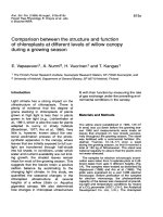
Figure 3 indicates the coincidences of analyzing software
reactions and voluntary awaking jaw movements. It was
realized that small muscle activity (under 5 %MVC) were
easily smeared with noises and the number of events went
to exceptional numbers.
Statistical Analysis
One-way ANOVA and Tukey HSD test were used to estab-
lish significance for variables on each of the three types of
bruxism activity, grinding, clenching, and tapping. Statis-
tical significance was evaluated at P < 0.05. The statistical
analyses were carried out using the Statistical Package for
SPSS (version 13.0).
Block diagram of data recording and analyzing systemFigure 2
Block diagram of data recording and analyzing system. Tapping activity could be separated from raw data based on
rhythmic, sharp and short integral EMG activity and Y axis movements. The clenching activity was separated from grinding
activity based on the long continuous muscle bursts with no or small deviation of the Y axis, and residual grinding activity
showed long continuous muscle bursts with mandibular movement in the Y axis.
EMG
ACC
Noise Elimination
Tapping
Clenching
Grinding
Original Data from EMG Original Data from ACC
50 Hz notch filter
60 Hz high pass filter
Smoothing
Absolute value integration
Ajusting the infra-threshold level to skim off supernatant waves
Noise Elimination
50 Hz notch filter
10 Hz high pass filter
Smoothing
200 Hz re-sampling
Amplitude of vibration from Y-axis mandibular accelerometer
Tapping
Mandibular vertical movement with EMG activity,
but not lateral movement
Clenching
Remaining EMG activity with lateral mandibular movement
Grinding
Head & Face Medicine 2009, 5:7 />Page 5 of 10
(page number not for citation purposes)
Results
Using the newly developed system, Bruxism was assigned
to three types; grinding, clenching and tapping. The distri-
bution of different patterns of bruxism activity showed
that clenching and grinding activities were more predom-
inant, whereas tapping activity was not highly prevalent
during sleep (Table 1, 2). Muscle activities (%MVC) were
greater in grinding (59.6%) than in clenching (35.6%),
while tapping activity was very low (4.9%). Calculation of
occurrence of events and length of event also indicated
that clenching and grinding were the predominant brux-
ism activities (Table 2). Sleep bruxism was constituted by
32.3% of grinding, 43.3% of clenching, and 24.4% of tap-
ping activities based on the count of events; whereas
56.8% of grinding, 37.4% of clenching, and 5.8% of tap-
ping were registered based on the length of events per
hour.
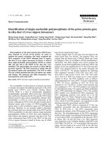
Fig. 4 shows the distribution of masseter-muscle activity
(%MVC) and percent activity of grinding, clenching and
tapping in each volunteer. A wide variation in masseter-
muscle activity (%MVC) was observed. Subjects with
higher muscle activity, such as volunteers 17 and 18,
tended to show a relatively high grinding activity, while
clenching and tapping activities were relatively low. In
contrast, subjects with lower muscle activity (%MVC),
such as volunteers 1 and 2, showed relatively high tapping
activity.
Comparisons of the duration of bruxism-events demon-
strated that individuals who had high muscle activity
(%MVC) also tended to show long event duration similar
to volunteers 18 and 19, whereas individuals with moder-
ate muscle activity (%MVC) showed relatively long event
duration such as volunteers 14 (Fig. 5).
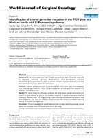
Fig. 6 shows the relationship between the masseter-mus-
cle activity (%MVC) and bruxism-event duration. The
majority of volunteers are plotted in the lower left quad-
rant, indicating that the muscle activity (%MVC) and
bruxism-event duration were not as high as the average
values, 55.1 ± 58.4 (%MVC) and 108.0 ± 90.4(sec/hour),
respectively. Seventy-nine percent of volunteers were
within one standard deviation, while the values of volun-
teers 14, 17, 18 and 19 were out of the average range.
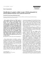
Characterization of different patterns of bruxism activitiesFigure 3
Characterization of different patterns of bruxism activities. Combined analysis of EMG and ACC showed that tapping
was a rhythmic muscle activity with Y-axis movement, clenching was strong muscle activity with no Y-axis movement, and
grinding was muscle activity with X and Y movement.
Table 1: Distribution of muscle activity (%MVC) in different
types of sleep bruxism
Muscle activity (%MVC)
Mean s.d. Min-Max (%)
Grinding 32.8 37.7 3.3 – 115 (59.5)
Clenching 19.7 23.4 0.3 – 106 (35.6)
Tapping 2.6 3.4 0.3 – 15.5 (4.9)
Total 55.1 58.4 (100)
Head & Face Medicine 2009, 5:7 />Page 6 of 10
(page number not for citation purposes)
Discussion
The definition of bruxism has evolved to include different
behavioral mandibular movements such as grinding,
clenching, and tapping. In this study, we developed a new
analyzing system of bruxism and analyzed the behavior of
sleep bruxism in 19 volunteers. The new analyzing system
of bruxism has two major advantages. First, the combined
system of EMG and Acc provides clear and easy distinction
between real bruxism activity and other activities, such as
the noise from body movements. Second, ACC analysis
offers an effective and reliable way to differentiate the
grinding, clenching and tapping activities. ACC packaging
itself is very small and light (5 mm long, 5 mm wide, 2
mm thickness, under 1 g weight). Precise data can be gath-
ered naturally.
The combined analysis of EMG and ACC provided dis-
tinctive patterns: rhythmic muscle activity with Y-axis
movement as tapping type, strong muscle activity with no
Y-axis movement as clenching type, and muscle activity
with XY movement as grinding-type bruxism. The brux-
ism pattern in individuals during sleep varied widely with
a combination of different mandibular movements. We
still do not know how and when the different types of
bruxism occur. Some individuals showed higher EMG
activity than maximum voluntary clenching. This was also
unexpected and it is not clear why such strong activity
occurs.
Our study indicates that two types of bruxism were domi-
nant, grinding and clenching. There was tendency that
higher muscle activity was in grinding than that in clench-
ing, especially in volunteers who brux strongly, although
the length and events of clenching and grinding were not
significantly different.
The results show that individual muscle activity (%MVC)
had a wide distribution from 223.0 %MVC to 7.20 %MVC
(Fig. 4). It was also demonstrated that muscle activity pre-
dominantly consisted of grinding and clenching activities.
Tapping activity in bruxism was low relative to the grind-
ing and clenching activities. Although we were still unable
to fully define which level of bruxism activity can be con-
sidered as a diagnostic parameter to distinguish between
the normal range of bruxism activity and bruxer or non-
physiological activity, a normal range of bruxism activity
can be proposed in which the average masseter-muscle
activity (%MVC) and bruxism-event duration are 55.1 ±
58.5 (%MCV) and 108.0 ± 90.4 (sec/hr), respectively. Sev-
enty-nine percent of the volunteers were included within
these ranges.
Whereas the duration of tooth contact during parafunc-
tional activity is fleeting in nature, an average episode of
sleep bruxism may last as long as 4–5 seconds with the
average rate of both grinding and clenching activities
about 40 seconds per hour (Table 2). The more severe the
sleep bruxism, the longer the teeth stay in contact with rel-
atively high muscle activity (Fig. 6), resulting in larger sus-
tained forceful muscle contraction.
Conclusion
The innovative bruxism-analyzing system developed
using EMC and ACC easily differentiates the three differ-
ent bruxism patterns: grinding, clenching, and tapping.
Sleep bruxism activity predominantly consisted of clench-
ing and grinding, which varied between individuals. Sev-
enty-nine percent of the volunteers were included within
average ranges of 55.1 ± 58.4 (% MCV) and 108.0 ± 90.4
(sec/hr).
Competing interests
The authors declare that they have no competing interests.
Authors' contributions
HY collected the data from volunteers at the sleep labora-
tory and participated in the analysis of raw data of EMG,
EEG, and ACC. KS participated in the development of new
analyzing system of sleep bruxism using EMG and ACC.
KT participated in collecting the data from the sleep labo-
ratory together with HY and helped to construct research
design. SS participated in the design of the study and coor-
dinated the drafting of the manuscript. All authors have
read and approved the final manuscript.
Table 2: Distribution of event number, event length in different types of sleep bruxism
Event number (/hour) Event length (sec/hour)
Mean s.d. Min-Max (%) Mean s.d. Min-Max (%)
Grinding 6.5 3.4 1.8–15.5 (32.3) 61.3 45.3 17.0–160.0 (56.8)
Clenching 8.7 4.7 1.5–18.9 (43.3) 40.4 51.7 1.60–211.5 (37.4)
Tapping 4.9 3.6 0–10.4 (24.4) 6.3 3.5 1.2–12.7 (5.8)
Total 20.1 (100) 108.0 90.4 (100)
Head & Face Medicine 2009, 5:7 />Page 7 of 10
(page number not for citation purposes)
Distribution of muscle activity (%MVC) into the different patterns of bruxismFigure 4
Distribution of muscle activity (%MVC) into the different patterns of bruxism. Variation of muscle activity (%MVC)
in volunteers was observed. There was a tendency that subjects who had higher muscle activity showed relatively high grinding
activity and lower muscle activity (%MVC) subjects showed relatively high clenching or tapping activities.
1
2
3
4
5
6
7
8
9
10
11
12
13
14
15
16
17
18
19
Masseter muscle activity (%MVC)
Volunteer
Clenching Activity (%)
0 10 20 30 40 50 60 70 80 90
Grinding Activity (%)
0 10 20 30 40 50 60 70 80 90
Tapping Activity (%)
0 5 10 15 20 25 30 35
1
2
3
4
5
6
7
8
9
10
11
12
13
14
15
16
17
18
19
Volunteer
0 30 60 90 120 150 180 210 240
Head & Face Medicine 2009, 5:7 />Page 8 of 10
(page number not for citation purposes)
Distribution of bruxism event length into the different patterns of bruxismFigure 5
Distribution of bruxism event length into the different patterns of bruxism. There was a tendency for subjects who
had long bruxism event duration to show increasing grinding event duration and decreasing clenching and tapping event dura-
tions.
1
2
3
4
5
6
7
8
9
10
11
12
13
14
15
16
17
18
19
Bruxism Event Length (Sec.)
Volunteer
0 10 20 30 40 50 60 70 80 90
Grinding Event Length (%)
0 10 20 30 40 50 60 70 80
Clenching Event Length (%)
0 10 20 30
Tapping Event Length (%)
1
2
3
4
5
6
7
8
9
10
11
12
13
14
15
16
17
18
19
Volunteer
0 500 1000 1500 2000 2500 3000
Head & Face Medicine 2009, 5:7 />Page 9 of 10
(page number not for citation purposes)
Consent
Written informed consent was obtained from our volun-
teers for publication of this clinical report and the accom-
panying images. A copy of the written consent is available
for review by the Editor-in-Chief of this journal.
Acknowledgements
This work was performed at the Research Institute of Occlusion Medicine
and Research Center of Brain and Oral Science, Kanagawa Dental College
and supported by a grant-in-aid for Open Research from the Ministry of
Education, Culture, Sports, Science and Technology-Japan.
References
1. Kato T, Thie N, Montplaisir J, Lavigne G: Bruxism and orofacial
movement disorder. Dent Clin North Am 2001, 45:657-684.
2. Muhlemann H: Ten years tooth mobility measurements. J Per-
iodontol 1960, 31:110-122.
3. Persson R: Assessment of tooth mobility using small loads II.
Effect of oral hygiene procedures. J Clin Periodont 1980,
7:506-515.
4. Arnold M: Bruxism and the occlusion. Dental Clin North Am 1981,
25:395-407.
5. Molina OF, Dos Santos J: The prevalence of some joint disor-
ders in craniomandibular disorder (CMD) and bruxers as
compared to CMD non bruxer patients and controls. J Crani-
omand Pract 1999, 17:17-29.
6. Bream M, Lambrechts P, Vanherle G: Stress-induced cervical
lesion. J Prosthet Dent 1992, 67:718-722.
7. Coleman T, Grippo J, Kinderknecht K: Cervical dentin hypersen-
sitivity. Part II: Associations with abfractive lesions. Quintes-
sence Int 2000, 31:466-465.
8. McCoy G: Dental compression syndrome: a new look at an
old disease. J Oral Implantol 1999, 25:35-49.
9. Onodera K, Kawagoe T, Protacio-Quismundo C, Sasaguri K, Sato S:
The use of a BruxChecker in the evaluation of different
occlusal schemes based on individual grinding patterns. J
Craniomand Pract 2006, 24:292-299.
10. Lavigne GJ, Rompre PH, Montplaisir JY: Sleep bruxism: validity of
clinical research diagnostic criteria in a controlled polysom-
nographic study. J Dent Res 1996, 75:546-552.
11. Takeuchi H, Kurahashi TA: Piezoelectric film-based intrasplint
detection method for bruxism. J Prosthet Dent 2001, 86:195-202.
12. Reding GR, Zepelin H, Robinson JE, Robinson JRJ, Zimmerman S,
Smith V: Nocturnal teeth-grinding: all-night psychophysiogic
studies. J Dent Res 1968, 47:786-797.
13. Fuchs P: The muscular activity of the chewing apparatus dur-
ing night sleep. An examination of healthy subjects and
patients with functional disturbances. J Oral Rehabil 1975,
2:35-48.
14. Kobayashi Y, Takeda Y, Ishihara H: The influence of experimental
occlusal interference on psychoendocrine responses. J Dent
Res 1985, 63(Special Issue):746.
15. Ware JC, Rugh JD: Destructive bruxism: sleep stage relation-
ship. Sleep 1988, 11:172-181.
Relationship between the muscle activity (%MVC) and the bruxism length (sec/hour) durationFigure 6
Relationship between the muscle activity (%MVC) and the bruxism length (sec/hour) duration. Majority of the
volunteers were displayed in the lower left quadrant which means that muscle activity (%MVC) and bruxism event duration
were not as high as in the volunteers.
Bruxism Length (sec / hr)
Masseter muscle activity (%MVC)
0
50
100
150
200
250
0 50 100 150 200 250 300
Vol#17
Vol#18
Vol#19
108.0±90.4
55.1±58.4
Publish with Bio Med Central and every
scientist can read your work free of charge
"BioMed Central will be the most significant development for
disseminating the results of biomedical research in our lifetime."
Sir Paul Nurse, Cancer Research UK
Your research papers will be:
available free of charge to the entire biomedical community
peer reviewed and published immediately upon acceptance
cited in PubMed and archived on PubMed Central
yours — you keep the copyright
Submit your manuscript here:
/>BioMedcentral
Head & Face Medicine 2009, 5:7 />Page 10 of 10
(page number not for citation purposes)
16. Sjoholm TT, Polo OJ, Alihanka JM: Sleep movements in teeth
grinders. J Craniomand. Disord 1992, 6:184-191.
17. Velly-Miguel AM, Montplasir J, Rompre PH, Lund JP, Lavigne GJ:
Bruxism and other orofacial movements during sleep. J Crani-
omand Disord Facial Oral Pain 1992, 6:71-81.
18. Rugh JD, Solberg WK: Electromyographic studies of bruxist
behavior before and during treatment. J Oral Rehabil 1975,
2:215-223.
19. Solbelg WK, Clark GT, Rugh JD: Nocturnal electromyographic
evaluation of bruxism patients under going short term splint
therapy. J Oral Rehabil 1975, 2:215-223.
20. Pierce CJ, Gale EN: A comparison of different treatments for
nocturnal bruxism. J Dent Res 1988, 67:597-601.
21. Ikeda T, Nishigawa K, Kondo K, et al.: Criteria for the detection
of sleep-associated bruxism in humans. J Orofac Pain 1996,
10:270-282.
22. Holgren K, Sheikholeslam A, Riise C: Effect of a full-arch maxil-
lary occlusal splint on parafunctional activity during sleep in
patients with nocturnal bruxism and signs and symptoms of
craniomandibular disorder. J Prosthet Dent 1993, 69:293-297.
23. Pierce CJ, Gale EN: Methodological considerations concerning
the use of bruxcore plates to evaluate nocturnal bruxism. J
Dent Res 1989, 68:1110-1114.
24. Takeuchi H, Ikeda T, Clark GT: Development of new detecting
system for bruxism. J Dent Res 1996, 75:341.