báo cáo khoa học: " Oral bisphosphonate-related osteonecrosis of the jaws in rheumatoid arthritis patients: a critical discussion and two case reports" potx
Bạn đang xem bản rút gọn của tài liệu. Xem và tải ngay bản đầy đủ của tài liệu tại đây (2.62 MB, 7 trang )
CAS E REP O R T Open Access
Oral bisphosphonate-related osteonecrosis of the
jaws in rheumatoid arthritis patients: a critical
discussion and two case reports
Nicolau Conte-Neto
1*†
, Alliny S Bastos
1†
, Luis C Spolidorio
2†
, Rosemary AC Marcantonio
1†
and
Elcio Marcantonio Jr
1†
Abstract
Background: Bisphosphonate-related osteonecrosis of the jaw (BRONJ) is a clinical condition characterized by the
presence of exposed bone in the maxillofacial region. Its pathogenesis is still undetermined, but may be associated
with risk factors such as rheumatoid arthritis (RA). The aim of this paper is to report two unpublished cases of
BRONJ in patients with RA and to conduct a literature review of similar clinical cases with a view to describe the
main issues concerning these patients, including demographic characteristics and therapeutic approaches applied.
Methods: Two case reports of BRONJ involving RA patients were discussed
Results: Both patients were aging female taking alendronate for more than 3 years. Lesions were detected in
stage II in posterior mandible with no clear trigger agent. The treatment applied consisted of antibiotics, oral rinses
with chlorhexidine, drug discontinuation and surgical procedures. Complete healing of the lesions was achieved.
Conclusions: This paper brings to light the necessity for rheumatologists to be aware of the potential risk to their
patients of developing BRONJ and to work together with dentists for the prevention and early detection of the
lesions. Although some features seem to link RA with oral BRONJ and act as synergistic effects, more studies
should be developed to support the scientific bases for this hypothesis.
Background
Bisphosphonates (BPs) areaclassofdrugscommonly
prescribed for bone diseases due to their osteoclast i nhi-
bition property. This class of drugs has been widely used
for osteoporosis and corticosteroid-induced osteoporosis
in patients with rheumatoid arthritis (RA). However,
reports of bone n ecrosis induced by bisphosphonates
(BRONJ) have generated great concern regarding the side
effects of these drugs. Although RA has been considered
a risk factor fo r this kind of osteonecrosis [1,2] , the rela-
tionship between these diseases has not, until now, been
completely elucidated.
The aim of this paper is to report two unpublished
cases of B RONJ in non-neoplastic patients with RA and
to conduct a literature review of similar clinical cases
with a view to describing the main issues related to
these patients, including demographic characteristics
and therapeutic approaches.
Case 1
A 58-year-old woman presented herself at a private dental
clinic in Decembe r, 2008, complaining about an intense
spontaneous pain in the mandibular right side after a pros-
thesis replacement in an implant area that was installed
sixteen years previously. The review of the patient’s medi-
cal history revealed that she started a therapy with Fosa-
max
®
(alendronate sodium) 70 mg, once a week for the
treatment of rheumatoid arthritis in 2004. The patient had
no history of smoking, radiotherapy, infectious process or
trauma in the maxillo-facial region, and the dental implant
presented normally until the symptoms began.
Upon clinical examination, a mild erythema was evi-
dent in the mucosa surrounding the di stally right dental
* Correspondence:
† Contribu ted equally
1
UNESP - Univ. Estadual Paulista, School of Dentistry, Department of
Diagnosis and Surgery, Division of Periodontology, Rua Humaitá, 1680,
14801-903 Araraquara, SP/Brazil
Full list of author information is available at the end of the article
Conte-Neto et al. Head & Face Medicine 2011, 7:7
/>HEAD & FACE MEDICINE
© 2011 Conte-Neto et al ; licensee BioMed Central Ltd. This is an Open Access article distributed under the terms of the Creative
Commons Attribution License ( which permits unrestricted use, distribution, and
reproduction in any medium, provided the original work is prope rly cited.
implant, without clinical evidence of purulent discharge,
gingival recession or bone exposure. However, probing
revealed increasing depth values and detachment of the
mucosa from the periimplantar bone with biological seal
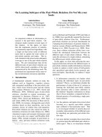
loss was observed (Figure 1). A computed tomography
(CT) was requested and showed a substantial radiolu-
cency around th e involved dental implant, featuring loss
of the crestal bone. (Figure 2)
Periimplantitis was the primary hypothesis considered
at that time, but BRONJ was also considered. The initial
treatment plan was mouth-rinsing with chlorhexidine
0.12% four times a day and antibiotic therapy with Clin-
damycin 300 mg twice a day for 10 days, since the
patient had allergy for b-lactam antibiotics. Surgical
decontamination of the implant surface was also
planned; however, upon mucosal flap incision, there was
no indication of any exposition of implant threads, but
there was a large zone of necrotic bone forming a
sequestrum area (Figure 3a). Therefore, it was opted to
removal of the implant with sequestrectomy and debri-
dement (Figure 3b) until a bleeding bone was observed
(Figure 3c). An interrupted suture was made with 4-0
silk in an attempt to close the wound primarily without
tension. After medical consensus, alendronate was
suspended.
The bone specimen obtained was fixed, processed and
paraffin embedded for histologica l analysis. Hematoxylin
and eosin (H&E) staining was used for histological
observation by light microscopy. The results revealed
necrotic lamellar bone fragments with chronic and acute
inflammatory cells, as well bacterial colonies (Figure 4).
In addition, the serum C-terminal cross-linking telo-
peptide of collagen (CTX) test to evaluate the bone
reabsorption status was solicited and revealed normal
values (250 pg/mL), but this exam was performed only
4 months after surgical treatment. The healing
progressed uneventfully and the patient displayed no
symptoms at 8 months of postoperative time.
Observations i n the clinical examination showed that
the soft tissue was with normal aspect and without any
signs of inflammatory or infectious processes (Figure 5)
Case 2
A 68-year-old woman was admitted to a private dental
clinic in October 2009, complaining about cold tooth
sensation in the region of left mandibular second pre-
molar. Review of the patient’s m edical history revealed
that since 2003, she had been taking 2.5 mg of metho-
trexate six times a week and 70 m g of Fosamax
®
(alen-
dronate sodium) once a week for the treatment of
rheumatoid arthritis. Besides, she also reported steroids
use during twenty years. The patient had no history of
radiotherapy, infectious process or trauma in the maxil-
lofacial region but did have a history of smoking.
During clinical examination a detachment of the
marginal gingival (Figure 6a) associated with an
increased probing depth value at the region of left man-
dibular second premolar (Figure 6b) was observed and
was associated with a mild mobi lity without painful
symptoms, purulent discharge and bone exposure. On
Figure 1 Clinical aspect of the BRONJ lesion. Mucosal erythema
surrounding the distally right implant associated with an increase
on probing depth values with no gingival recession or bone
exposure.
Figure 2 Imaging aspect of the BRONJ lesion. Computed tomography showing a radiolucency with aspect of loss of crestal bone around the
right distally implant.
Conte-Neto et al. Head & Face Medicine 2011, 7:7
/>Page 2 of 7
periapical radiographic analysis, t here was bone loss
associated with ost eosclerosis around the involved tooth
(Figure 7a). At that time, mouth-r insing with chlorhexi-
dine 0.12% was prescribed and, af ter medical consensus,
alendronate suspension was recommended. Further-
more, the serum C-terminal cross-linking telopeptide of
collagen (CTX) test was solicited to evaluate the bone
reabsorption status which revealed values of 33 pg/mL.
Two weeks later, during clinical examination, bone
exposure was detected on the vestibular side of the left
mandibular second premolar and on the disto-lingual
side of the edentulous alveolar bone surrounded by
inflamed soft tissue without evidence of purulent dis-
charge or pain symptoms (Figure 8). However, the lesions
progressed very quickly and, the patient complained of
painful symptoms and increased tooth mobility few days
later. Bone necrosis as sociated with mucosa ulceration
involving part of the jugal mucosa was also observed
(Figure 9a). On periapical radiographic analysis, it was
observed increased bone loss around the involved tooth
(Figure 7b) which was confirmed on computed tomogra-
phy (CT) since an osteolysis area was observe d around
the left mandibular second premolar associated with an
intense bone sclerosis (Figure 10). Given these observa-
tions, a diagnosis of BRONJ could be established.
The management of the case included the tooth
extraction and bone debridement under local anesthesia
(Figure 9b and 9c), and mouth ri nses with chlorhexidine
plus antibiotic therapy with Clavulin 500 mg three times a
day was prescribed. Within fourteen days, the formation
of granu lation tissue co uld be noted on the surgical area
with no signs of inflammation or infection (Figure 11a).
After two months, the debrided region was covered by
normal mucosa with no painful symptoms (Figure 11b).
Discussion
Rheumatoid arthritis is a systemic autoimmune disease
characterized by progressive joint destruction and a vari-
ety of systemic manifestations resulting from chronic
inflammation [3], which has been considered a risk fac-
tor for the development of BRONJ [1,2]. Although no
scientific link has been established between BRONJ and
RA, some relevant factors that could link these diseases
should be discussed. These factors include inflammatory
alterations and drug s prescribed for these patients,
including steroids and immunosuppressive agents, such
as methotrexate [4], that seem to play a relevant role in
the development of oral BRONJ.
The relevance of steroids and methotre xate in BRONJ
pathogenesis still remains not fully understood. How-
ever, considering that the main disease theories are
based on the suppression of bone remodeling, the angio-
genesis-inhibitory properties of t he bisphosphonate and
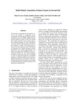
Figure 3 Surgical approach of the BRONJ lesion. A) Surgical exposition of the distally right implant showing a large bone sequestrum around
the dental implant; B) Sequestrectomy of the bone necrosis around the dental implant; C) Surgical area after the debridement showing a bone
bleeding surface associated with the dental implant removal.
Figure 4 Histological aspects of bone samples. A) H & E stained
section showing bone necrosis (Original magnification × 40); B)
Gram stained section showing gram negative and positive bacteria
(Original magnification × 100)
Figure 5 Clinic al asp ects of the BRONJ lesions after treatment.
Post operatory of 9 month showing a mucosa with normal aspect
without signals of inflammatory process or bone exposure
Conte-Neto et al. Head & Face Medicine 2011, 7:7
/>Page 3 of 7
the infectious process [5] are facto rs that could be
related to BRONJ; howev er, none of these theories have
been completely accepted.
Hypothetical factors linked with BRONJ include a pos-
sible excessive suppression of bone turnover and jaw
angiogenesis resulting from the association between
bisphosphonates and steroids, s ince these d rugs also
reduce bone remodeling [6] and angiogenesis [7]. In
addition, the immunosuppressive effects of steroids and
methotrexate [8] could leave these patients more prone
to infections.
In this discussion, observations that s upport and at the
same ti me argue against this hypothetical association are
made, especially in relation to steroid treatment. First of
all, although a large number of patients with RA that
develop oral BRONJ have a history of steroids and meth-
otrexate intake [4,9-12] (as in case 2), this disease also
occurs among patients with R A without the use of these
drugs [9,13,14] (as in case 1). Second, it is well known
that steroids can induce bone nec rosis, but this necrosis
differs from BRONJ because the steroids affect predomi-
nantly long bone s and almost never produce bone expo-
sure [15]. Finally, animal models of BRONJ have been
proposed to test the association of bisphosphonate and
steroids [16].
Recent tendencies included BPs among the most
frequently prescribed drugs in rheumatologic practice
[17] especially due to the high efficiency of BPs to be a
protection against generalized bone loss [18]. In this way,
patients with RA have been taking BPs to the pr evention
and treatment of osteoporosis which is a common feature
in RA for several reasons including: post-menopausal
women are the main risk group for RA and are at risk for
accentuated bone loss; steroid therapy is often prescribed
for the treatment of RA; physical inactivity is characteris-
tic of RA due to disease activity; and bone loss due to dis-
ease inflammatory mechanisms, such as systemic elevated
cytokines [19]. For these reasons, it is reasonable to
believe that the incidence of BRONJ will increase as a
result of the long-term use of BPs.
Regarding the link between inflammation and BRONJ, it
is well known that ext raarticular structures a lso can be
affected by the inflammatory process in RA [20]. Consid-
ering that this disease is char acteriz ed by pers istent high
levels of proinflammatory cytokines [21] and accumulation
of inflammatory cells [20], a link factor can be hypothe-
sized based on the observations made by Lesclous et al.
[22], who stated that BRONJ is associated with inflamma-
tion an d that the clinica l extension of the lesions is asso-
ciated with the number of inflammatory cells.
According to t he cases reported in literature, patients
with RA who develop BRONJ lesions aft er oral adminis-
tration of BPs are usually women, above 60 years old,
who have taken alendronate for more than 3 years. The
mandible is the most common site of BRONJ in these
patients. The cases reported here are in agreement with
this profile, except that the patient described in case 1 is
younger than 60 years old. Pazianas et al. [23] have
made the interesting observation that these features
have exactly the same charact eristic s for patient s with-
out RA that develop oral BRONJ.
Most of the oral BRONJ cases in patients with or
without RA a re triggered by invasive dental procedures,
such as extractions and dental implants. However, other
Figure 6 Initial clinical aspects of the BRONJ lesion. A)
Detachment of the marginal gingival at the vestibular and distal
side of # 35; B) Probing in the vestibular side of #35 showing
increased probing depth values.
Figure 7 Radiographic progressio n of bone loss in the BRON J
lesion. A) Periapical radiographic showing bone loss associated
with osteosclerosis around the #35; B) Periapical radiography
showing increased bone loss around the #35.
Figure 8 Clinical progression of the BRONJ lesions. A) Bone
exposure of the #35 on the vestibular side; B) Bone exposure on
the disto-lingual side of the edentulous alveolar bone surrounded
by inflamed soft tissue.
Conte-Neto et al. Head & Face Medicine 2011, 7:7
/>Page 4 of 7
cases of BRON J can be spontaneo us [1,10,12] as seen in
the cases reported in the present paper. However, some
concerns should be disc ussed. In case 1, although no
apparent precipitant factor was present, trauma may
have been a trigger event [24]. An eventual occlusal
overload on the prosthesis m ight have contributed to
BRONJ, because pain symptoms appeared soon after the
prosthesis replacement.
Another relevant factor is seen in case 2. Although
there w as no previous dentistry procedure, the patient
had periodontal disease. Periodontal disease has been
considered by some authors to be a trigger event [25]
due to the fact that this disease could increase the
potential quantity of BPs released. However, this theory
is still controversial [26]. An interesting observation is
that individuals with rheumatoid arthritis are more likely
to experience moderate to severe periodontal disease
compared to their healthy counterparts [27]. This clini-
cal association between the two diseases might be due
to a common underlying pathobiology of periodontitis
and rheumatoid arthritis [28].
The main clinical aspects of patients with RA who
develop oral BRONJ include bone exposure, edema, pain
and purulent discharge [9 -11,13,29,30]. These feature s
represent stage 2, as described by Ruggiero et al. [26], and
indicate the lack of early at tention to these patients in
initial stages because these stages include nonspecific
signals and symptoms in the oral cavity with no clinical
evidence of bone exposure. In case 2, lesions progressed
rapidly generating a great concern since in advanced
stages of BRONJ lesions, paresthesia, fistula formation and
pathologic fracture can also be present [9], although these
features are more common in neoplasic patients [29].
According R uggiero et al. [26], one of the diagnosis
criteria of BRONJ is the presence of exposed bone in
the maxillofacial region persisting for more than 8
weeks. Although most patients with RA have some kind
of bone exposure, this BRONJ definition has been
revised, due to some contrary observations. First, even
advanced cases can also occur with no bone exposure in
oral cavity [1]. Second, there is a lack of knowledge
about early clinical features and their progression
toward frank BRONJ [9]. This is well-illustrated in case
2, which shows the complete evolution of a BRONJ
lesion in which it was possible to identify an early soft
tissue necrosis and increased probing depth values that
Figure 9 Imaging aspect of the BRONJ lesion. A) Computed tomography showing an irregular radiolucency at the left side of the mandible
and a persistent alveolus of a molar that was extracted at least 10 years previously; B) Osteolysis around the left mandibular second premolar.
Figure 10 Clinical progression of the BRONJ lesions. A) Increasing of the bone necrosis around the #35 associated with a mucosal ulceration
involving part of the jugal mucosa; B) Exposed bone area after the #35 extraction; C) Surgical area after bone debridement
Conte-Neto et al. Head & Face Medicine 2011, 7:7
/>Page 5 of 7
progressed to exposed bone area. Another concern
about this case is that the distinction of early stages of
BRONJ from other diagnoses, such as localized reacuti-
zation of chronic periodontitis, may be difficult [13].
The appropriate management of patients with BRONJ
remains undefined and no widely accepted treatment
protocol exists. Although it has been stated th at surgical
procedures may achieve better outcomes in non-neo-
plastic patients [29], Marx et al. [25] state that surgical
procedures are not effective on patients with BRONJ
and that these procedures lead to further exposed bone,
worsening of the symptoms and a greater risk of patho -
logic fracture. These effects of surgery indicate long-
term an tibiotics and chlorhexidine 0 .12% as treatment.
Theliteraturehasshownthattreatmentofthelesions
in patients with RA using this approach along with the
discontinuation of the RA drugs have mostly positive
outcomes, including the complete healing of the lesion s
[10,12 ,14]. In contrast, surgical therapy literature shows
more divided outcomes, including b oth p ositive [1,30]
and poor outcomes [9,4,24]. In the cases reporte d in
this paper, surgical therapy was chosen, and excellent
outcomes were achieved.
The assessment of the risk of BRONJ for patients tak-
ing BPs is a challenge. Marx et al. (2007) report use of C-
terminal cross-linking telopeptide of type I collagen
(CTX) test as an indicator of the risk of BRONJ, suggest-
ing that values of less than 100 pg/mL represent a high
risk and more than 150 pg/mL a low risk. In this report
were found both normal values for CTX test (250 pg/mL
in case 1) as abnormal values (33 pg/mL in case 2). How-
ever, the patient CTX test in case 2 would be normal if
the scale purposed by Lehrer et al. (2008) is considered
where values ranging 32 from 580 pg/ml are considered
to be normal. Moreover, normal serum bone m arkers
also can be found in patients with BRONJ still using BPs
[31].Otherrelevantpointisthatpatient1justdidthe
exam 4 month after the drug suspension and after surgi-
cal treatment, which ma y contributed for this normal
values, as after the drug interruption there is a gradually
improvement in the values of CTX test [10,31].
We acknowledge that a limitation of the present paper
is the fact that it presents two BRONJ clinical cases in
RA patients. Therefore, we cannot validate any hypoth-
esis that could explain a definite association of synergis-
tic actions of both RA and BRONJ. More studies should
be developed with rigorous case ascertainment criteria,
as we ll as appropriate documentation of risk factors and
modifiers to support scientific bases for this hypothesis.
However, the present paper helps to highlight the
need for a change in clinical practice or diagnostic/prog-
nostic approaches related t o BRONJ. Considering that
BPs are among the most frequently prescribed drugs in
rheumatologic practice [17], associated with the lack of
knowledge about this disease among rheumatologists in
many countries, it is reasonable to expect an increased
tendency in the number of BRONJ reports involving RA
patients. This fact shows the clear necessity for the
improvement in the epidemiological vigilance systems of
Public Health Entities, a s well as a better coordination
of safety-related pharmacovigilance initiatives.
Conclusions
Although some features seem to link RA with oral
BRONJ and act as synergistic effects, more studies
should be developed to support the scientific bases for
this hypot hesis. In addition, most pat ients with RA and
oral BRONJ are diagnosed in stage 2, which indicates
the necessity for rheumatologists to be aw are of the
potential risk to their patients of developing BRONJ and
to work together with dentists for the prevention and
early detection of the lesions.
Consent
Written informed consent was obtained from the
patients for publication of these case reports and any
accompanying images. A copy of the written consent
form is available for review by the Editor-in-Chief of
this journal.
Author details
1
UNESP - Univ. Estadual Paulista, School of Dentistry, Department of
Diagnosis and Surgery, Division of Periodontology, Rua Humaitá, 1680,
14801-903 Araraquara, SP/Brazil.
2
UNESP - Univ. Estadual Paulista, School of
Dentistry, Department of Physiology and, Pathology, Division of Pathology,
Rua Humaitá, 1680, 14801-903 Araraquara, SP/Brazil.
Authors’ contributions
NCN performed one surgery under the supervison of the corresponding
author, analyzed the records, reviewed all patients’ data and designed the
case report. ASB drafted the manuscript and helped in writing the text. LCS
and RACM drafted the manuscript and reviewed it critically. EMJ performed
one of the surgical procedures and reviewed the manuscript. All authors
read and approved the final manuscript.
Figure 11 Clinical aspects of lesions after 2 months of
treatment. A) Fourteen days after surgical debridement showing
the formation of granulation tissue on the surgical area; B) Two
months after surgical debridement showing a normal mucosa
coverage of the involved area.
Conte-Neto et al. Head & Face Medicine 2011, 7:7
/>Page 6 of 7
Authors’ Information
NCN is a PhD student from Implantology program at Araraquara School of
Dentistry and ASB is a PhD student from Periodontology program at
Araraquara School of Dentistry. LCS is a professor and the chairman of the
Department of Physiology and Pathology, Division of Pathology at
Araraquara School of Dentistry. EMJ and RACM are professors and chairmen
of the Department of Diagnosis and Surgery, Division of Periodontology at
Araraquara School of Dentistry.
Competing interests
The authors declare that they have no competing interests.
Received: 27 January 2011 Accepted: 27 April 2011
Published: 27 April 2011
References
1. Park W, Kim NK, Kim MY, Rhee YM, Kim HJ: Osteonecrosis of the jaw
induced by oral administration of bisphosphonates in Asian population:
five cases. Osteoporos Int 2010, 21:527-33.
2. Malden N, Beltes C, Lopes V: Dental extractions and bisphosphonates: the
assessment, consent and management, a proposed algorithm. Br Dent J
2009, 206:93-8.
3. Alamanos Y, Drosos AA: Epidemiology of adult rheumatoid arthritis.
Autoimmun Rev 2005, 4:130-6.
4. Santos C, Alegre C: Osteonecrosis maxilar, bifosfonatos y artritis
reumatoide. Med Clin 2008, 130:37.
5. Allen MR, Burr DB: The pathogenesis of bisphosphonate-related
osteonecrosis of the jaw: so many hypotheses, so few data. J Oral
Maxillofac Surg 2009, 67:61-70.
6. Patschan D, Loddenkemper K, Buttgereit F: Molecular mechanisms of
glucocorticoid induced osteoporosis. Bone 2001, 29 :498-505.
7. Greenberger S, Boscolo E, Adini I, Mulliken JB, Bischoff J: Corticosteroid
suppression of VEGF-A in infantile hemangioma-derived stem cells. N
Engl J Med 2010, 362:1005-13.
8. Jain A, Witbreuk M, Ball C, Nanchahal J: Influence of steroids and
methotrexate on wound complications after elective rheumatoid hand
and wrist surgery. J Hand Surg Am 2002, 27:449-55.
9. Yarom N, Yahalom R, Shoshani Y, Hamed W, Regev E, Elad S: Osteonecrosis
of the jaw induced by orally administered bisphosphonates: incidence,
clinical features, predisposing factors and treatment outcome.
Osteoporos Int 2007, 18:1363-70.
10. Marx RE, Cillo JE Jr, Ulloa JJ: Oral bisphosphonate-induced osteonecrosis:
risk factors, prediction of risk using serum CTX testing, prevention, and
treatment. J Oral Maxillofac Surg 2007, 65:2397-410.
11. Barros SY: Is your knowledge up-to-date? Bisphosphonate-related
osteonecrosis of the jaw. Int J Dent Hyg 2008, 6 :376-7.
12. Junquera L, Gallego L, Cuesta P, Pelaz A, de Vicente JC: Clinical
experiences with bisphosphonate-associated osteonecrosis of the jaws:
analysis of 21 cases. Am J Otolaryngol 2009, 30:390-5.
13. Elad S, Gomori MJ, Ben-Ami N, Friedlander-Barenboim S, Regev E, et al:
Bisphosphonate-related osteonecrosis of the jaw: clinical correlations
with computerized tomography presentation. Clin Oral Investig 2010,
14:43-50.
14. Lo JC, O’Ryan FS, Gordon NP, Yang J, Hui RL, et al: Prevalence of
osteonecrosis of the jaw in patients with oral bisphosphonate exposure.
J Oral Maxillofac Surg 2010, 68:243-53.
15. Zigic TM, Marcous C, Hungerford DS: Corticosteroid therapy associated
with ischemic necrosis of bone in systemic lupus erythematosis. Am J
Med 1985, 79:596-604.
16. Sonis ST, Watkins BA, Lyng GD, Lerman MA, Anderson KC: Bone changes in
the jaws of rats treated with zoledronic acid and dexamethasone before
dental extractions mimic bisphosphonate-related osteonecrosis in
cancer patients. Oral Oncol 2009, 45:164-72.
17. Yip RML: Bisphosphonates and Osteonecrosis of the Jaw. Hong Kong Bull
Rheum Dis 2008, 8:19-25.
18. Breuil V, Euller-Ziegler L: Bisphosphonate therapy in rheumatoid arthritis.
Joint Bone Spine 2006, 73:349-54.
19. Joffe I, Epstein S: Osteoporosis associated with rheumatoid arthritis:
Pathogenesis and management. Seminars in Arthritis and Rheumatism
1991, 20:256-272.
20. Bartold PM, Marshall RI, Haynes DR: Periodontitis and rheumatoid arthritis:
a review. J Periodontol 2005, 76:2066-74.
21. Snyderman R, McCarty GA: Analogous mechanisms of tissue destruction
in rheumatoid arthritis and periodontal disease. In Host-Parasite
Interaction in Periodontal Disease. Volume 1 1 edition. Edited by: Genco RJ,
Mergenhagen SE. Washington, DC: American Society for Microbiology;
1982:354-362.
22. Lesclous P, Abi Najm S, Carrel JP, Baroukh B, Lombardi T, et al:
Bisphosphonate associated osteonecrosis of the jaw: a key role of
inflammation? Bone 2009, 45:843-52.
23. Pazianas M, Miller P, Blumentals WA, Bernal M, Kothawala P: A review of
the literature on osteonecrosis of the jaw in patients with osteoporosis
treated with oral bisphosphonates: prevalence, risk factors, and clinical
characteristics. Clin Ther 2007, 29:1548-58.
24. Sedghizadeh PP, Stanley K, Caligiuri M, Hofkes S, Lowry B, Shuler CF: Oral
bisphosphonate use and the prevalence of osteonecrosis of the jaw: an
institutional inquiry. J Am Dent Assoc 2009, 140:61-6.
25. Marx RE, Sawatari Y, Fortin M, Broumand V: Bisphosphonate-induced
exposed bone (osteonecrosis/osteopetrosis) of the jaws: risk factors,
recognition, prevention, and treatment. J Oral Maxillofac Surg 2005,
63:1567-75.
26. Ruggiero SL, Dodson TB, Assael LA, Landesberg R, Marx RE, Mehrotra B:
American Association of Oral and Maxillofacial Surgeons. J Oral Maxillofac
Surg 2009, 67:2-12.
27. Mercado F, Marshall RI, Klestov AC, Bartold PM: Is there a relationship
between rheumatoid arthritis and periodontal disease? J Clin Periodontol
2000, 27:267-72.
28. Modi DK, Chopra VS, Bhau U: Rheumatoid arthritis and periodontitis:
biological links and the emergence of dual purpose therapies. Indian J
Dent Res 2009, 20:86-90.
29. Favia G, Pilolli GP, Maiorano E: Osteonecrosis of the jaw correlated to
bisphosphonate therapy in non-oncologic patients: clinicopathological
features of 24 patients. J Rheumatol 2009, 36:2780-7.
30. Malden NJ, Pai AY: Oral bisphosphonate associated osteonecrosis of the
jaws: three case reports. Br Dent J 2007, 203:93-7.
31. Kunchur R, Need A, Hughes T, Goss A: Clinical investigation of C-terminal
cross linking telopeptide test in prevention and management of
bisphosphonate-associated osteonecrosis of the jaws. J Oral Maxillofac
Surg 2009, 67:1167-73.
doi:10.1186/1746-160X-7-7
Cite this article as: Conte-Neto et al.: Oral bisphosphonate-related
osteonecrosis of the jaws in rheumatoid arthritis patients: a critical
discussion and two case reports. Head & Face Medicine 2011 7:7.
Submit your next manuscript to BioMed Central
and take full advantage of:
• Convenient online submission
• Thorough peer review
• No space constraints or color figure charges
• Immediate publication on acceptance
• Inclusion in PubMed, CAS, Scopus and Google Scholar
• Research which is freely available for redistribution
Submit your manuscript at
www.biomedcentral.com/submit
Conte-Neto et al. Head & Face Medicine 2011, 7:7
/>Page 7 of 7