báo cáo khoa học: " Effects of enamel matrix derivative and transforming growth factor-b1 on human osteoblastic cells" pot
Bạn đang xem bản rút gọn của tài liệu. Xem và tải ngay bản đầy đủ của tài liệu tại đây (2.84 MB, 9 trang )
RESEARC H Open Access
Effects of enamel matrix derivative and
transforming growth factor-b1 on human
osteoblastic cells
Daniela B Palioto
1*
, Thaisângela L Rodrigues
1
, Julie T Marchesan
1
, Márcio M Beloti
2
, Paulo T de Oliveira
2
and
Adalberto L Rosa
1
Abstract
Background: Extracellular matrix proteins are key factors that influence the regenerative capacity of tissues. The
objective of the present study was to evaluate the effects of enamel matrix derivative (EMD), TGF-b1, and the
combination of both factors (EMD+TGF-b1) on human osteoblastic cell cultures.
Methods: Cells were obtained from alveolar bone of three adult patients using enzymatic digestion. Effects of
EMD, TGF-b1, or a combination of both were analyzed on cell proliferation, bone sialoprotein (BSP), osteopontin
(OPN) and alkaline phosphatase (ALP) immunodetection, total protein synthesis, ALP activity and bone-like nodule
formation.
Results: All treatments significantly increased cell proliferation compared to the control group at 24 h and 4 days.
At day 7, EMD group showed higher cell proliferation compared to TGF-b1, EMD + TGF-b1 and the control group.
OPN was detected in the majority of the cells for all groups, whereas fluorescence intensities for ALP labeling wer e
greater in the control than in treated groups; BSP was not detected in all groups. All treatments decreased ALP
levels at 7 and 14 days and bone-like nod ule formation at 21 days compared to the control group.
Conclusions: The exposure of human osteoblastic cells to EMD, TGF-b1 and the combination of factors in vitro
supports the development of a less differentiated phenotype, with enhanced proliferative activity and total cell
number, and reduced ALP activity levels and matrix mineralization.
Introduction
Periodontal regeneration is a complex series of cell and
tissue events that include cell adhesion, migration, and
extracellular matrix (ECM) protein synthesis and secre-
tion. Phenotypic expression depends on cell interactions
with ECM proteins, which regulate cell signaling events
and ultimately gene expression[1]. The ECM prot eins
are, therefore, key factors that influence the regenerative
capacity[2]. However, to date, it remains undefined
which factors would determine the maximum rege nera-
tive capacity.
Enamel matrix derivative (EMD) has been used in var-
ious clinical applications aiming to promote periodontal
tissue regeneration. The rationale for such application is
based on the expression of enamel matrix protei ns dur-
ing the initial phases of root formation, which has been
associated with cementoblast differenti ation[3,4]. In
addition, the use of EMD in various experimental and
clinical protocols has been demonstrated to positively
affect not only new cementum formation but also bone
regeneration[5-8]. However, some controversial results
in terms of new bone formation has also been desc ribed
in the literature[9].
Despite clinical evidences supporting a positive effect
of EMD on periodontal regeneration and in vitro obser-
vations on how EMD affects PDL fibroblasts[10] and
osteoblast functions[11], it is still to be clarified the
mechanisms by which EMD stimulates different period-
ontal cell types and differentiat ion stages. It seems to be
well determined that EMD upregulates proliferation of
* Correspondence:
1
Department of Oral Maxillofacial Surgery and Periodontology, School of
Dentistry of Ribeirão Preto - University of São Paulo, Av. do Café s/n, 14040-
904 Ribeirão Preto, SP, Brazil
Full list of author information is available at the end of the article
Palioto et al. Head & Face Medicine 2011, 7:13
/>HEAD & FACE MEDICINE
© 2011 Palioto et al; licensee BioMed Central Ltd. Thi s is an Open Access article distributed under the terms of the Creative Commons
Attribution License (http://creativecomm ons.org/licenses/by/2.0), which permits unrestricted use, distribution, and reproductio n in
any medium, provided the original work is prop erly cited.
PDL fibroblasts [10,12,13], cementoblasts[14], follicle
cells[15], and osteoblasts[16]. The controversial results
are, indeed, focused on how, and if so, EMD promotes
cell differentiation in various cell types. For instance,
while the addi tion of EMD in MG63 cell cultures results
in the upregulation of osteocalcin and TGF-b1[17], it
does not affect cell differentiation in other osteoblastic
cell lines[18].
Althought Gestrelius et al. [12] demonstrated that
EMD has no growth factors in its composition, others
have shown that EMD may act as a natural and efficient
drug delivery system for growth factors including TGF-
b1[19]. Additionaly, EMD can stimulate the production
of TGF-b1 by cells[17]. Indeed, PDL cells express high
levels of endogenous TGF-b1 on the presence of EMD
[20-22], raising the hypothesis that the action of EMD
would be mediated by growth factors found in its com-
position or in the culture medium modif ied by cells
under EMD exposure[15].
The interactions between growth factors and precur-
sor c ells are key factors in the process of periodontal
healing and regeneration[23] and the association of
growth factors seems to s ynergistically affect the regen-
erative proc ess[24-27]. Because the eff ects of the asso-
ciation of E MD with growth factors and other proteins
are still little explored, and considering that TGF- b1
regulates various cellular activities and has been
demonstrated to affect osteoblastic cell behavior, the
present study aimed to evaluate the effects of EMD,
exogenous TGF-b1 and the associatio n of such factors
on key parameters of the de velopment of the osteo-
genic phenotype in human alveolar bone-derived cell
cultures.
Materials and methods
Cell culture
Human alveolar bone fragments (explants) were
obtained from adult healthy donors (ranging from 15 to
25 years old), using palatal/lingual and/or interradicular
alveolar bone associated with either premolars or third
molars extracted for orthodontic reasons, with clinically
healthy periodontium. Osteoblastic cells were obtained
from these explants by enzymatic digestion using col-
lagenase type II (Gibco - Life Technologies, Grand
Island, NY) as described by Mailhot and Borke[28].
Importantly, to avoid contamination with periosteal,
periodontal ligament, and gingival cells, bone fragments
were scrapped and the first 2 digestions were discarded.
Primary cells were cultured in a-minimum essential
medium (a-MEM - Gibco), supplemented with 10%
fetal bovine serum (FBS - Gibco), 50 μg/mL gentamicin
(Gibco), 0.3 μg /mL fungizone (Gibco), 10
-7
M dexa-
methasone (Sigma, St. Louis, MO), 5 μg/m L ascorbic
acid (Gibco), and 7 mM b-glycerophosphate (Sigma).
Such osteogenic culture condition supports the develop-
ment of the osteoblastic phenotype[29,30].
Subconfluent cells in primary culture were harvested
after treatment with 1 mM ethylenediamine tetraacetic
acid (EDTA - Gibco) and 0.25% trypsin (Gibco) and
subcultured cells under osteogenic culture condition
were used in all experiments. The progression of the
subcultured cells and the acquisition of the osteoblastic
phenotype have been well characterized by the work of
de Oliveira et al. [31]. During the culture period, cells
were incubated at 37°C in a humidified atmosphere of
5% CO
2
and 95% air; the medium was changed every
three or four days. All experiments were performed
using three different sets of subcultures, and each
experiment conducted in quadruplicate. All patients
were informed about the study’ s purpose before they
consented to participate. The local Research Ethics
Committee approved the protocol.
Treatments
Emdogain gel (EMD - Biora, Malmo, Sweden) was dis-
solved in acidic water, pH 5.9, whereas TGF-b1(Sigma
Chemical Co., St. Louis, MO, USA) was dissolved in
acetonitrile plus trifluoracetic acid (Sigma). Both solu-
tions were aliquoted and stored at -70°C. Two concen-
trations had to be chosen because the osteoblastic cell
subculture would not allow a more extensive experi-
mental design than the one proposed herein. Thus,
based on previous studies[10,32], treatment with EMD
and TGF-b1 was performed at concentrations of 100
μg/mL and 5 ng/mL, respectively. Four experimental
conditions were established: 1) medium supplemented
with 10% FBS (control); 2) 100 μg/mL EMD in medium
supplemented with 10% FBS (EMD group); 3) 5 ng/mL
TGF-b1 i n medium supplemented with 10% FBS (TGF-
b1); 4) combination of 100 μg/mL EMD and 5 ng/mL
TGF-b1 in medium supplemented with 10% FBS (EMD
+TGF- b1 group). T he final pH for all groups was in the
7.2-7.4 range. A negative control was not possible
because culture medium with either no FBS or a mini-
mum co ncentration of FBS did not support the progres-
sion of the osteoblastic cell cultures (data not shown).
Cell growth assay
The c ell growth assay was performed using a modified
method of Coletta et al. (1998)[33]. Osteoblastic cells
were plated in a 24-well culture plate (Corning Inc., NY,
USA) at a density of 20,000 cells/well in 1 mL of a-
MEM supplemented with 10% FBS (Gibco), 50 μg/mL
gentamicin (Gibco), 0.3 μg/mL fungizone (Gibco), 10
-7
M dexamethasone (Sigma), 5 μg/mL ascorbic acid
(Gibco), and 7 mM b-glycerophosphate (Sigma). The
cells were allowed t o attach an d spread for 24 h, and
then washed with PBS and cultured in serum-free a-
Palioto et al. Head & Face Medicine 2011, 7:13
/>Page 2 of 9
MEM for an additional 24 h. After treatments with the
four experimental conditions for four and seven days,
cells were enzymatically (1 mM EDTA, 1.3 mg/mL col-
lagenase type II, and 0.25% trypsin - Gibco) detached.
Aliquots of these so lutions were incubated for 5 min
with the same volume of trypan blue and directly
counted in a hemocytometer (Fisher Scientific, Pitts-
burgh, PA, USA). For each time point, total cell number
(×10
4
/well) was determined, which included trypan
blue-stained cells.
Bromodeoxyuridine-labeling (BrdU) index
Effect of EMD, TGF-b1 and the combination of both on
osteoblastic cells proliferation was assessed by direct
counting of cell number and BrdU incorporation into
DNA. The BrdU is detecting in the tissue through pri-
mary antibodies. These primary antibodies are then
labeled with a secondary antibody tagged with a sub-
strate for diaminobenzidine ( DAB, Nunc International,
Naperville, IL, USA)[34]. The substitution of an endo-
genous DNA base, thymidine, with the BrdU analogue
ensures specific labeling of only the dividing cells during
S-phase (DNA synthesis). Osteoblastic cells were plated
on 8-well glass culture cha mber slides (Nunc Interna-
tional, Naperville, IL, USA) at a density of 20,000 cells/
well in 500 μlofa-MEM supplemented with 10% FBS
(Gibco), 50 μg/mL gentamicin (Gibco), 0.3 μg/mL fungi-
zone (G ibco), 10
-7
M dexametha sone (Sigma), 5 μg/mL
ascorbic acid (Gibco), and 7 mM b-glycerophosphate
(Sigma), and were incubated at 37°C and 5% CO
2
.Fol-
lowing 2 4 h of serum starvation, cells were exposed to
the four experimental culture conditions for 24 h. After
treatment, cells were incubated with B rdU (diluted
1:1,000) for 1 h under the same conditions, washed in
PBS and fixed in 70% ethanol for 15 min. BrdU incor-
poration in proliferating cells was revealed using immu-
nohistochemistry (AmershanPharmaciaBiotechInc.,
Piscataway, NJ). Briefly, the anti-5-bromo-2’-deoxyuri-
dine monoclonal antibody, diluted 1:100 in nuclease
with deionized water, were added to the wells and incu-
bated for 1 h. The wells were then washed three times
with 500 μL of PBS and the peroxidase anti-mouse
IgG2a (15:1,000) were added to the wells and i ncu bated
for 1 h. After another washing step, the reaction was
developed with 0.6 mg/mL of 3,3’-diaminobenzidine tet-
rahydrochloride (Sigma) containing 1% H
2
O
2
and 1%
DMSO for 5 min at 37°C. The cells were then stained
with Crazzi hematoxylin and examined under trans-
mitted light microscopy. The BrdU labeling ind ex,
expressed as the percentage of cells labeled with BrdU,
was determined by counting 1,500 cells using an image
analysis system (Kontron 400, Zeiss, Eching bei Munich,
Germany).
Fluorescence labeling
For immunofluorescence labeling of noncollagenous
matrix proteins, cells were treated with the four experi-
mental culture conditions for five days. At day 5, cells
were fixed for 10 min at room temper ature (RT) using
4% paraformaldehyde in 0.1 M phosphate buffer (PB),
pH 7.2. After washing in PB, they were processed for
immunof luorescence labeling[31]. In addition, cell adhe-
sion and spreading were morphologically evaluated by
direct fluorescence with fluoro phore-conjugated probes.
Briefly, cells were permeabilized with 0.5% Triton X-100
in PB for 10 min followed by blocking with 5% skimmed
milk in PB for 30 min. Primary monoclonal antibodies
to bone sialoprotein (anti -BSP,1:200,WVID1-9C5,
Developmental Studies Hybridoma Bank, Io wa City, IA,
USA), alkaline phosphatase (anti-ALP, 1:100, B4-78,
Developmental Studies Hybridoma Bank), and osteopon-
tin (anti-OPN, 1:800, MPIIIB10-1, Developmental Stu-
dies Hybridoma Bank) were used , followed by a mixture
of Alexa Fluor 594 (red fluorescence)-conjugated goat
anti-mouse secondary antibody (1:200, Molecular
Probes) and Alexa Fluor 488 (green fluorescence)-conju-
gated phalloidin (1:200, Molecular Probes), which labels
actin cytoskeleton. Replacement of the primary mono-
clonal antibody with PB was used as control. All anti-
body incubations were performed in a humidified
environment for 60 min at RT. Between each incubation
step, the samples were washed three times (5 min each)
in PB. Before mounting for microscope observation,
samples were briefly washed with dH
2
O and cell nuclei
stained with 300 nM 4’ , 6-diamidino-2-phenylindole,
dihydrochloride (DAPI, Molecular Probes) for 5 min.
After mounting with an antifade kit (Prolong, Molecular
Probes), the samples were examined under epifluores-
cence using a Leica DMLB light microscope (Leica, Ben-
sheim, Germany), with N Plan (X2.5/0.07, X10/0.25,
X20/0.40) and HCX PL Fluotar (X40/0.75, X100/1.3)
objectives, outfitted with a Leica DC 300F digital cam-
era. The acquired digital images were processed with
Adobe Photoshop software (versio n 7.0.1, Adobe
Systems).
Total protein synthesis
Osteoblastic cells were plated in 24-well culture plates at
a density of 20,000 cells/well in 2 mL of a-MEM sup-
plemented with 10% FBS (Gibco), 50 μg/mL gentamicin
(Gibco), 0.3 μg /mL fungizone (Gibco), 10
-7
M dexa-
methasone (Sigma), 5 μg/mL ascorbic acid (Gibco), and
7mMb-glycerophosphate (Sigma) at 37°C in a humidi-
fied atmosphere with 5% CO
2
. Following serum starva-
tion, cells were exposed to the four experimental culture
conditions described previously for seven and fourteen
days. Media was changed and suppl emented every three
Palioto et al. Head & Face Medicine 2011, 7:13
/>Page 3 of 9
or four days. Total protein content was determined
using a modification of the Lowry method. Briefly, pro-
teins were extracted from each well with 0.1% sodium
lauryl sulphate (Sigma) for 30 min, resulting in a lysates
of the cells, and mixed 1:1 with Lowry solution (Sigma)
for 20 min at RT. The resulting solution was diluted in
Folin and Ciocalteau’ s phenol reagent (Sigma) for 30
minatRT.Absorbancewasmeasuredat680nmusing
a spectrophotometer (Cecil CE3021, Cambridge, UK).
The total protein content was calculated from a stan-
dard curve and expressed as μg/mL.
Alkaline phosphatase activity
Osteoblastic cells were plated in 24-well culture plates at
a density of 20,000 cells/well in 2 mL of a-MEM sup-
plemented with 10% FBS (Gibco), 50 μg/mL gentamicin
(Gibco), 0.3 μg/mL fungizone (Gibco), 10-7 M dexa-
methasone (Sigma), 5 μg/mL ascorbic acid (Gibco), and
7mMb-glycerophosphate (Sigma) at 37°C in a humidi-
fied atmosphere with 5% CO
2
. Following serum starva-
tion, cells were exposed to the four experimental culture
conditions described previously for seven and fourteen
days. Media was changed and suppl emented every three
or four days. Alkaline phosphatase (ALP) was extracted
from each well with 0.1% sodium lauryl sulphate
(Sigma) for 30 min, resulting in a lysates of the cells
ALP activity was measured as the release of thy-
molphthalein from thymolphthalein monophosphate
using a commercial kit (Labtest Diagnostica, MG, Bra-
zil). Briefly, 50 μl thymolphthalein monophosphate was
mixed with 0.5 ml 0.3 M diethanolamine buffer, pH
10.1, and left for 2 min at 37°C. The solution was then
added to 50 μl of the lysates obtained from each well
for 10 min at 37°C. For color development, 2 ml 0.09 M
Na
2
CO
3
and 0.25 M NaOH were added. After 30 min,
absorbance was measured at 590 nm and ALP activity
was calculated from a standard curve using thy-
molphthalein to give a range from 0.012 to 0.4 μmol
thymolphthalein/h/ml. Data were expressed as ALP
activity normalized for total protein content at 7 and 14
days.
Mineralized bone-like nodule formation
Osteoblastic cells were plated in 24-well culture plates at
a density of 20,000 cells/well in 2 mL of a-MEM sup-
plemented with 10% FBS (Gibco), 50 μg/mL gentamicin
(Gibco), 0.3 μg /mL fungizone (Gibco), 10
-7
M dexa-
methasone (Sigma), 5 μg/mL ascorbic acid (Gibco), and
7mMb-glycerophosphate (Sigma) at 37°C in a humidi-
fied atmosphere with 5% CO
2
. Following serum starva-
tion, cells were exposed to the four experimental culture
conditions described previously with differentia-
tion medium for 21 days. Media was changed and
supplemented every three or four days. At day 21, cul-
tures were washed in PBS and fixed with 10% formalde-
hyde in PBS, pH 7.2, for 16 h at 4°C. The samples were
then dehydrated in a graded series of ethanol and
stained with 2% Alizarin red S (Sigma), pH 4.2, for 8
min at RT. Using a n inverted light microscope (X10
objective; Carl Zeiss, Jena, Germany), equipped with a
digital camera (Canon EOS Digital Rebel Camera, 6.3
Megapixel CMOS sensor, Canon USA Inc., Lake Suc-
cess, NY, USA), the formation of minera lized areas was
analyzed. Ten microscopic fields in ea ch sample were
randomly selected and the mineralized area was mea-
sured as a percentage area of the well using an image
analyzer (Image Tool; University of Texas Health
Science Center, San Antonio, TX, USA).
Statistical analysis
Data represent the pooled results of three independent
experiments. Each experiment was conducted using cells
ofasingledonor.Allexperimentswereperformedin
quadruplicate for each set of subculture. All results are
presented a s mean ± standard deviation, and the non-
parametric Kruskal-Wall is test for independent samples
was used for statistical analyses. If the result of the
Kruskal-Wallis test was significant (P<0.05), the
Fischer’s test for multiple comparisons, computed on
ranks rather than data, was performed[35].
Results
Effect of EMD, TGF-b1 or both on cell proliferation and
total cell number
Nuclear immunoreactivi ty f or BrdU was clearly noticed
in osteoblastic cells under all treatments. Both treat-
ments and their combination affected the proliferation
at the first 24 hours of experiments compared to the
control (EMD, P <0.001;TGF-b1, P <0.001;EMD+
TGF-b1, P < 0.05) (Figure 1). In addition, treatment
with EMD significantly increased total cell number
compared t o TGF-b1(P < 0.05) and the combination
of the factors (P < 0.001). Treatments with EMD, TGF-
b1andEMD+TGF-b1 significantly increased total cell
number at day 4 compared to the control (P < 0.001, P
< 0.01, and P < 0.001, respectively); the treatment with
only EMD resulted in higher values compared to the
TGF-b1treatment(P < 0.001) and the combination o f
the factors (P < 0.01), whereas total cell number f or
EMD+TGF-b1 was significantly higher compared to
TGF-b1(P < 0.01) . O n day 7, no statistical differences
among TGF-b1, EMD+TGF-b1 and control groups
were detected. However, all these groups showed a sig-
nificantly lower number of cells compared to the E MD
group (control, P < 0.01; TGF-b1, P <0.05;EMD+
TGF-b1, P < 0.01) (Figure 2).
Palioto et al. Head & Face Medicine 2011, 7:13
/>Page 4 of 9
Cellular morphology and indirect immunofluorescence for
localization of noncollagenous matrix proteins
Epifluorescence of actin cytoskeleton labeling revealed
that cells were adherent and spread, showing a polygonal
elongated morphology, with focal areas of multilayer for-
mations (Figure 3A-D). Indirect immunofluorescence
using a primary antibody anti-OPN showed that such
protein was expressed in the majority of cells, mostly in
the perinuclear area suggestive of Golgi apparatus, and in
a dot pattern throughout the cytoplasm. No differences
in terms of OPN labeling pattern and fluorescence inten-
sities among control and EMD, TGF-b1 e EMD+TGF-b1
groups were noticed; for all groups, no extracellular OPN
labeling was detected (Figure 3A-D). Immunolabeling for
ALP was more intense for control than for the treated
groups, with a labeling pattern characterized by punctate
deposits throughout the cell surface and cytoplasm
(Figure 3E-H). At day 5, no bone sialo protein labeling
was detected for all groups (data not shown).
Effects of EMD, TGF-b1 or both on total protein synthesis,
ALP activity, and mineralized matrix formation
Total protein synthesis was not significantly affected by
the treatments (P > 0.05) (Figure 4); however, a tendency
for greater values of total protein was clearly seen at day
7 for all treated groups and for the EMD group at day
14. ALP activity was negatively affected by EMD, TGF-b1
and EMD+TGF-b1 treatments compared t o the control
both at days 7 and 14. On day 14, the treatments with
EMD and EMD+TGF-b1 exhibited lower ALP activity
than TGF-b1group(P <0.01andP < 0.001, respectively)
(Figure 5). At day 21, matrix mineralization was signifi-
cantly higher for the control group compared to EMD (P
< 0.05), TGF-b1(P < 0.001) and EMD+TGF-b1groups
(P < 0.01) (Figures 6 and 7).
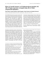
Figure 1 Effect of EMD, TGF-b1 and the combination of both
factors on cell proliferation by means of BrdU-labeling at 24 h
post-treatment.*P < 0.05; **P < 0.01; ***P < 0.001.
Figure 2 Effect of EMD, TGF-b1 and the combination of both
factors on cell growth. All treatments showed an increase in cell
proliferation. The EMD proliferation rate was higher than the
positive control at days 4 and 7. *P < 0.05; **P < 0.01; ***P < 0.001.
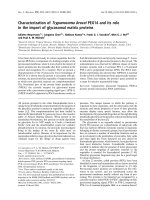
Figure 3 Epifluorescence at day 5 post-treatment with the
factors. (A-D) Immunolabeling for osteopontin (OPN, red
fluorescence) was mainly cytoplasmic, in perinuclear area and in
punctate deposits. Cell-associated green fluorescence reveals actin
cytoskeleton (Alexa Fluor 488-conjugated phalloidin), whereas blue
fluorescence indicates cell nuclei (DAPI - DNA staining). No major
differences were noticed among groups in terms of labeling pattern
and fluorescence intensity for OPN. (E-H) Immunolabeling for
alkaline phosphatase (ALP, red fluorescence) was more intense for
the positive control compared to the treated groups.
Palioto et al. Head & Face Medicine 2011, 7:13
/>Page 5 of 9
Discussion
The exposure of human osteoblastic cells to EMD, TGF-
b1andEMD+TGF-b1 resulted in early increased cell
proliferation, and reduced ALP activity and matrix
mineralization. The present results are corroborated by
several works that observed EMD stimulation of the
proliferative capacity of both osteoblastic cells[14,16, 36]
and PDL fibroblasts[10,12,13,20,22]. In contrast to PDL
fibroblast response to EMD, which shows signs of
matrix mineralization when EMD are used even at ear-
lier time points[13], osteoblastic cell cultures seem to be
inhibited in terms of osteogenic differentiation. Interest-
ingly, the associat ion of EMD and exogenous TGF- b1
did not alter the osteogenic potential of the cultures.
Although the results of the present study point toward
the development of a less differentiated osteoblastic phe-
notype when cells were exposed to EMD, TGF-b1or
EMD+TGF-b1, no morphologic differences were observed
among the groups. Cell morphology was considered within
the typical features of human alveolar bone-de rived cells
cultured on plain conventional substrates, showing an
elongated polygonal shape[31,37,38]. None of the treat-
ments supported the development and progression of ste-
late-like shaped cells, with thin and elongated cytoplasmic
extensions, which could be indicative of less differentiated
phenotypes[31].
The total protein content showed a tendency to be
increased during the initial periods of cultures for all
the treatments comparing to control, which could be
due to the increased number of cells at the end of the
proliferative phase. It has been demonstrated in various
cell types that EMD seems to augment total protein
production and collagen content[12,18].
It has been well-established that t here is an inverse
relationship between cell proliferation and cell differen-
tiation for the osteoblast lineage; as the proliferative
capacity increases, the cell differentiation decreases.
Indeed, full expression of the osteoblast pheno type leads
to terminal cell cycle exit[39,40]. In the present study,
two multifunctional noncollagenous matrix proteins
(OPN and BSP) with a role in t he matrix mineral ization
process were used as osteoblastic cell differentiation
markers[41-44]. OPN had a similar distribution and
fluorescence intensities in cultures of all groups at day 5
post-treatments, which is in agreement with the biphasic
pattern of expression (at days 5 and 14)[42] and sup-
ports the interpretation of the presence of less differen-
tiated osteoblastic cells[45]. Since BSP is a marker of
initial osteoblast differentiation, the absence of BSP
labeling at day 5 post-treatment and in control cultures
could indicate that none of the treatments were able to
promote the early expression of this matrix protein.
Based on published data, the effect of EMD in osteo-
blastic cells seems to be dependent on cell type and cul-
ture con dition and to act in a dose-dependent manner
[46,47]. Hama et al. [48], working with fetal rat calvarial
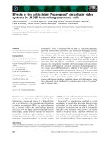
Figure 4 Total protein content at 7 and 14 days. The values (μg/
mL) are expressed as mean ± SD of representative results of three
separate experiments in cell cultures established from three
different patients, performed in quadruplicate for each treatment.
There were no statistically significant differences among groups (P >
0.05).
Figure 5 ALP activity at 7 and 14 days. The results are expressed
as μmol thymolphthalein/h/mg protein. The values are expressed as
mean ± SD of representative results of three separate experiments
in cell cultures established from three different patients, performed
in quadruplicate for each treatment. *P < 0.05; **P < 0.01; ***P <
0.001.
Figure 6 Alizarin red S stained areas of osteoblastic cell
cultures treated with 100 μg/mL EMD, 5 ng/mL TGF-b1, and
100 μg/mL EMD plus 5 ng/mL TGF-b1, at 21 days. Percentage of
stained areas was significantly higher for non-treated cultures. *P <
0.05; **P < 0.01; ***P < 0.001.
Palioto et al. Head & Face Medicine 2011, 7:13
/>Page 6 of 9
cells, has reached similar results as the ones found in
the present study. EMD decreased, in a dose dependent
manner, osteocalcin and core binding proteins expres-
sion, ALP activity, and bone-like nodule formation.
They also sought to determine the possible role of TGF-
b1 on these effects by inhibiting i ts expression. Treat-
ment wit h TGF-b1 antibody partly restored t he inhibi-
tory effect of EMD on ALP activity. Conversely, in our
work human osteo blastic cells were sensitized with exo-
genous TGF-b1 and the same inhibitory effect on osteo-
blastic differentiation was noticed.
Although the roles of ALP during the process of matrix
mineralization are still not fully clarified, it has been pro-
posed that such enzyme generates the phosphate needed
for hydroxyapatite formation. In addition, ALP has also
been hypothesized to hydrolyze pyrophosphate, a minera-
lization inhibitor, in order to facilitate mineral precipita-
tion and growth[31]. In the present study, a significant
decrease in ALP activity at days 7 and 14 post-treatment
with EMD, TGF-b1 or EMD+TGF-b1 was associated
with reduced ALP immunodetection, a finding that is
consistent with increased cell proliferation and reduced
osteogenic potential of the cultures[31]. Indeed, signifi-
cantly reduced mineralization levels were detected for all
treated groups compared to control. The treatments
likely delayed or limited the matrix mineralization pro-
cess due to the lower levels of ALP activity.
TGF-b1 has been recognized as a molecule that acts on
the proliferative c apacity of osteoblastic cells but not on
osteoblast activities, which include osteoid matrix
production and mineralization. McCauley & Somerman
[49] demonstrated that TGF-b1 inhibits the formation of
mineralized nodules in vitro. In addition, TGF-b1
expressed by platelets in fracture sites or by osteoclasts
during bone remodeling may stimulate the f ormation of
an osteoid matrix with no minera l phase, which could be
possibly related to the lower levels of ALP activity[50].
Fina lly, considering that the use of EMD and TGF-b1
has been proposed as a strategy to support periodontal
tis sue regeneration, the present in vitro results show an
inhibitory effect on cell differentiation and cell-mediated
matrix mineralization when human osteoblastic cells are
exposed to either EMD, TGF-b1 or the combination of
both. Although it is difficult to extrapolate the in vitro
find ings to the in vivo situation, we may speculate from
these results that new bone formation in the context
of periodontal regeneration could not be as prominent as
dental cementum and periodontal ligament regeneration.
Conclusion
Within the limits of the present study, the exposure of
human osteoblastic cells to EMD, TGF-b1 and the com-
bination of factors in vitro support s the development of
a less differentiated phenot ype, with enhanced prolifera-
tive activity and total cell number, and reduced ALP
activity levels and matrix mineralization.
Acknowledgements
The authors thank Mr. Roger R. Fernandes and Ms. Junia Ramos, from Cell
Culture Laboratory, School of Dentistry of Ribeirão Preto, University of São
Figure 7 Light microscopy of Alizarin red S stained-osteoblastic cell cultures: (A) control group; (B) 100 μg/mL EMD; (C) 5 ng/mL TGF-
b1; (D) 100 μg/mL EMD plus 5 ng/mL TGF-b1. Phase contrast, ×10 objective.
Palioto et al. Head & Face Medicine 2011, 7:13
/>Page 7 of 9
Paulo, Ribeirão Preto, SP, Brazil, for their helpful technical assistance, and
Luciana Prado Maia, from the Department of Oral and Maxillofacial Surgery
and Periodontology, School of Dentistry of Ribeirão Preto, University of São
Paulo, Ribeirão Preto, SP, Brazil, for the contribution in the manuscript
preparation. The mouse monoclonal anti-human bone ALP antibody (B4-78),
developed by Jerry A. Katzmann, and anti-rat osteopontin (MPIIIB10-1) and
bone sialoprotein (WVID1-9C5) antibodies, developed by Micha el Solursh
and Ahnders Franzen, were obtained from the Developmental Studies
Hybridoma Bank developed under the auspices of the NICHD and
maintained by the Department of Biological Sciences of the University of
Iowa (Iowa City, IA 52242).
Author details
1
Department of Oral Maxillofacial Surgery and Periodontology, School of
Dentistry of Ribeirão Preto - University of São Paulo, Av. do Café s/n, 14040-
904 Ribeirão Preto, SP, Brazil.
2
Department of Morphology, Stomatology and
Physiology, School of Dentistry of Ribeirão Preto - University of São Paulo,
Av. do Café s/n, 14040-904 Ribeirão Preto, SP, Brazil.
Authors’ contributions
DBP designed the research. MMB and ALR established the cell culture
protocol. TLSR, JTM and MMB performed the research. DBP and PTO
analysed the data. DBP and PTO wrote the manuscript. All authors read and
approved the final manuscript.
Competing interests
The authors declare that they have no competing interests.
Received: 6 December 2010 Accepted: 18 July 2011
Published: 18 July 2011
References
1. Nathan C, Sporn M: Cytokines in context. J Cell Biol 1991, 113:981-986.
2. Wirthlin MR: Growth substances: potential use in periodontics. J West Soc
Periodontol Periodontal Abstr 1989, 37:101-125.
3. Hammarstrom L: Enamel matrix, cementum development and
regeneration. J Clin Periodontol 1997, 24:658-668.
4. Robinson C, Brookes SJ, Shore RC, Kirkham J: The developing enamel
matrix: nature and function. Eur J Oral Sci 1998, 106(Suppl 1):282-291.
5. Heijl L, Heden G, Svardstrom G, Ostgren A: Enamel matrix derivative
(EMDOGAIN) in the treatment of intrabony periodontal defects. J Clin
Periodontol 1997, 24:705-714.
6. Sculean A, Donos N, Blaes A, Lauermann M, Reich E, Brecx M: Comparison
of enamel matrix proteins and bioabsorbable membranes in the
treatment of intrabony periodontal defects. A split-mouth study.
J Periodontol 1999, 70:255-262.
7. Pontoriero R, Wennstrom J, Lindhe J: The use of barrier membranes and
enamel matrix proteins in the treatment of angular bone defects. A
prospective controlled clinical study. J Clin Periodontol 1999, 26:833-840.
8. Kawana F, Sawae Y, Sahara T, Tanaka S, Debari K, Shimizu M, Sasaki T:
Porcine enamel matrix derivative enhances trabecular bone
regeneration during wound healing of injured rat femur. Anat Rec 2001,
264:438-446.
9. Stenport VF, Johansson CB: Enamel matrix derivative and titanium
implants. J Clin Periodontol 2003, 30:359-363.
10. Palioto DB, Coletta RD, Graner E, Joly JC, de Lima AF: The influence of
enamel matrix derivative associated with insulin-like growth factor-I on
periodontal ligament fibroblasts. J Periodontol 2004, 75:498-504.
11. He J, Jiang J, Safavi KE, Spangberg LS, Zhu Q: Emdogain promotes
osteoblast proliferation and differentiation and stimulates
osteoprotegerin expression. Oral Surg Oral Med Oral Pathol Oral Radiol
Endod 2004, 97:239-245.
12. Gestrelius S, Andersson C, Lidstrom D, Hammarstrom L, Somerman M: In
vitro studies on periodontal ligament cells and enamel matrix derivative.
J Clin Periodontol 1997, 24:685-692.
13. Rodrigues TL, Marchesan JT, Coletta RD, Novaes AB Jr, Grisi MF, Souza SL,
Taba M Jr, Palioto DB: Effects of enamel matrix derivative and
transforming growth factor-beta1 on human periodontal ligament
fibroblasts. J Clin Periodontol 2007, 34:514-522.
14. Tokiyasu Y, Takata T, Saygin E, Somerman M: Enamel factors regulate
expression of genes associated with cementoblasts. J Periodontol 2000,
71
:1829-1839.
15.
Hakki SS, Berry JE, Somerman MJ: The effect of enamel matrix protein
derivative on follicle cells in vitro. J Periodontol 2001, 72:679-687.
16. Jiang J, Goodarzi G, He J, Li H, Safavi KE, Spangberg LS, Zhu Q: Emdogain-
gel stimulates proliferation of odontoblasts and osteoblasts. Oral Surg
Oral Med Oral Pathol Oral Radiol Endod 2006, 102:698-702.
17. Schwartz Z, Cochran DL: Osteoblast phenotypic expression is modulated
by enamel matrix protein (abstract). J Dent Res 1999, 79:517.
18. Jiang J, Fouad AF, Safavi KE, Spangberg LS, Zhu Q: Effects of enamel
matrix derivative on gene expression of primary osteoblasts. Oral Surg
Oral Med Oral Pathol Oral Radiol Endod 2001, 91:95-100.
19. Kawase T, Okuda K, Yoshie H, Burns DM: Anti-TGF-beta antibody blocks
enamel matrix derivative-induced upregulation of p21WAF1/cip1 and
prevents its inhibition of human oral epithelial cell proliferation.
J Periodontal Res 2002, 37:255-262.
20. Van Der Pauw MT, Van Den Bos T, Everts V, Beertsen W: Enamel matrix-
derived protein stimulates attachment of periodontal ligament
fibroblasts and enhances alkaline phosphatase activity and transforming
growth factor 1 release of periodontal ligament and gingival fibroblasts.
J Periodontol 2000, 71:31-43.
21. Lyngstadaas SP, Lundberg E, Ekdahl H, Andersson C, Gestrelius S: Autocrine
growth factors in human periodontal ligament cells cultured on enamel
matrix derivative. J Clin Periodontol 2001, 28:181-188.
22. Okubo K, Kobayashi M, Takiguchi T, Takada T, Ohazama A, Okamatsu Y,
Hasegawa K: Participation of endogenous IGF-I and TGF-beta 1 with
enamel matrix derivative-stimulated cell growth in human periodontal
ligament cells. J Periodontal Res 2003, 38:1-9.
23. Lekic P, McCulloch CA: Periodontal ligament cell population: the central
role of fibroblasts in creating a unique tissue. Anat Rec 1996, 245:327-341.
24. Lynch SE, Williams RC, Polson AM, Howell TH, Reddy MS, Zappa UE,
Antoniades HN: A combination of platelet-derived and insulin-like
growth factors enhances periodontal regeneration. J Clin Periodontol
1989, 16:545-548.
25. Rutherford RB, Ryan ME, Kennedy JE, Tucker MM, Charette MF: Platelet-
derived growth factor and dexamethasone combined with a collagen
matrix induce regeneration of the periodontium in monkeys. J Clin
Periodontol 1993, 20:537-544.
26. Dennison DK, Vallone DR, Pinero GJ, Rittman B, Caffesse RG: Differential
effect of TGF-beta 1 and PDGF on proliferation of periodontal ligament
cells and gingival fibroblasts. J Periodontol 1994, 65:641-648.
27. Giannobile WV, Finkelman RD, Lynch SE: Comparison of canine and non-
human primate animal models for periodontal regenerative therapy:
results following a single administration of PDGF/IGF-I. J Periodontol 1994,
65:1158-1168.
28. Mailhot JM, Borke JL: An isolation and in vitro culturing method for
human intraoral bone cells derived from dental implant preparation
sites. Clin
Oral Implants Res 1998, 9:43-50.
29. Rosa AL, Beloti MM: Effect of cpTi surface roughness on human bone
marrow cell attachment, proliferation, and differentiation. Braz Dent J
2003, 14:16-21.
30. Coelho MJ, Fernandes MH: Human bone cell cultures in biocompatibility
testing. Part II: effect of ascorbic acid, beta-glycerophosphate and
dexamethasone on osteoblastic differentiation. Biomaterials 2000,
21:1095-1102.
31. de Oliveira PT, de Oliva MA, Maximiano WM, Sebastiao KE, Crippa GE,
Ciancaglini P, Beloti MM, Nanci A, Rosa AL: Effects of a mixture of growth
factors and proteins on the development of the osteogenic phenotype
in human alveolar bone cell cultures. J Histochem Cytochem 2008,
56:629-638.
32. Martelli-Junior H, Cotrim P, Graner E, Sauk JJ, Coletta RD: Effect of
transforming growth factor-beta1, interleukin-6, and interferon-gamma
on the expression of type I collagen, heat shock protein 47, matrix
metalloproteinase (MMP)-1 and MMP-2 by fibroblasts from normal
gingiva and hereditary gingival fibromatosis. J Periodontol 2003,
74:296-306.
33. Coletta RD, Almeida OP, Graner E, Page RC, Bozzo L: Differential
proliferation of fibroblasts cultured from hereditary gingival fibromatosis
and normal gingiva. J Periodontal Res 1998, 33:469-475.
Palioto et al. Head & Face Medicine 2011, 7:13
/>Page 8 of 9
34. Kee N, Sivalingam S, Boonstra R, Wojtowicz JM: The utility of Ki-67 and
BrdU as proliferative markers of adult neurogenesis. J Neurosci Methods
2002, 115:97-105.
35. Conover WJ: Some methods based on ranks. In Practical nonparametric
statistics 2 edition. Edited by: Conover WJ. New York: Wiley; 1980:213-343.
36. Jiang J, Safavi KE, Spangberg LSW, Zhu Q: Enamel matrix derivative
prolongs primary osteoblast growth (abstract). J Dent Res 2000, 79:344.
37. Beloti MM, De Oliveira PT, Schwartz Filho HO, Rosa AL, Nanci A: Influence
of a nanostructured titanium surface on cultured human osteoblastic
cells. Eur Cell Mater 2005, 10:STE1.
38. Beloti MM, de Oliveira PT, Gimenes R, Zaghete MA, Bertolini MJ, Rosa AL: In
vitro biocompatibility of a novel membrane of the composite poly
(vinylidene-trifluoroethylene)/barium titanate. J Biomed Mater Res A 2006,
79:282-288.
39. Stein GS, Lian JB, Stein JL, Van Wijnen AJ, Montecino M: Transcriptional
control of osteoblast growth and differentiation. Physiol Rev 1996,
76:593-629.
40. Thomas DM, Johnson SA, Sims NA, Trivett MK, Slavin JL, Rubin BP,
Waring P, McArthur GA, Walkley CR, Holloway AJ, et al: Terminal osteoblast
differentiation, mediated by runx2 and p27KIP1, is disrupted in
osteosarcoma. J Cell Biol 2004, 167:925-934.
41. Ganss B, Kim RH, Sodek J: Bone sialoprotein. Crit Rev Oral Biol Med 1999,
10:79-98.
42. Sodek J, McKee MD: Molecular and cellular biology of alveolar bone.
Periodontol 2000 2000, 24:99-126.
43. Tye CE, Rattray KR, Warner KJ, Gordon JA, Sodek J, Hunter GK, Goldberg HA:
Delineation of the hydroxyapatite-nucleating domains of bone
sialoprotein. J Biol Chem 2003, 278:7949-7955.
44. Tye CE, Hunter GK, Goldberg HA: Identification of the type I collagen-
binding domain of bone sialoprotein and characterization of the
mechanism of interaction. J Biol Chem 2005, 280:13487-13492.
45. de Oliveira PT, Zalzal SF, Irie K, Nanci A: Early expression of bone matrix
proteins in osteogenic cell cultures. J Histochem Cytochem 2003,
51:633-641.
46. Schwartz Z, Carnes DL Jr, Pulliam R, Lohmann CH, Sylvia VL, Liu Y,
Dean DD, Cochran DL, Boyan BD: Porcine fetal enamel matrix derivative
stimulates proliferation but not differentiation of pre-osteoblastic 2T9
cells, inhibits proliferation and stimulates differentiation of osteoblast-
like MG63 cells, and increases proliferation and differentiation of normal
human osteoblast NHOst cells. J Periodontol 2000, 71:1287-1296.
47. Heng NH, N’Guessan PD, Kleber BM, Bernimoulin JP, Pischon N: Enamel
matrix derivative induces connective tissue growth factor expression in
human osteoblastic cells. J Periodontol
2007, 78:2369-2379.
48. Hama H, Azuma H, Seto H, Kido J, Nagata T: Inhibitory effect of enamel
matrix derivative on osteoblastic differentiation of rat calvaria cells in
culture. J Periodontal Res 2008, 43:179-185.
49. McCauley LK, Somerman MJ: Biologic modifiers in periodontal
regeneration. Dent Clin North Am 1998, 42:361-387.
50. Wrana JL, Maeno M, Hawrylyshyn B, Yao KL, Domenicucci C, Sodek J:
Differential effects of transforming growth factor-beta on the synthesis
of extracellular matrix proteins by normal fetal rat calvarial bone cell
populations. J Cell Biol 1988, 106:915-924.
doi:10.1186/1746-160X-7-13
Cite this article as: Palioto et al.: Effects of enamel matrix derivative and
transforming growth factor-b1 on human osteoblastic cells. Head & Face
Medicine 2011 7:13.
Submit your next manuscript to BioMed Central
and take full advantage of:
• Convenient online submission
• Thorough peer review
• No space constraints or color figure charges
• Immediate publication on acceptance
• Inclusion in PubMed, CAS, Scopus and Google Scholar
• Research which is freely available for redistribution
Submit your manuscript at
www.biomedcentral.com/submit
Palioto et al. Head & Face Medicine 2011, 7:13
/>Page 9 of 9