Báo cáo y học: " Rice black-streaked dwarf virus P6 self-interacts to form punctate, viroplasm-like structures in the cytoplasm and recruits viroplasm-associated protein P9-1" docx
Bạn đang xem bản rút gọn của tài liệu. Xem và tải ngay bản đầy đủ của tài liệu tại đây (1.37 MB, 15 trang )
RESEARCH Open Access
Rice black-streaked dwarf virus P6 self-interacts to
form punctate, viroplasm-like structures in the
cytoplasm and recruits viroplasm-associated
protein P9-1
Qian Wang, Tao Tao, Yanjing Zhang, Wenqi Wu, Dawei Li, Jialin Yu, Chenggui Han
*
Abstract
Background: Rice black-streaked dwarf virus (RBSDV), a member of the genus Fijivirus within the family Reoviridae,
can infe ct several graminaceous plant species including rice, maize and wheat, and is transmitted by planthoppers.
Although several RBSDV proteins have been studied in detail, functions of the nonstructural protein P6 are still
largely unknown.
Results: In the current study, we employed yeast two-hybrid assays, bimolecular fluorescence complementation
and subcellular localization experiments to show that P6 can self-interact to form punctate, cytoplasmic viroplasm-
like structures (VLS) when expressed alone in plant cells. The region from residues 395 to 659 is necessary for P6
self-interaction, whereas two polypeptides (residues 580-620 and 615-655) are involved in the subcellular
localization of P6. Furthermore, P6 strongly interacts with the viroplasm-associated protein P9-1 and recruits P9-1 to
localize in VLS. The P6 395-659 region is also important for the P6-P9-1 interaction, and deleting any region of P9-1
abolishes this heterologous in teraction.
Conclusions: RBSDV P6 protein has an intrinsic ability to self-interact and forms VLS without other RBSDV proteins
or RNAs. P6 recruits P9-1 to VLS by direct protein-pr otein interaction. This is the first report on the functionality of
RBSDV P6 protein. P6 may be involved in the process of viroplasm nucleation and virus morphogenesis.
Background
Rice black-streaked dwarf virus (RBSDV), an important
pathogen that belongs to the genus Fijivirus in the
family Reoviridae, causes rice black-streaked dwarf and
maize rough dwarf diseases, which lead to severe yield
losses of crops in southeast Asian countries [1-4]. The
virusistransmittedtograminaceousplantspeciesvia
the planthopper Laodelphax striatellus in a persistent,
circulative manner [4-6]. Typical symptoms caused by
RBSDV include stunting, darkening of leaves and white
tumours or black-streaked swellings along the vei ns on
the back of the leaves, leaf blades and sheaths. Micro-
scopy of ultrathin sections has s hown that the virions
are restricted to the phloem tissues in infected plants
and that viroplasms, virus crystals and tubular structures
are abundantly synthesized in both i nfected plants and
insect cells [1,4,7,8].
The RBSDV virion is an icosahedral, double-lay ered
particle with a diameter of 75-80 nm and consists of ten
genomic dsRNA segments [9-12]. Protein sequence ana-
lysis suggested that S1 encodes a putative 168.8-kDa
RNA-dependent RNA polymerase. S2 and S4 encode a
core protein and an outer-shell B-spike protein, respec-
tively [8,11,12]. The protein encoded by S3 is assumed
to have some guanylyltransferase activity [13]. Proteins
translated from S8 and S10 are the components of the
major capsid and outer capsid, respectively [8,14,15].
Both S7 and S9 encode nonstructural proteins. S7 ORF1
P7-1 and S9 ORF1 P9-1 are components of the tubular
structures and viroplasm produced in infected cells,
respectively [8]. Recent studies have demonstrated that
P9-1, an a-helical protein with a molecular mass of 40
* Correspondence:
State Key Laboratory for Agro-biotechnology and Ministry of Agriculture Key
Laboratory for Plant Pathology, China Agricultural University, Beijing 100193,
P. R. China
Wang et al. Virology Journal 2011, 8:24
/>© 2011 Wang et al; licensee BioMed Central Ltd. This is an Open Access article distributed under the terms of the Creative Commons
Attribution License (http://c reativecommons.org/licenses/by/2.0), which permits unrestricted use, distribution, and rep roduction in
any medium, provided the original work is properly cited.
kDa, self-interacts to form dimers, and it is proposed to
be the minimal viral component required for viroplasm
formation [16]. P6 is a large nonstructural protein con-
taining 792 amino acids with a molecular mass of 89.6
kDathatistranslatedfromS6,whichis2645bpin
length and contains a single long OR F. It is synthesized
abundantly in RBSDV-i nfected plants and viruliferous
planthoppers [17]. However, further characterization
and elucidation of the functions of P6 have not yet been
reported.
In this study, we investigated the homologous interac-
tion P6-P6 using a yeast two-hybrid (YTH) assay and
bimolecular fluorescence complementation assay (BiFC)
and determined the subcellular localization of P6 and
P6 derivatives using two different fluorescent markers.
P6 self-interacts and forms l arge discrete viroplasm-like
structures (VLS) in plant cytoplasm. The minimal region
of P6 necessary for P6 self-i nteraction in vivo is com-
posed of amino acids residing between positions 395
and 659. The exact residues in this region that greatly
affect the subce llular distri bution of P6 were also deter-
mined. Furthermore, a strong interaction between P6
and the viroplasm-associated protein P9-1 was apparent
from YTH analyses and co-expression experiments.
These results might provide deeper understanding of
the process of viroplasm formation of RBSDV.
Results
P6 forms punctate, cytoplasmic viroplasm-like structures
in vivo and self-interacts in YTH system
To determine the subcellular localization of P6, the plas-
mid expressing P6 fused with green fluorescent protein
(GFP) at its C terminus (P6-GFP) was introduced into
onion epidermal cells by particle bombardment. Confo-
cal fluorescence microscopy analysis indicated that
abundant, punctate viroplasm-like fluorescent foci were
observed in the cytoplasm of the onion cells. The bright
discrete foci were of different sizes and scattered in the
cytoplasm. No apparent fluorescence was visualized in
the nuclei. As a negative control, free GFP resulted in a
diffuse pattern of fluorescence that was both nuclear
and cytoplasmic, which indicated that the moiety GFP
does not affect the localization of P6-GFP (Figure 1A).
Identical results were observed when the proteins were
expres sed in the protoplasts of Nicotiana. benthamiana
(Additional file 1, Figure S1). This demonstrated that P6
tends to aggrega te to form structures that resemb le the
matrix of the viroplasm when expressed in the absence
of other RBSDV proteins, and led us to speculate that
P6 might self-associate and be involved in the formation
of the viroplasm.
Subsequently, a YTH assay was perfo rmed to find out
whether P6 had an intrinsic ability to self-interact in
vivo. Combinations of plasmids expressing bait protein
BD-P6 and prey protein AD-P6 were transformed into
Y187 and AH109 strains, respectively. Making sure
there was no transcriptional activation or toxicity of
BD-P6 for yeast strains, western blot analysis was car-
riedouttoverifythatbothBD-P6andAD-P6were
expressed in the yeast (data not shown). Cotransforma-
tion and yeast mating assays showed that independent
yeast colonies containing pGADT7-P6 and pGBKT7-P6
grew well and turned blue in the b-galactosidase colony-
lift filter assay (data not shown), indicating that there
were strong interactions between P6 molecules. In con-
trast, no growth was observed for the negative controls
(Figure 1B). This suggested that P6 has an inherent abil-
ity to self-interact and is able to form VLS when
expressed alone in plant cells.
YTH assays indicate the centrally located region spanning
residues 395 to 659 is necessary for P6 self-interaction
As there was not much information available from the
literature about P6, protein sequence analysis wa s p er-
formed. BLAST searches indicated that the region
approximately inclusive of residues 400 to 675 exhib-
ited limited conservation of amino-acid sequence with
the ATPase domain of structural maintenance of chro-
mosomes proteins (SMCs), which play an essential role
in chromosome segregation, condensation and organi-
zation [18].
In order to determine the region necessary for P6-P6
self-interaction, we sequentially constructed a collection
of truncation derivatives that express BD-P6
98-792
,BD-
P6
274-792
,BD-P6
274-703
,BD-P6
395-703
,BD-P6
395-659
,AD-
P6
1-449
,AD-P6
341-792
,AD-P6
271-703
,AD-P6
274-703
,AD-
P6
395-703
and AD-P6
395-659
, based on the protein
sequence analysis results. Homologous binding capabil-
ities between P6 and these deletions were investigated
via the YTH assay. Schematic representation of the dif-
ferent P6 truncations is shown in Figure 2A.
The YTH analysis indicated that a centrally loc ated
domain between positions 395 and 659 was required for
P6-P6 interaction. All truncations harbouring this region
were able to interact with intact P6. However, as their N
and C termini appro ached this region, the ab ilities of
the P6 m utants to associate with intact P6 decreased.
Varying interaction abilities were indicated by the rates
of yeast growth on the selective medium. When the
deletion comprised exactly the region from positions
395 to 659, the interaction with P 6 w as very weak, and
the colo nies transform ed with pGADT7-P6
395-659
/
pGBKT7-P6 or pGADT7-P6/p GBKT7- P6
395-659
showed
obvious growth inhibition and the streaks turned dark
red. Mutant P6
1-449
, in w hich most of the central and
C-terminal region was deleted, showed complete
Wang et al. Virology Journal 2011, 8:24
/>Page 2 of 15
inability to interact with P6 (Figure 2B). Binding capabil-
ities between these deletions were also investigated, and
the results demonstrated that, even when both the N
and C termini were absent, the deletions had some abil-
ity to associate with each other (data not shown). The
results suggested that the region from residues 395 to
659 is necessary to sustain the P6 self-interaction and
that further truncation might abolish this interaction.
Transient expression experiments of P6 derivatives
indicate residues 395 to 659 are important for P6 self-
interaction
Recombinant plasmids that can express P6
274-792
,P6
395-
703
and P6
395-659
, fused in-fr ame to the N terminus of
GFP (P6
mutant
-GFP) or the C terminus of DsRed2
(DsRed-P6
mutant
), were constructed and their subcellular
localization was determined. Plasmids expressing
P6-GFP GFP
A
B
SD/AHW
L
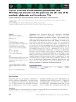
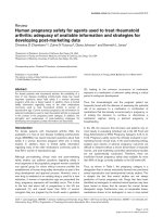
Figure 1 P6 forms punctate, cytoplasmic VLS in the onion epidermal cells and self-interacts in YTH system. (A) Subcellular localization of
RBSDV P6 fused to GFP and free GFP in onion epidermal cells. Punctata VLS of different sizes were prevalently formed in the onion cells
expressing P6-GFP, while diffuse GFP fluorescence was observed in the nucleus and cytoplasm of the cells expressing free GFP. The results were
observed 16-24 h after particle bombardment. Bars, 50 μm. (B) Yeast colonies containing pGBKT7-P6/pGADT7-P6 grew well on the selective
medium as did yeast colonies containing pGBKT7-T/pGADT7-p53, which was used as the positive control, whereas yeast transformed with
pGBKT7-P6/pGADT7 or pGBKT7/pGADT7-P6 used as negative controls were unable to grow.
Wang et al. Virology Journal 2011, 8:24
/>Page 3 of 15
P6
mutant
-GFP were delivered into onion epidermal cells
via biolistic bombardment, whereas those expressing
DsRed-P6
mutant
were introduced into epidermal cells of
N. benthamiana leaves by agroinfiltration assay [19].
Biolistic bombardment experiments indicated that
P6
274-792
-GFP mostly formed large bright discrete foci in
the cytoplasm of onion cells, but low levels of diffuse
cytoplasmic fluorescence were also observed. P6
395-703
-
GFP expression r esulted in the formation of irregular
aggregate-like structures, and minor levels of diffuse
GFP signals were also observed at the peripheries of
the nuclei, P6
395-659
-GF P res ulted in very few (generally
less than five) discrete and bright foci in the cytoplasm
(Figure 3A). Similar results were obtained when these
mutants fused with DsRed2 were expressed in the epi-
dermal cells of tobacco leaves (Figure 3B) or tobacco
protoplasts (A dditional file 2, Figure S2). Numerous dis-
persed punctate VLS were detected in the tobacco cells
expressing DsRed-P6
274-792
, and the expression of
DsRed-P6
395-703
and DsRed-P6
395-659
resulted in
amounts of irregular aggregate-like foci. Weak and
uniform red fluorescence signals were present in the
cells expressing free DsRed2.
Generally, the fluorescence distribution patterns of the
three mutants (P6
274-792
,P6
395-703
and P6
395-659
)indi-
cated that the 395-659 region is important for P6 local i-
zationandthatself-assembly is possible outside of the
P6 native environment. The results also suggested that
residues on both sides of the 395-659 region might be
engaged in the process, based on the numbers and the
size of the fluorescent foci.
Bimolecular fluorescence complementation assay
confirms that P6 molecules self-interact in planta
In order to determine whether P6 molecules self-inter-
act in planta, bimolecular fluorescence complementa-
tion assays were carried out (Figure 4). One pair of
combinations that can express P6
274-703
fused either to
YN or YC was constructed and then delivered into N.
benthamiana leaves via agroinfiltration. As expected, co-
expression of P6
274-703
-YN and P6
274-703
-YC induced
strong recovered YFP signals, which formed numerous
tiny fluorescent sites or irregular aggregate-like struc-
tures in the cytoplasm. No YFP signals were detected
for the negative controls following the co-expression of
P6
274-703
-YN/YC or P6
274-703
-YC/YN. The BiFC assay
provided strong evidence that the truncated mutant
P6
274-703
participates in self-interaction so that recov-
ered YFP signals are de tected easily in the tobacco cells.
From these results, we can confirm that P6 molecules
have the ability to self-interact in planta.
Polypeptides consisting of residues 580 to 620 and 615
to 655 are involved in VLS formation
In light of the results above, it is evident that P6
395-659
,
which only constitutes one-third of the entire P6 pro-
tein, is essential to P6 self-interaction. It is possible that
some s pecific elements in this fragment are responsible
for the VLS formation. A P6 motif prediction using My-
Hits scan
showed that three puta-
tive motifs might have relat edness to this interacting
region. These three putative motifs are designated pumi-
lio RNA-bindi ng repeat profile, sialic-acid binding
micro nemal adhesive repeat and intra-flagellar transport
protein 57, and they correspond to P6 residues 40 1-439,
584-608 and 624-654, respectively. In addition, the sec-
ondary structure prediction demonstrated that a puta-
tive coiled-coil motif might reside in the region from
residues 550 to 640. To determine which motifs might
be involved in VLS formation, corresponding derivatives
that express P6
△403-440
-GFP, P6
△580-620
-GFP, P6
△615-655
-
GFP, DsRed-P6C
△403-440
, Ds Red-P6C
△580-620
and DsRed-
P6C
△ 615-655
were constructed and their subcellular
localization was investigated. It is noteworthy that we
341-792 ++ ND
271-703 ++ ND
395-703 + ++
395-659 + +
1-792 +++ +++
274-703 ++ ND
274-792 +++ ++
1-449 - ND
98-792 +++ ND
a.a. P6-P6 VLS
interaction formation
P6
400 675
A
B
AD-P6
1-449 341-792 271-703 274-703 395-703 395-659
BD-P6
AD-P6
1-792 98-792 274-792 274-703 395-703 395-659
BD-P6
SD/AHW
L
SD/AHW
L
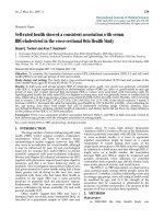
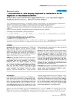
Figure 2 Mapping of the P6 region involved in P6 self-
interaction. (A) Schematic representation of P6 and P6 truncations
in the study. The full-length P6 (spanning residues 1 to 792) and P6
truncations are indicated by open bars. The P6 domain
(approximately from position 400 to 675) homologous to SMC
ATPase is indicated by the gray bar and the deleted regions by the
dashed lines. The numbers denote P6 amino acid positions. The
ability of P6 truncations to interact with intact P6 in YTH assays is
indicated in the middle (+, positive; -, negative). The VLS-forming
abilities of the different P6 derivatives are shown on the right (+ +
+, abundant and large VLS; + +, moderate in size and number; +,
few in number; -, negative with diffuse distribution; ND, not
determined). (B) Homologous interaction between intact P6 and P6
deletions in YTH assays. All truncations harbouring this region were
able to interact with intact P6. As their N and C termini approached
this region, the interaction ability was decreasing.
Wang et al. Virology Journal 2011, 8:24
/>Page 4 of 15
did create several plasmids aiming to express intact P6
fused with DsRed2 but failed to detect the fused protein
for unknown reasons. Previous results showed that
DsRed-P6
274-792
was sufficient to induce inclusion
bodies, so we created the corresponding mutants
(DsRed-P6C
△ 403-440
,DsRed-P6C
△ 580-620
and DsRed-
P6C
△615-655
) ba sed on this abridged construction. Sche-
matic representation of the different P6 deletion deriva-
tives is shown in Figure 5. As described earlier, plasmids
expressing P6
mutant
-GFP were bombarded into onion
P6
274-792
-GFP P6
395-703
-GFP P6
395-659
-GFP GFP
A
DsRed
-
P6
274
-
792
DsRed
-
P6
395
-
703
DsRed
-
P6
395
-
659
DsRed2
B
DsRed
-
P6
274
792
DsRed
-
P6
395
703
DsRed
-
P6
395
659
DsRed2
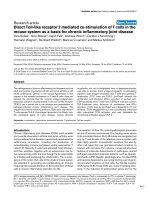
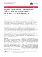
Figure 3 Distribution of P6 truncated versions in planta. (A) Subcellular localization of P6 truncations fused with GFP and free GFP in onion
epidermal cells. GFP was excited at 488 nm and emission was measured at 550-590 nm. Bars, 50 μm. (B) Subcellular localization of P6
truncations fused with DsRed2 and free DsRed2 in the epidermal cells of N. benthamiana leaves. DsRed2 was excited at 543 nm and emission
was measured at 570-600 nm. Bars, 20 μm. The fluorescence and merged images are depicted in the upper and lower panels, respectively.
Wang et al. Virology Journal 2011, 8:24
/>Page 5 of 15
P6
274-703
-NE/P6
274-703
-CE NE/P6
274-703
-CE P6
274-703
-NE/CE
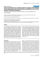
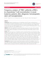
Figure 4 BiFC visualization of P6
274-703
interaction in agrobacterium-infiltrated N. benthamiana leaves. Co-expression of P6
274-703
-YN and
P6
274-703
-YC induced strong recovered YFP signals in the cytoplasm, and no YFP signals were detected for the negative controls following the
co-expression of P6
274-703
-YN/YC or P6
274-703
-YC/YN. YFP was excited at 488 nm and emission was measured at 550-590 nm. The fluorescent and
bright field images are depicted in the upper and lower panels, respectively. Bars, 20 μm.
P
rote
i
n expresse
d
P6-GFP
1
395 659 792
GFP
P6
Ƹ
403-440
-GFP
P6
Ƹ
580-620
-GFP
P6
Ƹ
615-655
-GFP
DsRed-P6
274-792
GFP
GFP
GFP
DsRed2
DsRed-P6C
Ƹ
403-440
DsRed-P6C
Ƹ
580-620
DsRed-P6C
Ƹ
615-65
5
DsRed2
DsRed2
DsRed2
Figure 5 Schematic representation of P6 deleted versions fused with GFP or DsRed2. The full-length P6 and its deleted versions are
indicated by open bars and the deleted regions by dashed lines. The numbers denote P6 amino acid positions. P6 395-659 fragment is
indicated by the gray bar and the three predicted motifs designated pumilio RNA-binding repeat profile, sialic-acid binding micronemal adhesive
repeat and intra-flagellar transport protein 57 are indicated by the checkered, black, and hatched boxes, respectively. GFP and DsRed2 are
indicated by the green and red bars, respectively.
Wang et al. Virology Journal 2011, 8:24
/>Page 6 of 15
cells, while those expressing DsRed-P6
mutant
were intro-
duced into tobacco leaves by agroinfiltration assay.
Confocal fluorescence microscopy showed that P6
△403-
440
-GFP accumulated to form numerous punctate bright
foci in the cytoplasm, indistinguishable from those
induced by P6-GFP. In contrast, P6
△ 580-620
-GFP and
P6
△ 615-655
-GFP distributed throughout the cytoplasm
displaying a weaker fluorescence pattern, compared to
free GFP, and the fluorescence signals were always
visualized at the periphery of the nuclei. Similar results
were obtained when P6 mutants were fused with
DsRed2. Numerous dispersed punctate aggregates were
detected in th e tobacco cells expressing DsRed-P6C
△403-
440
, whereas weak and uniform DsRed2 signals were pre-
sent in the cells expressing either DsRed-P6C
△580-620
or
DsRed-P6C
△615-655
. The results are shown in Figure 6.
To sum up, two polypeptide chains, comprising resi-
dues 580 to 620 and 615 to 655, are implicated in VLS
formation, and loss of them alters the subcellular locali-
zation of P6.
YTH assays demonstrate P6 interacts with P9-1
Immunoelectron microscopy revealed that antibodies
against P9-1 reacted with viroplasm in infected cells [8].
Based on our findings above, P6 likely participates in
viroplasm formation. This prompted us to further
explore the relationship between P6 and P9-1 via a YTH
assay. A plasmid that can express BD-P9-1 was con-
structed and transformed into Y187 strain. Interestingly,
the results showed that there is an intimate association
between P9-1 and P6 (Figure 7A). Yeast colonies con-
taining b oth pGBKT7-P9-1 and pGADT7-P6 grew well
on the selective medium, whereas yeast transformed
with pGBKT7-P9-1 and pGADT7, which was used as a
negative control, was unable to grow. This result indi-
cated that P6 interacts with P9-1 in vivo.
P9-1 cannot form inclusion-like structures when
expressed alone
Two plasmids that express P9-1-GFP and DsRed-P9-1
were constructed and bombarded into onion epidermal
cells to de termine P9-1 subcellular local ization. Fluores-
cence microscopy indicated that both P9-1-GFP and
DsRed-P9-1 resulted in a pattern of diffuse and uniform
fluorescence distr ibution in the cytoplasm and nuclei of
onion cells, which was a little weaker than that of free
GFP or DsRed2 controls (Figure 8A). Our results are
incon sistent with the conclusion of Zhang et al that P9-
1 alone aggregates to form inclusion bodies [ 16]. The
same results were obtained when the plasmids were
delivered into tobacco protoplasts via polyethylene gly-
col (PEG) transfection method or introduced into epi-
dermal cells of tobacco leaves by agroinfiltration assay
(Additional file 3, Figure S3). Therefore, we consider
that P9-1 has a widespread distribution but no ab ility to
aggregate in the cytoplasm when expressed in plant cells
on its own.
Colocalization experiments indicate P6 relocalizes the
distribution of P9-1 and recruits P9-1 to VLS
Co-expression experiments were developed to investi-
gate potential P6-P9-1 interactions (Figure 8B). We
introduced two plasmids expressing P6-GFP and DsRed-
P9-1 into onion cells by cobombardment. Contrary to
the case when DsRed-P9-1 was expressed alone, when
P6-GFP and DsRed-P 9-1 were co -expressed, a striking
relocalization of red fluorescence emerged. DsRed-P9-1
displayed a nearly complete coincidence with t he intra-
cellular distribution of P6-GFP. The two proteins were
colocalized and exclusively presented in discrete punc-
tate VLS, identical to those formed by P6-GFP alone,
and no diffuse green or red fluorescent signals were
observed in the cytoplasm or the nuclei. Control combi-
nations were also investigated to rule out the possibility
that GFP or DsRed2 expression might have some aber-
rant effects on the DsRed-P9-1 or P6-GFP distribution.
The colocalization of P6-GFP and DsRed-P9-1 con-
firmed that P6 has a dramatic effect on the d istribution
of P9-1 and that it is caused by the direct association
between these two proteins.
YTH assays confirm residues 395 to 659 of P6 are
necessary for P6-P9-1 heterologous interaction
Further YTH analyses were performed to examine the
regions o f P6 crucial for P6- P9-1 heterologous interac-
tion. P6 AD-fused deletions, including AD-P6
1-449
,AD-
P6
341-792
,AD-P6
274-703
,AD-P6
271-703
,AD-P6
395-703
and
AD-P6
395-659
, were tested and all P6 deletions except
AD-P6
1-449
were able to interact with P9-1. Transfor-
mants expressing BD-P9-1 and AD-P6
1-449
showed no
growth on the selective medium, whereas those contain-
ing other combinations grew well (Figure 7B). The
results indicated that the region located between amino
acids 395 and 659 is indispensable for P6-P9-1
interaction.
YTH assays indicate deletion mutants of P9-1 do not
interact with P6
We also investigated P9-1 regions crucial for P6-P9-1
interaction. A dozen P9-1 BD-fused deletions that
express fusions BD-P9-1
1-197
,BD-P9-1
1-207
,BD-P9-1
1-
248
,BD-P9-1
76-347
,BD-P9-1
167-347
,BD-P9-1
198-347
,BD-
P9-1
208-347
and BD-P9-1
76-207
were constructed. YTH
results indicated that all deletions completely lost the
ability to interact with P6 (Figure 7C). It is supposed
that minor changes in the protein sequence might affect
the properties and protein structure of P9-1 and thereby
abrogate P6-P9-1 interaction.
Wang et al. Virology Journal 2011, 8:24
/>Page 7 of 15
P6
Ƹ
403-440
-GFP P6
Ƹ
580-620
-GFP P6
Ƹ
615-655
-GFP
DsRed
-
P6C
Ƹ
403-440
DsRed
-
P6C
Ƹ
580-620
DsRed
-
P6C
Ƹ
615-65
5
DsRed
P6C
DsRed
P6C
DsRed
P6C
Figure 6 Transient expression results of P6 deleted derivatives. The upper two panels indicate the distribution of P6 deletions fused with
GFP expressed in the onion epidermal cells, showing that P6
△580-620
-GFP and P6
△615-655
-GFP have a diffuse fluorescence pattern while P6
△403-440
-
GFP forms numerous VLS. GFP was detected with excitation at 488 nm and emission capture at 550-590 nm. Bars, 20 μm. The lower two panels
indicate the distribution of DsRed2-fused P6 deletions expressed in the epidermal cells of N. benthamiana leaves. Similarly, Both DsRed-P6C
△580-
620
and DsRed-P6C
△615-655
show a diffuse and weak red fluorescence distribution whereas DsRed-P6C
△403-440
forms VLS. Red fluorescence was
detected with excitation at 543 nm and emission capture at 570-600 nm. Bars, 50 μm.
Wang et al. Virology Journal 2011, 8:24
/>Page 8 of 15
Discussion
Compared to animal reoviruses, most events in the Fiji-
virus life cycle, such as virus entry, replication, and
packaging and particle assembly and systemic move-
ment, are poorly understood, as are the functions of
proteins encoded by the viral g enome. In this study, we
investigated the uncharacterized protein P6 of RBSDV, a
member of th e Fijivirus ge nus, by employing the related
experiments in protein-protein interactions.
YTH analysis a nd/or subcellular localizati on experi-
ments showed that P6 interact (Figure 2B) and establish
punctate VLS when solely expressed in plant cells
(Figure 1; Additional file 1, Figure S1), and BiFC assays
also indicated that the truncated version P6
274-703
(equivalent to one-third of the whole P6 protein) is able
to interact intimately to form aggregate-like structures
(Figure 4). These results, which clearly demonstrated
that P6 has a strong ability to self-assemble, prompted
us to question whether P 6 is capable of forming multi-
meric structures. Multimerization of viral proteins
always plays an essential role in the virus cycle [20-22].
In Reoviridae, the viroplasm determinants, such as
NSP2 and NSP5 of rotaviruses, μNS and sNS of orthor-
eoviruses,NS2oforbiviruses,andPns12ofricedwarf
virus, all share this characteristic to assemble into
higher-order complexes to recruit other viral proteins or
RNAs [23-29]. The self-interaction of RBSDV P6 might
be prerequisite for its multimerization and subsequently
for its biological functions.
The coiled-coil region might be involved in P6-P6
interactions. Coiled-coil motifs are increasingly recog-
nized as key determinants in both intra- and inter-mole-
cular interactions. In o ur experiments, the P6 region
spanning residues 365 to 659, which is predicted to har-
bour a coiled-coil structure and show some sequence
homology with the ATPase domain of SMCs, is crucial
for VLS formation. Deleting two peptide chains (aa 580-
620 and aa 624-654) abolishes VLS formation (Figure 6),
which suggests that loss of this region might have a pro-
nounced effect in altering the context of the whole pro-
tein and perturb the correct folding of the co iled-coil
domain and thereby inhibit molecular interactions. On
the basis of the different rates of yeast growth in the
YTH assay and the different numbers of fluorescent foci
formed in transient expression experiments, we con-
clude that, whereas the central region spanning residues
365 to 659 is identified as important for P6-P6 or P6-
P9-1 interactions, the amino acid sequences near to this
region might also affect these interactions by changing
the stability of the newly-built protein complexes.
A strong interaction between P6 and P9-1 was detected
in our experim ents. The two proteins are both expressed
at high levels in infected plants and viruliferous insects,
as detected by using antibodies against them [8,17]. Pre-
vious experiments indicatedthattheviroplasmmatrix
was densely and evenly immunolabelled with antibodies
against P9-1 [8]. Although corresponding electron micro-
scopy results have not been obtained for P6, the ability of
P6 to form VLS and the heterologous interaction
between P6 and P9-1, as well as the localization of P9-1
in hosts, hint that P6 m ight associate with viroplasm and
playaroleintheviroplasmnucleation.Itisnoteworthy
that orthoreovirus μNS, which plays an essential role in
the process of viroplasm formation, is able to assemble
into globular VLS when expressed alone and recruit
another viroplasm-associated protein sNS to the VLS
[24,30,31]. This is quite similar to our results.
Despite the lack of detectable protein sequence
homology with an imal reovirus protein s, P6 posse sses
A
A
SD/AHWL
AD-P6
1-449 341-792 271-703
B
274-703 395-703 395-659
BD-P9-1
AD-P6
SD/AHWL
C
BD-P9-1
1-197 1-207 1-248 76-347
BD-P9-1
167-347 198-347 208-347 76-207
AD-P6 SD/AHW
L
Figure 7 Investigation of P6-P9-1 interaction in YTH system. (A)
Yeast colonies containing pGBKT7-P9-1/pGADT7-P6 grew well on
the selective medium, whereas the yeast transformed with pGBKT7-
P9-1 and pGADT7, used as a negative control, was unable to grow.
(B) Yeast colonies expressing BD-P9-1 with AD-P6
341-792
, AD-P6
274-
703
, AD-P6
271-703
, AD-P6
395-703
, or AD-P6
395-659
grew well on the
selective medium, but those expressing BD-P9-1 with AD-P6
1-449
did
not. The numbers denote P6 amino acid positions. (C) Yeast
colonies expressing AD-P6 with any of the P9-1 mutants fused with
BD domain showed no growth on the selective medium. The
numbers denote P9-1 amino acid positions.
Wang et al. Virology Journal 2011, 8:24
/>Page 9 of 15
GFPP9-1-GFP DsRed2DsRed-P9-1
A
B
P6-GFP/
DsRed-P9-1
GFP/
DsRed-P9-1
P6-GFP/
DsRed2
GFP/
DsRed2
a b c d
Figure 8 P6 is able to recruit P9-1 to VLS in onion epidermal cells. (A) Subcellular localization of P9-1 fused with GFP or DsRed2. P9-1-GFP
and DsRed-P9-1 were distributed diffusely in the onion cells and were unable to form inclusion bodies. (B) Co-expression of P6-GFP and DsRed-
P9-1 in onion epidermal cells. Detection of green (lane a) and red (lane b) fluorescence was achieved with excitation at 488 nm and 543 nm,
respectively; co-localization of green and red fluorescence is indicated in yellow (lane c); superposition of the green and red fluorescence images
as well as the bright field image is shown on the right (lane d). The co-expression results indicate that P6 was able to relocate the distribution of
P9-1, that both proteins were present exclusively in the discrete and punctate foci, and that expression of DsRed2 or GFP had no aberrant
effects on DsRed-P9-1 or P6-GFP distribution. Bars, 50 μm.
Wang et al. Virology Journal 2011, 8:24
/>Page 10 of 15
some common features with their viroplasm determi-
nants. Being expressed at high level in hosts, posse ssing
the ability to form VLS and recruiting the viroplasm-
associated protein P9-1, P6 protein prediction showed
that the P6 fragment located between amino acids 400
and 675 has a low homology with the SMC ATPase
domain, whereas the region from positions 404 to 439 is
likely to be a pumilio RNA-binding repeat profile, which
indicates that P6 might be involved in ATP hydrolysis
and binding of RNA. Generally, viroplasm determinants
are often inferred to possess NTP-hydrolysis and RNA-
binding a ctivities to assist in the process of RNA repli-
cation, especially in Reoviridae [32,33]. It is necessary to
do further work to elucidate the biochemical and bio-
physical properties of P6 and to determine whether P6
functions according to a mechanism that is similar to
other viroplasm determinants of reoviruses in the pro-
cess of viroplasm formation.
RBSDV P9-1 wa s previously reported to form inclu-
sion bodies when tagged with GFP at its C-terminus
and expressed in Arabidopsis protoplasts [16], which is
contrary to our findings. We investigated the P9-1 dis-
tribution by two different transient expression strategies.
Whenever the protein is fused with GFP at its C termi-
nus or DsRed2 at its N terminus and expressed in onion
(Figure 8A) or tobacco epidermal cells or tobacco proto-
plasts (Additional file 3, Figure S3), P9-1 has a diffuse
distribution pattern in the cytoplasm and cannot form
aggregates. Consistent with the conclusion reached by
Zhang et al [16], we confirmed that P9-1 self-interacts
in the YTH system and forms stable dimers in vitro
(data not shown). P9-1 itself might not be the nucleating
factor in plant c ells for it is located in the VLS only
when coexpressed with P6.
Being highly homologous to RBSDV P9-1 (64.5% iden-
tity) [34], Mal de Rio Cuarto vi rus (MRCV) P9-1, which
was detected in viroplams in both infected plants and
planthoppers [35], was found to be sufficient for the for-
mation of viral inclusion body (VIB)-like structures
when expressed in Spodoptera frugiperda Sf9 cells [36].
There might be distinct mechanisms involved in viro-
plasm formation in insect and plant hosts. These ques-
tions need to be addressed in future work.
Conclusions
This is the first report on the functionality of RBSDV
nonstructural protein P6, which previously was comple-
tely uncharacterized. Our results showed RBSDV P6
self-interacts and forms punctate cytoplasmic VLS when
expressed alone. Furthermore, P6 strongly interacts with
the viroplasm-associated protein P9-1 and recruits P9-1
to localize in VLS. The P6 and P9-1 regions necessary
for these homologous or heterologo us interactions were
also determin ed, as well as the exact residues essential
for P6 VLS formation. Results presented here might
provide clues for understanding the viroplasm nuclea-
tion of RBSDV and allow us to gain further insigh t into
the relationship between P6 and P9-1 in the virus life
cycle.
Methods
General
Healthy N. benthamiana plants were grown at 23 °C
under 1,000 lumens with a 16-hour daylight regimen.
Agrobacterium tumefaciens strain EHA105 was grown
on LB agar containing 50 g/ml rifampin. The yeast
strains, Saccharomyces cerevisiae AH109 and Y187, and
the yeast vectors, pGBKT7 and pGADT7, as well as the
positive control plasmids, pGBKT7-T and pGADT7-
p53, were used for YTH analyses (Clontech). The bi nary
expression vectors pGDR and pGDp19 used to express
Tomato bushy stunt virus (TBSV) p19 fo r suppressing
gene silencing were obtained as generous gifts from Pro-
fessor Andrew O. Jackson of the University of California
at Berkeley, USA, while another plasmid, pEGFP (Clon-
tech) which harboured EGFP segment, was kindly pro-
vided by Professor Zaifeng Fan, China Agricultural
University,PRChina.BiFCvectors,pSPYNE-35Sand
pSPYCE-35S, were kindly provided by Professor Jörg
Kudla, Universität Müneter, Germany. Both RBSDV S6
(GenBank: AY144570) and S9 (GenBank: AF536564)
full-length cDNA clones were maintained in o ur lab
[11].
Construction of recombinant plasmids
To generate transient expression v ector pGFPI, pBI221
was digested with HindI II/ XbaIandSacI/EcoRI respec-
tively, and the liberated CaMV 35S promoter and nos
terminator were ligated to pUC18-T corresponding
clone sites to obtain an intermediate vector. EGFP
encoding the autofluorescent protein was ampl ified
using primers EGFP-1/EGFP-2 from plasmid pEGFP
(clontech). PCR products were digested with KpnI/SacI,
and ligated into the KpnI/SacI- digested pUC18-T inter-
mediate to generate pGFPI. Only the XbaIandKpnI
can be used t o express GFP-tagged protein in this vec-
tor. To generate expression recombinants for GFP-
tagged P6, full-length of P6 ORF was amplified using a
pair of primers PS6-1/PS6-6 (Table 1). PCR products
were ligated to pMD19-T to obtain pMD19-T-S6.
pMD19-T-S6 was digested wit h BamHI/XhoI, and
ligated into BamHI/XhoI-digested pSPYNE-35S. The
clone was then cut by XbaI/KpnI and the liberated frag-
ment was ligated into the XbaI/KpnI-digested pGFPI to
yield pS6GFPI, which can express P6-GFP. P6 deletion
and truncation fragments were produced through PCR
amplification, using the primers shown in Table 1, and
PCR products were ligated to pMD19-T or self-ligated
Wang et al. Virology Journal 2011, 8:24
/>Page 11 of 15
to obtain intermediates selected for further use. The
intermediates containing P6 truncation fragments
(P6
274-792
,P6
395-703
,P6
395-659
) were digested with XbaI/
KpnI and the liberated fragments were ligated into
XbaI/KpnI-digested pGFPI to yield vectors expressing
P6 truncations fused with GFP (P6
274-792
-GFP, P6
395-703
-
GFP, P6
395-659
-GFP). As in the construction of pS6GFPI,
similar strategies were used to obtain three P6 deletion
derivatives ( expressing P6
△403-440
-GFP, P6
△580-620
-GFP,
P6
△615-655
-GFP) and pS9-1GFPI (expressing P9-1-GFP).
For expression of the DsRed2-fused proteins, intermedi-
ates containing P6 truncation fragments (P6
274-792
,
P6
395-703
,P6
395-659
) and those containing P6 deletion
fragments (P6C
△403-440
,P6C
△580-620
,P6C
△615-655
)were
digested with XhoI/BamHI and HindIII/SalI, respec-
tively, and the liberated fragments were ligated to the
corresponding XhoI/BamHI- and HindIII/SalI-treated
pGDR to generate vectors expressing P6 truncations and
Table 1 Primers used for PCR amplification
Primer Sequence (5′®3′)
a
Locations
b
and modifications
PGFP-F ggtacc ATGGGTAAAGGAGAAGAAC 1aa;KpnI
PGFP-R gagctc TTATTTGTATAGTTCATC full-length reverse primer with stop codon; SacI
PS6-1-F CG ggatcc ATGTCTGCCC 1aa; BamHI
PS6-4-F CTAG ccatgg GA ATGTCTGCCCACCTGACCAATTTAG 1aa; NcoI
PS6-5-R CG ggatcc TTACTCAGAGCTTAGTTGCCAGAGG full-length reverse primer with stop codon; BamHI
PS6-6-R CCG ctcgag CTCAGAGCTTAGTTGCC full-length reverse primer without stop codon; XhoI
PS6-8-R CCG ctcgag ATCAGCTACTTCGTCAG 449aa; XhoI
PS6-9-F CG ggatcc AC ATGTCTGCCCACCTG 1aa; BamHI
PS6-10-F CCG ctcgag ccatgg AAGCTTCTGATGTCCAG 274aa; XhoI, NcoI
PS6-11-F CCG ctcgag ccatgg ACTTGATTAATCATGCC 395aa; XhoI, NcoI
PS6-12-R CG ggatcc ggtacc ATCTCCAAAGTTAGCATCTAC 703aa; BamHI, KpnI
PS6-15-R CG ggatcc ggtacc CGTTTCATTAGCAGATGTTTTG 659aa; BamHI, KpnI
PS6-16-R TCC cccggg GAACAGATCGGCATGATTAATC 403aa; SmaI
PS6-17-F TCC cccggg GTGAATGATTTAACTGACGAAG 440aa; SmaI
PS6-18-R CATG gggccc GTCTTTCTCTTTTAGTAAAGAACAG 615aa; ApaI
PS6-19-F CATG gggccc TCTGCTAATGAAACGAATGATG 655aa; ApaI
PS6-20-R CATG gggccc GGCAATCTGTTCTTTAGCTTGTC 580aa; ApaI
PS6-21-F CATG gggccc GAGAACGAAATGTTGAAGGAACAG 620aa; ApaI
PS6-24-F GC tctaga ccatgg ACGTACTCAACCTGTCCAA 98aa; XbaI, NcoI
PS6-25-F GC tctaga ccatgg AAGCTTCTGATGTCCAGTC 274aa; XbaI, NcoI
PS6-26-R CCG ctcgag ggtacc CTCAGAGCTTAGTTGCCAGAG full-length reverse primer without stop codon; XhoI KpnI
PS9-5-F CTAG ccatgg GA ATGGCAGACCAAGAGCG 1aa; NcoI
PS9-6-R CG ggatcc AACGTCCAATTTCAAGG full-length reverse primer without stop codon; BamHI
PS9-9-F CG ggatcc ATGGCAGACC AAGAGCG 1aa; BamHI
PS9-10-R CCG ctcgag AACGTCCAATTTCAAGG full-length reverse primer without stop codon; XhoI
PS9-11-F CCG gaattc TCTCATCTCCCTAACC 76aa; EcoRI
PS9-12-R CG ggatcc CAAATACATTAAAAAGCC 207aa; Bam
HI
PS9-13-F
CCG gaattc GGTGAAAATCCAAACTC 208aa; EcoRI
PS9-14-R CG ggatcc GTGATTAACTTCTTTATTTG 248aa; BamHI
PS9-15-F CCG ctcgag CT ATGGCAGACCAAGAGCG 1aa; XhoI
PS9-16-R ggtacc ggatcc TCAAACGT CCAATTTCAAG full-length reverse primer, KpnI, BamHI
PS9-17-F gaattc gtcgac ATGGCAGACCAAGAGC 1aa, EcoRI, SalI
PS9-18-F gaattc gtcgac ATGTCGTTGTTGCCAAT 167aa, EcoRI, SalI
PS9-19-F gaattc gtcgac ATGTATATAAAAGGCTT 198aa, EcoRI, SalI
a
Introduced restriction endonuclease sites are in lower case. Two extra nucleotides (italicized) were added to allow in-frame expression of fusion proteins of
interest.
b
Numbered according to P6 amino acid sequence. The F or R designation in the primer names denotes whether the primer is a forward (5 ′ ) or reverse (3′)
primer, respectively.
Wang et al. Virology Journal 2011, 8:24
/>Page 12 of 15
P6 deletions fused with DsRed2. A vector expressing
DsRed-P9-1 was constructed similarly to those expres-
sing P6 truncation fused with DsRed2.
To generate yeast plasmids for the two-hybrid assay,
P6 ORF was amplified using primers PS6-4/PS6-5. PCR
products were digested with NcoI/Ba mHI, and then
ligated into the same sites of pGADT7 to generate
pGADT7-S6. Vectors pGADT7-S9-1 and pGBKT7-S9-1
were created using similar strategies. P6 ORF was ampli-
fied using primers PS6-6/PS6-9 and the P CR products
were ligated to pMD19-T. The clone was digested with
BamHI/Sa lI, and P6 BamHI/SalI-fragments were ligated
into the corr esponding sites of pGBKT7 to obtain
pGBKT7-S6. P6 NcoI/BamHI-fragments excised from
the pMD19-T intermediates (containing P6
274-703
,P6
395-
703
,P6
395-659
) were ligated in to NcoI/BamHI sites of
pGADT7 and pGBKT7 to generate vectors expressing
truncations fused with AD or BD. pGADT7-S6 was
digested with EcoRI/BamHI and EcoRV, and the liber-
ated fragments were ligated into pGADT7 EcoRI/BamHI
and SmaI to obtain constructs expressing AD-P6
341-792
and AD-P6
271-703
.P6BamHI/XhoI-fragment from the
intermediate harbouring P6
1-449
was inserted into the
corresponding sites of pET30a, and then the clone was
digested with NcoI/XhoI. The liberated fragment was
cloned into the Nco I/XhoI-digested pGADT7 to obtain
constructs expressing AD-P6
1-449
.P6NcoI/XhoI-frag-
ments excised from the pMD19-T intermediates (con-
taining P6
98-792
and P6
274-792
) were ligated into the
same sites of pGBKT7 to generate vectors expressing
truncations BD-P6
98-792
and BD-P6
274-792
. Similar strate-
gies were used to generate the constructs expressing P9-
1 mutants fused with BD.
To obtain construction binary vectors for BiFC, the
intermediate which contained P6
274-703
was digested
with XbaI/Bam HI, and the P6
274-703
fragment was then
inserted into the same sites of pSPYNE-35S and
pSPYCE-35S to generate vectors expressing P6
274-703
-
NE and P6
274-703
-CE.
The primers used in the e xperiments are shown in
Table 1. All clones derived from the PCR products were
verified by sequencing, and the recombinant plasmids
were confirmed by restriction analyses.
YTH and b-galactosidase assays
Yeast transformations were conducted using the small-
scale lithium acetate method. Two-hybrid assays were
performed using the Matchmaker GAL4 Two-Hybrid
System3 (Clontech), according to the manufacturer’ s
protocols. Cotransformants were plated on synthetic
defined (SD) minimal medium minus adenine, histidine,
leucine, and tryptophan (SD/-Ade/-His/-Leu/-Trp), and
positive yeast colonies that could grow on the auxo-
trophic medium were lysed in liquid nitrogen and then
tested for b-galactosidase activity as mentioned in the
b-galactosidase colony-lift filter assay.
Transient expression of protein in onion cells
To introduce plasmid DNA into onion epidermal cells,
particle bombardment was conducted using a helium-
driven particle accelerator PDS-1000/He (Bio-Rad). 2-5
μgplasmidDNAin5μL distilled water were mixed
with 8 μL of a 60 mg/mL 1.0-μm-diameter gold particle
solutio n, 20 μL of 2.5 M CaCl
2
, and 8 μL of 0.1 M fresh
prepared spermidine. The re sultant suspension was
incubated for 10 min with intermittent mixing every 1
to 2 min at room temperature. The golden particles
coated with plasmid DNA were collected by 5-s pulse
centrifugation. After the supernatant was removed, the
pellet was washed with 100 μLof70%coldethanolfol-
lowed by the same volume of 100% cold eth anol, and
then suspended in 10 μL 100% ethanol. After being
dried on t he center of an aluminum foil rupture disk,
the gold particles were bombarded into onion cells
under a vacuum of 28 mm Hg with 6-cm target dis-
tances. The bombarded onion epidermal cells were cul-
tured on 0.6% agar with 2 ,4-D-free MS medium at
25 °C in darkness. Fluorescence signals were detected at
16 to 24 h after bombardment [37,38].
Subcellular localization of RBSDV P6 derivatives and BiFC
assay in N. benthamiana leaves
Different binary plasmids were transformed into A.
tumefaciens EHA105 by a freeze-thaw method. Cultures
of EHA105 harbouring a relevant binary plasmid were
grown in LB medium containing rifampicin (50 g/ml)
and kanamycin (100 g/ml) at 28 °C for 16 h. For expres-
sion of different fusions, EHA105 strains containing the
pGDR derivatives and pGDp19 plasmid were resus-
pended and adjusted to an OD
600
of 0.5:0.3 with infiltra-
tion medium (10 mM MES, pH 5.6, 10 mM MgCl
2
,150
mM acetosyringone). For the BiFC assay, Agrobacter ium
cultures containing the BiFC plasmids and the pGDp19
plasmid were resuspended at a fina l OD
600
of 0.5:0.5:0.3.
The cells were incubated at room temperature for 2 to
4 h, and then infiltrated into 5-6- week-old N. benthami-
ana leaves. Underside epidermal c ells of tobacco infil-
trated leaves were assayed for fluorescence 48-96 h after
infiltration [39].
Laser-scanning confocal microscopy
Fluorescence analysis was performed using a Nikon
ECLIPSE TE2000-E inverted fluorescence microscope
equipped with a Nikon D-ECLIPSE C1 spectral confocal
laser scanning system. GFP and YFP were both detected
with an excitation at 488 nm and emission capture at
550-590 nm. DsRed2 was excited at 54 3 nm using a
543-nm helium neon laser, and the emission was
Wang et al. Virology Journal 2011, 8:24
/>Page 13 of 15
captured at 570 to 600 nm [40]. For analysis of coex-
pression assays, multi-tracking was used to prevent
emission cross-talk between the channels.
Additional material
Additional File 1: Transient expression of P6 fused with GFP in N.
benthamiana protoplasts. Tobacco protoplasts were isolated and
transfected using a modified PEG method. Punctata VLS of different sizes
were prevalently formed in N. benthamiana protoplasts expressin g P6-
GFP, while diffuse GFP fluorescence was observed in the nucleus and
cytoplasm of the cells expressing free GFP. The results were observed 16
h after PEG transfection. Bars, 20 μm.
Additional File 2: Transient expression of P6 truncations fused with
DsRed2 in N. benthamiana protoplasts. DsRed-P6
274-792
, DsRed-P6
395-
703
and DsRed-P6
395-659
formed discrete bright aggregate-like structures
in the N. benthamiana protoplasts, while a weak and diffuse fluorescence
was also detected in the cytoplasm. Free DsRed2 resulted in a diffuse
pattern of fluorescence that was both nuclear and cytoplasmic. Bars, 20
μm.
Additional File 3: Transient expression of DsRed-P9-1 and P9-1-GFP
in N. benthamiana cells or protoplasts. The plasmids expressing
DsRed-P9-1 and P9-1-GFP were introduced into tobacco cells by agro-
infiltration assay or PEG transfection, respectively. Both DsRed-P 9-1 and
P9-1-GFP resulted in a pattern of diffuse and uniform fluorescence
distribution in the cytoplasm of N. benthamiana cells or protoplasts,
which indicated that P9-1 is unable to form aggregate-like structures
when expressed alone in tobacco cells. Bars, 20 μm.
Acknowledgements
We are grateful to Professor Andrew O. Jackson (Department of Plant and
Microbial Biology, University of California, Berkeley) and Sek-Man Wong
(National University of Singapore, Singapore) for providing valuable
suggestions. We also thank Professors Jörg Kudla (Universität Münster,
Germany) for providing BiFC vectors. This research was supported by the
National Basic Research Program (2006CB101903) and National Department
Public Benefit Research Funds (nyhyzx07-051, 2008ZX08003-001 and
2009ZX08003-010B).
Authors’ contributions
QW carried out most of the experiments and wrote the manuscript. TT and
WW anticipated the construction of the recombinants. YZ provided useful
advice and anticipated in the protein transient expression assays. CH, DL and
JY conceived of the study and participated in its design and coordination.
All authors read and approved the final manuscript.
Competing interests
The authors declare that they have no competing interests.
Received: 26 October 2010 Accepted: 18 January 2011
Published: 18 January 2011
References
1. Shikata E, Kitagawa Y: Rice black-streaked dwarf virus: its properties,
morphology and intracellular localization. Virology 1977, 77:826-842.
2. Bai FW, Yan J, Qu ZC, Zhang HW, Xu J, Ye MM, Shen DL: Phylogenetic
analysis reveals that a dwarfing disease on different cereal crops in
China is due to rice black streaked dwarf virus (RBSDV). Virus Genes 2002,
25:201-206.
3. Fang S, Yu J, Feng J, Han C, Li D, Liu Y: Identification of rice black-
streaked dwarf fijivirus in maize with rough dwarf disease in China. Arch
Virol 2001, 146:167-170.
4. Hibino H: Biology and epidemiology of rice viruses. Annu Rev Phytopathol
1996, 34:249-274.
5. Shikata E: Rice black-streaked dwarf virus. CMI/AAB description of plant
viruses 1974:135.
6. Uyeda I, Kimura I, Shikata E: Characterization of genome structure and
establishment of vector cell lines for plant reoviruses. Adv Virus Res 1995,
45:249-279.
7. Conti M, Lovisolo O: Tubular structures associated with Maize Rough
Dwarf Virus particles in crude extracts: electron microscopy study. J Gen
Virol 1971, 13:173-176.
8. Isogai M, Uyeda I, Lee BC: Detection and assignment of proteins encoded
by rice black streaked dwarf fijivirus S7, S8, S9 and S10. J Gen Virol 1998,
79:1487-1494.
9. Milne RG, Conti M, Lisa V: Partial purification, structure and infectivity of
complete maize rough dwarf virus particles. Virology 1973, 53:130-141.
10. Boccardo G, Milne RG: Plant reovirus group. CMI/AAB Descriptions of plant
viruses 1984, 294.
11. Wang ZH, Fang SG, Xu JL, Sun LY, Li DW, Yu JL: Sequence analysis of the
complete genome of rice black-streaked dwarf virus isolated from maize
with rough dwarf disease. Virus Genes 2003, 27:163-168.
12. Zhang HM, Chen JP, Adams MJ: Molecular characterisation of segments 1
to 6 of Rice black-streaked dwarf virus from China provides the
complete genome. Arch Virol 2001, 146:2331-2339.
13. Supyani S, Hillman BI, Suzuki N: Baculovirus expression of the 11
mycoreovirus-1 genome segments and identification of the
guanylyltransferase-encoding segment. J Gen Virol 2007, 88:342-350.
14. Liu H, Wei C, Zhong Y, Li Y: Rice black-streaked dwarf virus minor core
protein P8 is a nuclear dimeric protein and represses transcription in
tobacco protoplasts. FEBS Lett 2007, 581:2534-2540.
15. Liu H, Wei C, Zhong Y, Li Y: Rice black-streaked dwarf virus outer capsid
protein P10 has self-interactions and forms oligomeric complexes in
solution. Virus
Res 2007, 127:34-42.
16. Zhang C, Liu Y, Liu L, Lou Z, Zhang H, Miao H, Hu X, Pang Y, Qiu B: Rice
black streaked dwarf virus P9-1, an α-helical protein, self-interacts and
forms viroplasms in vivo. J Gen Virol 2008, 89:1770-1776.
17. Fang S, Wang Z, Han C, Li D, Yu J: Genomic segment 6 of Rice Black
Streaked Dwarf Virus encodes for a viral non-structural protein. Acta
Agriculturae Boreali-Sinica 2007, 22:5-8.
18. Hirano M, Hirano T: Hinge-mediated dimerization of SMC protein is
essential for its dynamic interaction with DNA. EMBO J 2002,
21:5733-5744.
19. Goodin MM, Dietzgen RG, Schichnes D, Ruzin S, Jackson AO: pGD vectors:
versatile tools for the expression of green and red fluorescent protein
fusions in agroinfiltrated plant leaves. Plant J 2002, 31:375-383.
20. Haas G, Azevedo J, Moissiard G, Geldreich A, Himber C, Bureau M,
Fukuhara T, Keller M, Voinnet O: Nuclear import of CaMV P6 is required
for infection and suppression of the RNA silencing factor DRB4. EMBO J
2008, 27:2102-2112.
21. Schuck P, Taraporewala Z, McPhie P, Patton J: Rotavirus nonstructural
protein NSP2 self-assembles into octamers that undergo ligand-induced
conformational changes. J Biol Chem 2001, 276:9679.
22. Nakai K, Okamoto T, Kimura-Someya T, Ishii K, Lim CK, Tani H, Matsuo E,
Abe T, Mori Y, Suzuki T, et al: Oligomerization of hepatitis C virus core
protein is crucial for interaction with the cytoplasmic domain of E1
envelope protein. J Virol 2006, 80:11265-11273.
23. Taraporewala Z, Chen D, Patton JT: Multimers formed by the rotavirus
nonstructural protein NSP2 bind to RNA and have nucleoside
triphosphatase activity. J Virol 1999, 73:9934-9943.
24. Becker MM, Peters TR, Dermody TS: Reovirus σNS and μNS proteins form
cytoplasmic inclusion structures in the absence of viral Infection. J Virol
2003, 77:5948-5963.
25. Wei T, Shimizu T, Hagiwara K, Kikuchi A, Moriyasu Y, Suzuki N, Chen H,
Omura T: Pns12 protein of Rice dwarf virus is essential for formation of
viroplasms and nucleation of viral-assembly complexes. J Gen Virol 2006,
87:429-438.
26. Jiang X, Jayaram H, Kumar M, Ludtke SJ, Estes MK, Prasad BV: Cryoelectron
microscopy structures of rotavirus NSP2-NSP5 and NSP2-RNA
complexes: implications for genome replication. J Virol 2006,
80:10829-10835.
27. Taraporewala ZF, Chen D, Patton JT: Multimers of the bluetongue virus
nonstructural protein, NS2, possess nucleotidyl phosphatase activity:
similarities between NS2 and rotavirus NSP2. Virology
2001,
280:22
1-231.
28. Butan C, Tucker P: Insights into the role of the non-structural protein 2
(NS2) in Bluetongue virus morphogenesis. Virus Res 2010, 151:109-117.
Wang et al. Virology Journal 2011, 8:24
/>Page 14 of 15
29. Taraporewala ZF, Patton JT: Nonstructural proteins involved in genome
packaging and replication of rotaviruses and other members of the
Reoviridae. Virus Res 2004, 101:57-66.
30. Broering TJ, Parker JSL, Joyce PL, Kim J, Nibert ML: Mammalian reovirus
nonstructural protein μNS forms large inclusions and colocalizes with
reovirus microtubule-associated protein μ2 in transfected cells. J Virol
2002, 76:8285-8297.
31. Miller CL, Broering TJ, Parker JSL, Arnold MM, Nibert ML: Reovirus σNS
protein localizes to inclusions through an association requiring the μNS
amino terminus. J Virol 2003, 77:4566-4576.
32. Vasquez-Del Carpio R, Gonzalez-Nilo FD, Riadi G, Taraporewala ZF, Patton JT:
Histidine triad-like motif of the rotavirus NSP2 octamer mediates both
RTPase and NTPase activities. J Mol Biol 2006, 362:539-554.
33. Gillian AL, Schmechel SC, Livny J, Schiff LA, Nibert ML: Reovirus protein
σNS binds in multiple copies to single-stranded RNA and shares
properties with single-stranded DNA binding proteins. J Virol 2000,
74:5939-5948.
34. Guzman FA, Distefano AJ, Arneodo JD, Hopp HE, Lenardon SL, del Vas M,
Conci LR: Sequencing of the bicistronic genome segments S7 and S9 of
Mal de Rio Cuarto virus (Fijivirus, Reoviridae) completes the genome of
this virus. Arch Virol 2007, 152:565-573.
35. Guzman FA, Arneodo JD, Saavedra Pons AB, Truol GA, Luque AV, Conci LR:
Immunodetection and subcellular localization of Mal de Rio Cuarto virus
P9-1 protein in infected plant and insect host cells. Virus Genes 2010,
41:111-117.
36. Maroniche GA, Mongelli VC, Peralta AV, Distefano AJ, Llauger G, Taboga OA,
Hopp EH, del Vas M: Functional and biochemical properties of Mal de Rio
Cuarto virus (Fijivirus, Reoviridae) P9-1 viroplasm protein show further
similarities to animal reovirus counterparts. Virus Res 2010, 152:96-103.
37. Kitajima A, Asatsuma S, Okada H, Hamada Y, Kaneko K, Nanjo Y, Kawagoe Y,
Toyooka K, Matsuoka K, Takeuchi M, et al: The rice α-amylase glycoprotein
is targeted from the Golgi apparatus through the secretory pathway to
the plastids. Plant Cell 2009, 21:2844-2858.
38. Goodin MM, Austin J, Tobias R, Fujita M, Morales C, Jackson AO:
Interactions and nuclear import of the N and P proteins of sonchus
yellow net virus, a plant nucleorhabdovirus. J Virol 2001, 75:9393-9406.
39. Walter M, Chaban C, Schutze K, Batistic O, Weckermann K, Nake C,
Blazevic D, Grefen C, Schumacher K, Oecking C, et al: Visualization of
protein interactions in living plant cells using bimolecular fluorescence
complementation. Plant J 2004, 40:428-438.
40. Deng M, Bragg JN, Ruzin S, Schichnes D, King D, Goodin MM, Jackson AO:
Role of the sonchus yellow net virus N protein in formation of nuclear
viroplasms. J Virol 2007,
81:5362-5374.
doi:10.1186/1743-422X-8-24
Cite this article as: Wang et al.: Rice black-streaked dwarf virus P6 self-
interacts to form punctate, viroplasm-like structures in the cytoplasm
and recruits viroplasm-associated protein P9-1. Virology Journal 2011
8:24.
Submit your next manuscript to BioMed Central
and take full advantage of:
• Convenient online submission
• Thorough peer review
• No space constraints or color figure charges
• Immediate publication on acceptance
• Inclusion in PubMed, CAS, Scopus and Google Scholar
• Research which is freely available for redistribution
Submit your manuscript at
www.biomedcentral.com/submit
Wang et al. Virology Journal 2011, 8:24
/>Page 15 of 15