Báo cáo y học: "HIV-1 Nef increases astrocyte sensitivity towards exogenous hydrogen peroxide" potx
Bạn đang xem bản rút gọn của tài liệu. Xem và tải ngay bản đầy đủ của tài liệu tại đây (627.98 KB, 7 trang )
RESEARCH Open Access
HIV-1 Nef increases astrocyte sensitivity towards
exogenous hydrogen peroxide
Sabine Masanetz
1
, Michael H Lehmann
1,2*
Abstract
Background: HIV-1 infected individuals are under chronic exposure to reactive oxygen species (ROS) considered to
be instrumental in the progression of AIDS and the development of HIV-1 associated dementia (HAD). Astrocytes
support neuronal function and protect them against cytotoxic substances including ROS. The protein HIV-1 Nef, a
progression factor in AIDS pathology is abundantly expressed in astrocytes in patients with HAD, and thus may
influence its functions.
Results: Endogenous expressed HIV-1 Nef leads to increased sensitivity of human astrocytes towards exogenous
hydrogen peroxide but not towards TNF-alpha. Cell death of nef-expressing astrocytes exposed to 10 μM hydrogen
peroxide for 30 min occurred within 4 h.
Conclusion: HIV-1 Nef may contribute to neuronal dysfunction and the development of HAD by causing death of
astrocytes through decreasing their tolerance for hydrogen peroxide.
Background
Both HIV-1 associated dementia (HAD) and a milder
form of HIV-1 associated cognitive disorder, known as
minor cognitive and motor disorder (MCMD) are fre-
quent complications of the acquired immunodeficiency
syndrome (AIDS) and are ch aracterized by neuronal
dysfunction and cell death caused by HIV-1 through
direct and indirect mechanisms [1-4].
Recently, a sophisticated inspection of brains from
HIV-1 infected patients confirmed that neurons are not
infected with HIV-1 and surprisingly revealed that astro -
cytes, the most abundant cell type in the brain, are exten-
sively infected. Additionally, this study elucidated that
infection of astrocytes with HIV-1 correlated with the
severity of neuropathology [5]. Astrocytes play an impor-
tant role in maintaining homeostasis, providing neuropro-
tection and regulating physiological activities within the
brain [6-8]. Therefore, astrogliosis and astrocyte death
occurring in HIV-infected individuals [9-12] may contri-
bute indirectly to neuronal dysfunction.
Even though HIV-1 is integrated in the astrocyte gen-
ome, it rarely replicates in this cell type in vivo, however,
regulatory proteins such as Nef are found to be abun-
dantly expressed [13-15]. The presence of HIV-1 Nef in
the brain is associ ated with astrogliosis and recruitment
of monocytes/macrophages [16], correlating with the
development of HAD [17].
Astrocytes stably transfected with HIV-1 Nef function
as appropriate cellular model systems for the purpose of
investigating basic mechanisms pertinent to the under-
standing of HAD pathogenesis. Using these cells, we pre-
viously showed that HIV-1 Nef induces CCL2/MCP-1
[18], thereby, providing an alternative hypothesis for the
occurrence of this chemokine at high concentrations in
the cerebrospinal fluid (CSF) of HIV-i nfected individuals
with HAD [19,20]. CCL2 plays an important role in the
cerebral infiltration of monocytes/macrophages in these
patients [21,22]. Infiltrated and activated monocytes/
macrophages, which are considered to be the effector
cells in cellular and tissue damage in AIDS, produce
cytotoxic substances such as reactive oxygen species
(ROS) and inflammatory cytokines [23,24]. Consequently,
HIV-1 infected and non-infected astrocytes are subjected
to an environment characterized,amongstothers,by
high concentrations of hydrogen peroxide and tumor
necrosis factor (TNF)-alpha. Therefore, the aim of this
study was to investigate the effect of HIV-1 Nef on the
* Correspondence:
2
Institute for Infectious Diseases and Zoonoses, Ludwig-Maximilians-
University Munich, 80539 Munich, Germany
Full list of author information is available at the end of the article
Masanetz and Lehmann Virology Journal 2011, 8:35
/>© 2011 Masanetz and Lehmann; licensee BioMed Central Ltd. This is an Open Access article distributed under the terms of the Creative
Commons Attribution License (http:/ /creativecommons.org/licenses/by/2.0), which permits unrestricted use, distribution, and
reproduction in any medium, provided the original work is properly c ited.
cellular viability of human astrocytes exposed to these
particular cytotoxic substances.
Results
Astrocytes stably transfected with HIV-1 nef are highly
sensitive to hydrogen peroxide induced cell death
Astrocytes fulfil a protective function for neurons through
elimination of ROS such as hydrogen peroxide [25]. Yet
astrocytes are more vulnerable to the effects of hydrogen
peroxide than neurons [26,27], but it is not known how
this is modulated by HIV-1 Nef. Therefore, the sensitivity
of human astrocytic U251MG-Nef cells towards hydrogen
peroxide was tested in comparison with the sensitivity of
U251MG-parental and U251MG-pNeo cells. Cells treated
with hydrogen peroxide at concentrations of 1 μMand
10 μM for 30 min were investigated after 24 h for viability
using AlamarBlue
®
reagent containing resazurin, a
non-toxic, oxidation-reduction indicator indicating mito-
chondrial metabolic activity. The analysis revealed that in
astrocytic cells stably expressing nef, hydrogen peroxide
significantly reduced the cell viability as compared to
mock-treated cells, hydrogen peroxide-treated U251MG-
parental cells and hydrogen peroxide-treated U251MG-
pNeo cells (Figure 1). Similar results were obtained at 48 h
(additional file 1).
Hydrogen peroxide rapidly induced cell death of
astrocytes stably transfected with HIV-1 nef
A light microscopic analysis was performed in order to
examine whether signs of cell death induced by hydro-
gen peroxide may be detected earlier than 24 h in nef-
expressing astrocytes. Indeed, in contrast to the control
cells, the previously flat-shaped nef-expressing astrocytes
had underg one a morphological alteration to being
roun d-shaped and almost completely detached from the
cell culture flask surface 3 h 30 min subsequent to
30 min treatment with hydrogen peroxide at a concen-
tration of 10 μM (Figure 2).
Translocation of the membrane phospholipid phos-
phatidylserine (PS) to the outer leaflet of the plasma
membrane occurs rapidly after exposure to a cytotoxic
agent a nd mostly indicates a point-of-no-return during
the cellular dying process . Using the Annexin V assay, it
was confirmed that hydrogen peroxide at a concentra-
tion of 10 μM severely affected cellular viability of
U251MG-Nef cells but had only a small effect on astro-
cytic U251MG-parental and U251MG-pNeo cells. PS
exposure on the cell surface in combination with posi-
tive PI staining indicating loss of plasma membrane
integrity, which is criterion to consider a cell as
dead [28], has been detected in about 75% of the nef-
expressing cells (Figure 3).
Astrocytes stably transfected with HIV-1 nef are as
sensitive to TNF-alpha induced cell death as non-
transfected cells
Previously, it has been shown that HIV-1 Nef protects
T cells against TNF-alpha induced apoptosis [29]. Con-
sequently, we tested whether HIV-1 Nef is also capable
of protecting astrocytes against TNF-alpha induced cell
death. Human astrocytic U251MG-parental, -pNeo and
-Nef cells were treated with TNF-alpha for 24 h and
their viabi lity was analysed using AlamarBlue
®
reagent.
Data revealed that TNF-alpha significantly reduced the
cell viability of each astrocytic cell type investigated here
to a similar degree including the stably nef-transfected
Mock
1.0
1.5
2.0
H
2
O
2
(1 μM)
1.0
1.5
2.0
H
2
O
2
(10 μM)
1.0
1.5
2.0
U251MG-parenta
l
U251MG-pNeo
U251MG-Nef
**
**
**
**
f
luorescence
1h 2h 3h 4h
0.0
0.5
1h 2h 3h 4h
0.0
0.5
1h 2h 3h 4h
0.0
0.5
In
cuba
ti
o
n tim
e
Relative
f
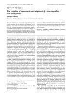
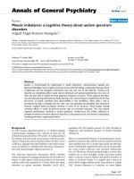
Figure 1 Hydrogen peroxide decreased the viability of nef-expressing astrocytes. U251MG-parental, -pNeo and -Nef cells were treated
with hydrogen peroxide for 30 min at indicated concentrations. Subsequently cells were washed twice with PBS and incubated in VLE-RPMI
1640 medium containing 10% FCS for a further 24 h. The medium was then exchanged and cell viability assay was performed as described in
the Material section. The times indicated are relative to the moment of adding AlamarBlue
®
reagent to the cell culture medium. The relative
fluorescence represents the ratio of the fluorescence intensity of study cells versus mock-treated cells at 1 h after start of the assay. Data
represent mean ± s.e.m. (n = 6); **, P < 0.01.
Masanetz and Lehmann Virology Journal 2011, 8:35
/>Page 2 of 7
cells (Figure 4). This result has been confirmed using
the Annexin V assay (additional file 2).
Discussion
Chronic oxidative st ress in HIV-inf ected patients plays
an important role in AIDS progression [30,31]. This
phenomenon is explained by a depletion of endogenous
antioxidant moieties and an increased production of
ROS. Oxidative stress, in particular, is thought to be a
cause of neuronal cell death in the brain of HIV-1
infected patients and believed to contribute to develop-
ment of HAD [32,33]. Moreover, ROS-induced astr ocyte
death is also thought to play a role in the occurrence of
HAD [26,27].
Here we show that a short exposure of exogenous
hydrogen peroxide to nef-expressing astrocytes led to
their rapid cell death. The early detection of a high num-
ber of propidium iodide/annexin V double positive cells
points to necrotic cell death [34], which was previously
suggested when astrocytes are subject ed to tertiary- butyl
hydroperoxide [35]. But it can not be final ly defin ed only
from this observation what kind of cell death exactly
occurred in our model. Also it depends on the concentra-
tion of hydrogen peroxide applied whether a cell dies in
an apoptotic or necrotic manner [36]. In this context it is
interesting to note that astrocytes are vulnerable to
hydrogen peroxide at concentrations ranging from
0.5 mM to 2.5 mM [27], values approximately a 1.000
fold higher than the concentration applied to induce
death of nef-expressing astrocytes herein. So it remains a
challenge for further studies to elucidate what HIV-1 Nef
precisely alters in the cell leading to increased sensitivity
to exogenous hydrogen peroxide. Intriguingly, it has been
shown during the preparation of this manuscript that
HIV-1 Nef in primary human astrocytes and in the brain
of mice increases oxidative stress [37], which is in line
with our finding.
Since HIV-1 Nef i s known to inhibit apoptosis of
T-cells [29,38,39] and m ono cytes/ma cropha ges [40,41],
it was somewhat surprising that TNF-alpha decreased
the viability of U251MG-Nef cells and U251MG-paren-
tal cells equally. Additionally, this finding is in contrast
to previously reported data demonstrating that HIV-1
Nef prevents TNF-alpha triggered apoptosis in astrocytic
U251M G cells [42]. This discrepancy may be due to the
U251MG-parental U251MG-pNEO U251MG-Nef
A
Mock
H
2
O
2
(1 ȝM)
H
2
O
2
(10 ȝM
)
r
op
idi
um
i
o
did
e
Annexin V
P
r
U251MG parental
B
75
100
U251MG
-
parental
U251MG-pNeo
U251MG-Nef
]
%
75
100
%
B
25
50
[Living cells
]
25
50
[dead cells]
Mock 1 10
0
[H
2
O
2
] μM
Mock 1 10
0
[H
2
O
2
] μM
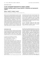
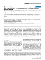
Figure 3 Hydrogen peroxide leads to rapid loss of the cell
membrane integrity in nef-expressing astrocytes. (A) U251MG-
parental, -pNeo and -Nef cells were treated with hydrogen peroxide
for 30 min at concentrations as indicated. Cells then were washed
twice with PBS, incubated in VLE-RPMI 1640 medium containing
10% FCS for a further 3 h 30 min and subsequently the annexin V
assay was performed as described in the Methods section. (B)
Summary of three independent experiments. Annexin V, PI double-
negative cells (living cells) and annexin V, PI double-positive cells
(dead cells) are shown.
U251M
G
-parental U251M
G
-Ne
f
H
2
O
2
Mock
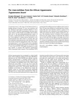
Figure 2 Hydrogen peroxide leads to rapid detachment of nef-
expressing astrocytes. U251MG-parental and -Nef cells were
treated with hydrogen peroxide (10 μM) for 30 min. Cells were
subsequently washed twice with PBS, incubated in VLE-RPMI 1640
medium containing 10% FCS for a further 3 h 30 min and
subsequently a light microscopic analysis of astrocytic cells was
performed. A Zeiss Axiovert 25 microscope (Carl Zeiss Jena GmbH,
Jena, Germany) was used. Original magnification, × 100.
Masanetz and Lehmann Virology Journal 2011, 8:35
/>Page 3 of 7
use of cells stably transfected with nef in our study,
which could clearly well simulate the long term effect of
HIV-1 Nef i n chronically infected ce lls [43] than cells
transiently transfected with nef. More over, involvement
of HIV-1 Nef in cell survival is subject to generally con-
troversy [44,45].
HIV-1 encodes a glutathione peroxidase [46], which has
been shown to protect the cell against exogenous and
endogenous ROS [47]. Consequently, what ever the reason
why HIV-1 Nef causes an increase of sensitivity towards
hydrogen peroxide, it is concei vable that the HIV-1 GPX
could counteract this action of HIV-1 Nef by detoxifying
hydrogen peroxide. Thereby HIV-1 GPX would prevent
the cytotoxic potential of HIV-Nef, which is considered as
a progression factor in AIDS [48-50] and known to induce
an AIDS-like disease in a mouse model [51,52]. Thus, this
could explain the paradoxical effect that functional HIV-1
GPXs are frequently found in long-term non-progressors
while non-functional HIV-1 GPXs are present in HIV-1
isolates from patients developing AIDS [47].
Conclusions
Besides other known direct and indirect effects of HIV-1
proteins, HIV-1 Nef may contribute to cellular and tis-
sue injury frequently detected in HIV-1 infected indivi-
duals, including various AIDS-associated diseases such
as HAD, by increasing the sensitivity of Nef-harboring
cells to hydrogen peroxide.
Methods
Cell culture
The human astrocytoma cell line U251MG was obtained
from M. Brenner (National Institutes of Health, Bethesda,
MD). The cell l ines U251MG-Nef
Bru
clone 4/4.2 stably
expressing nef from HIV-1
Bru
(GenBank accession num-
ber K02013) and U251MG-pNeo carrying only the neo-
mycin resistance gene were establishe d as reported [53].
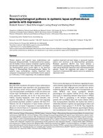
HIV-1 Nef expression was confirmed by immunoblotting
(Figure 5). Cells were routinely incubated at 37° under
5% CO2, and 90% humidity in VLE-RPMI 1640 medium
certified to contain < 0.01 endotoxin units/ml, and sup-
plemented with 10% fetal calf serum (FCS), 100 U/ml
penicillin as well as 100 μg/ml strept omycin (all from
Biochrom AG, Berlin, Germany). Before treatment
with hydrogen peroxide (Merck KgaA) or TNF-alpha
(BioSource International Inc., Camarillo, CA), cells were
seeded at a density of 1 × 10
5
cells/ml in 96-well flat bot-
tomed microtiter plates (BD Biosciences) for the cell
viability assay or in 12-well plates (Costar) for the
annexin V assay and incubated overnight in VLE-RPMI
1640 medium supplemented with 10% FCS.
Immunoblotting and immunodetection
Lysates of U251MG-parenta l, -pNeo and -Nef cells were
prepared by directly adding 1x SDS sample loading buffer
to the cells followed by sonication. Samples were sepa-
rated on a 4-20% tris-glycine gradient gel (Anamed,
Darmstadt, Germany) and blotted on a nitrocellulose
membrane. The blotted membranes were immunostained
using mouse anti-Nef 3E6 mAb provided by K. Krohn
through the National Institute for Biological Standards
and Control Centralised Facility for AIDS Reagents,
mouse anti-GAPDH mAb MAB347 (Chemicon Interna-
tional, Inc., Temecula, CA) and MFP488-conjugated goat
anti-mouse antibody (MoBiTec Gmb H, Göttingen,
Germany), and positive signals were detected by fluores-
cence scanning (excitatio n wavelength 488 nm, emission
filter 520BP40) using the Typhoon 9410 Fluorescence
Mock
2.0
TNF-alpha (100 ng/ml)
2.0
TNF-alpha
(
200 ng
/
ml
)
2.0
U251MG-pNeo
U251MG-parenta
l
U251MG-Nef
R
elative
f
luorescence
1h
2h
3h
4h
0.0
0.5
1.0
1.5
1h
2h
3h
4h
0.0
0.5
1.0
1.5
1h
2h
3h
4h
0.0
0.5
1.0
1.5
In
cuba
ti
o
n tim
e
R
1h
2h
3h
4h
1h
2h
3h
4h
1h
2h
3h
4h
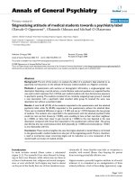
Figure 4 HIV-1 Nef does not modulate TNF-alpha decrease d viability of astrocytes. U251MG-paren tal, -pNeo and -Nef cells were treated
with TNF-alpha for 24 h at indicated concentrations. The medium was then exchanged and cell viability assay was performed as described in
the Material section. The times indicated are relative to the moment of adding AlamarBlue
®
reagent to the cell culture medium. The relative
fluorescence represents the ratio of the fluorescence intensity of study cells versus mock-treated cells at 1 h after start of the assay. Data
represent mean ± s.e.m. (n = 5).
Masanetz and Lehmann Virology Journal 2011, 8:35
/>Page 4 of 7
Scanner (GE Healthcare), and analyzed using Image-
Quant 5.2 software (Molecular Dynamics).
Cell viability assay
The AlamarBlue
®
reagent (Molecular Probes, Inc.,
Eugene, OR) containing the water soluble, non-toxic dye
resazurin (7-Hydroxy-3H -phenoxazin-3-one 10-oxide)
was used to quantify mitochondrial activity according to
the manufacturer’s recommendation. Briefly, 1/10th of
the volume of AlamarBlue
®
reagent was added directly to
the cells in culture medium. Using the Typhoon™ 9410
fluorescence scanner (GE Healthcare), fluorescence mea-
sureme nt was performed by applying an excitation wave-
length of 532 nm and an emission filter of 580BP30 nm.
Data were analyzed using ImageQuant™ TL software
(GE Healthcare). T he fluorescence intensity of medium
containing only AlamarBlue
®
was determined simulta-
neously and was subtracted from all values.
Annexin V assay
Phosphatidylserine on the cell surface was detected with
the Annexin V-FITC Apoptosis Detection Kit I (BD
Bioscie nces). Briefly, cells were plated and treated in 12-
well plates (Costar). Then cells were washed twice with
cold PBS and incubated in the dark for 15 min in 1 ×
binding buffer supplemented with annexin V-FITC. Pro-
pidium iodide (PI) was added to the cell suspension
immediately before analyzing with the BD FACSCanto™
flow cytometer (BD Biosciences). Data were evaluated
using FlowJo© software (Tree Star).
Statistical analysis
GraphPad Prism 4 (GraphPad S oftware , Inc., San Diego,
CA) was used for statistical analysis. The Mann-Whitney
test was used to compare the groups; a P value of less
than 0.05 was considered significant. Tests were per-
formed exactly and two-tailed.
Additional material
Additional file 1: Hydrogen peroxide significantly decreased the
viability of nef-expressing astrocytes. U251MG-parental and -Nef cells
were treated with hydrogen peroxide for 30 min at indicated
concentrations. Cells were subsequently washed twice with PBS,
incubated in VLE-RPMI 1640 medium containing 10% FCS for a further
48 h. The medium was then exchanged and cell viability assay was
performed as described in the Methods section. The relative fluorescence
represents the ratio of the fluorescence intensity of study cells versus
mock-treated. Data obtained after 4 h of starting the assay represent
mean ± s.e.m. (n = 6); **, P < 0.01.
Additional file 2: TNF-alpha equally induces PS externalization in
U251MG-parental and -Nef cells. Cells were treated with TNF-alpha for
4 h or with hydrogen peroxide for 30 min at concentrations as indicated.
Cells treated with hydrogen peroxide were washed twice with PBS,
incubated in VLE-RPMI 1640 medium containing 10% FCS for a further 3
h 30 min and subsequently the annexin V assay was performed as
described in the Methods section.
Acknowledgements
The authors wish to thank Susanne Kramer for providing astrocytic cells,
Nasim Kroegel, B.Sc., for reviewing the manuscript, and Volker Erfle for his
general support. This study was supported by an internal grant from the
Helmholtz Zentrum München.
Author details
1
Institute of Virology, Technical University of Munich/Helmholtz Zentrum
München, 81675 Munich, Germany.
2
Institute for Infectious Diseases and
Zoonoses, Ludwig-Maximilians-University Munich, 80539 Munich, Germany.
Authors’ contributions
MHL conceived and designed the experiments, SM performed the
experiments, SM and MHL analyzed the data, MHL wrote the paper. All
authors have read and approved the final manuscript.
Authors’ information
After receiving her M.Sc. in Molecular Biotechnology, SM moved to the
Physiology Weihenstephan, Technical University Munich, Freising, Germany
to work for her PhD.
MHL received his PhD in Biology from the Friedrich-Schiller-University of
Jena, Germany and currently holds a faculty position at the Institute for
Infectious Diseases and Zoonoses, Ludwig-Maximilians-Universität München,
Germany.
Competing interests
The authors declare that they have no competing interests.
Received: 6 September 2010 Accepted: 22 January 2011
Published: 22 January 2011
References
1. Gonzalez-Scarano F, Martin-Garcia J: The neuropathogenesis of AIDS. Nat
Rev Immunol 2005, 5:69-81.
o
ntal
U251MG-Nef
U251MG-pNe
o
U251MG-pare
GAPDH ʊ
H
IV-1 Nef ʊ
Figure 5 Detection of HI V-1 Nef by immunoblotting.Lysatesof
U251MG-parental, -pNeo and -Nef cells were prepared, separated
and blotted, and HIV-1 Nef and GAPDH have been detected as
described in the Methods section.
Masanetz and Lehmann Virology Journal 2011, 8:35
/>Page 5 of 7
2. Kaul M, Garden GA, Lipton SA: Pathways to neuronal injury and apoptosis
in HIV-associated dementia. Nature 2001, 410:988-994.
3. Minagar A, Commins D, Alexander JS, Hoque R, Chiappelli F, Singer EJ,
Nikbin B, Shapshak P: NeuroAIDS: characteristics and diagnosis of the
neurological complications of AIDS. Mol Diagn Ther 2008, 12:25-43.
4. Piacentini M, Kroemer G: Cell death pathways in retroviral infection. Cell
Death Differ 2005, 12(1):835-836.
5. Churchill MJ, Wesselingh SL, Cowley D, Pardo CA, McArthur JC, Brew BJ,
Gorry PR: Extensive astrocyte infection is prominent in human
immunodeficiency virus-associated dementia. Ann Neurol 2009,
66:253-258.
6. Perea G, Araque A: Communication between astrocytes and neurons: a
complex language. J Physiol Paris 2002, 96:199-207.
7. Chen Y, Swanson RA: Astrocytes and brain injury. J Cereb Blood Flow
Metab 2003, 23:137-149.
8. Bouzier-Sore AK, Merle M, Magistretti PJ, Pellerin L: Feeding active neurons:
(re)emergence of a nursing role for astrocytes. J Physiol Paris 2002,
96:273-282.
9. Epstein LG, Gendelman HE: Human immunodeficiency virus type 1
infection of the nervous system: pathogenetic mechanisms. Ann Neurol
1993, 33:429-436.
10. Sabri F, Titanji K, De Milito A, Chiodi F: Astrocyte activation and apoptosis:
their roles in the neuropathology of HIV infection. Brain Pathol 2003,
13:84-94.
11. Shi B, De Girolami U, He J, Wang S, Lorenzo A, Busciglio J, Gabuzda D:
Apoptosis induced by HIV-1 infection of the central nervous system. J
Clin Invest 1996, 98:1979-1990.
12. Petito CK, Roberts B: Evidence of apoptotic cell death in HIV encephalitis.
Am J Pathol 1995, 146:1121-1130.
13. Bagasra O, Lavi E, Bobroski L, Khalili K, Pestaner JP, Tawadros R,
Pomerantz RJ: Cellular reservoirs of HIV-1 in the central nervous system
of infected individuals: identification by the combination of in situ
polymerase chain reaction and immunohistochemistry. AIDS 1996,
10:573-585.
14. Tornatore C, Chandra R, Berger JR, Major EO: HIV-1 infection of subcortical
astrocytes in the pediatric central nervous system. Neurology 1994,
44:481-487.
15. Saito Y, Sharer LR, Epstein LG, Michaels J, Mintz M, Louder M, Golding K,
Cvetkovich TA, Blumberg BM: Overexpression of nef as a marker for
restricted HIV-1 infection of astrocytes in postmortem pediatric central
nervous tissues. Neurology 1994, 44:474-481.
16. Mordelet E, Kissa K, Cressant A, Gray F, Ozden S, Vidal C, Charneau P,
Granon
S: Histopathological and cognitive defects induced by Nef in the
brain. FASEB J 2004, 18:1851-1861.
17. Ranki A, Nyberg M, Ovod V, Haltia M, Elovaara I, Raininko R, Haapasalo H,
Krohn K: Abundant expression of HIV Nef and Rev proteins in brain
astrocytes in vivo is associated with dementia. AIDS 1995, 9:1001-1008.
18. Lehmann MH, Masanetz S, Kramer S, Erfle V: HIV-1 Nef upregulates CCL2/
MCP-1 expression in astrocytes in a myristoylation- and calmodulin-
dependent manner. J Cell Sci 2006, 119:4520-4530.
19. Conant K, Garzino-Demo A, Nath A, McArthur JC, Halliday W, Power C,
Gallo RC, Major EO: Induction of monocyte chemoattractant protein-1 in
HIV-1 Tat-stimulated astrocytes and elevation in AIDS dementia. Proc
Natl Acad Sci USA 1998, 95:3117-3121.
20. Cinque P, Vago L, Mengozzi M, Torri V, Ceresa D, Vicenzi E, Transidico P,
Vagani A, Sozzani S, Mantovani A, et al: Elevated cerebrospinal fluid levels
of monocyte chemotactic protein-1 correlate with HIV-1 encephalitis
and local viral replication. AIDS 1998, 12:1327-1332.
21. Eugenin EA, Osiecki K, Lopez L, Goldstein H, Calderon TM, Berman JW:
CCL2/monocyte chemoattractant protein-1 mediates enhanced
transmigration of human immunodeficiency virus (HIV)-infected
leukocytes across the blood-brain barrier: a potential mechanism of HIV-
CNS invasion and NeuroAIDS. J Neurosci 2006, 26:1098-1106.
22. Gonzalez E, Rovin BH, Sen L, Cooke G, Dhanda R, Mummidi S, Kulkarni H,
Bamshad MJ, Telles V, Anderson SA, et al: HIV-1 infection and AIDS
dementia are influenced by a mutant MCP-1 allele linked to increased
monocyte infiltration of tissues and MCP-1 levels. Proc Natl Acad Sci USA
2002, 99:13795-13800.
23. Brabers NA, Nottet HS: Role of the pro-inflammatory cytokines TNF-alpha
and IL-1beta in HIV-associated dementia. Eur J Clin Invest 2006,
36:447-458.
24. Williams KC, Hickey WF: Central nervous system damage, monocytes and
macrophages, and neurological disorders in AIDS. Annu Rev Neurosci
2002, 25:537-562.
25. Desagher S, Glowinski J, Premont J: Astrocytes protect neurons from
hydrogen peroxide toxicity. J Neurosci 1996, 16:2553-2562.
26. Robb SJ, Connor JR: An in vitro model for analysis of oxidative death in
primary mouse astrocytes. Brain Res 1998, 788:125-132.
27. Feeney CJ, Frantseva MV, Carlen PL, Pennefather PS, Shulyakova N,
Shniffer C, Mills LR: Vulnerability of glial cells to hydrogen peroxide in
cultured hippocampal slices. Brain Res 2008, 1198:1-15.
28. Kroemer G, El-Deiry WS, Golstein P, Peter ME, Vaux D, Vandenabeele P,
Zhivotovsky B, Blagosklonny MV, Malorni W, Knight RA, et al: Classification
of cell death: recommendations of the Nomenclature Committee on Cell
Death. Cell
Death Differ 2005, 12(2):1463-1467.
29. Geleziunas R, Xu W, Takeda K, Ichijo H, Greene WC: HIV-1 Nef inhibits
ASK1-dependent death signalling providing a potential mechanism for
protecting the infected host cell. Nature 2001, 410:834-838.
30. Baruchel S, Wainberg MA: The role of oxidative stress in disease
progression in individuals infected by the human immunodeficiency
virus. J Leukoc Biol 1992, 52:111-114.
31. Pace GW, Leaf CD: The role of oxidative stress in HIV disease. Free Radic
Biol Med 1995, 19:523-528.
32. Gray F, Adle-Biassette H, Chretien F, Lorin de la Grandmaison G, Force G,
Keohane C: Neuropathology and neurodegeneration in human
immunodeficiency virus infection. Pathogenesis of HIV-induced lesions
of the brain, correlations with HIV-associated disorders and
modifications according to treatments. Clin Neuropathol 2001, 20:146-155.
33. Mollace V, Nottet HS, Clayette P, Turco MC, Muscoli C, Salvemini D,
Perno CF: Oxidative stress and neuroAIDS: triggers, modulators and
novel antioxidants. Trends Neurosci 2001, 24:411-416.
34. Vermes I, Haanen C, Steffens-Nakken H, Reutelingsperger C: A novel assay
for apoptosis. Flow cytometric detection of phosphatidylserine
expression on early apoptotic cells using fluorescein labelled Annexin V.
J Immunol Methods 1995, 184:39-51.
35. Robb SJ, Connor JR: Nitric oxide protects astrocytes from oxidative stress.
Ann N Y Acad Sci 2002, 962:93-102.
36. Hampton MB, Orrenius S: Dual regulation of caspase activity by hydrogen
peroxide: implications for apoptosis. FEBS Lett 1997, 414:552-556.
37. Acheampong EA, Roschel C, Mukhtar M, Srinivasan A, Rafi M, Pomerantz RJ,
Parveen Z: Combined effects of hyperglycemic conditions and HIV-1 Nef:
a potential model for induced HIV neuropathogenesis. Virol J 2009, 6:183.
38. Wolf D, Witte V, Laffert B, Blume K, Stromer E, Trapp S, d’Aloja P,
Schurmann A, Baur AS: HIV-1 Nef associated PAK and PI3-kinases
stimulate Akt-independent Bad-phosphorylation to induce anti-
apoptotic signals. Nat Med 2001, 7:1217-1224.
39. Greenway AL, McPhee DA, Allen K, Johnstone R, Holloway G, Mills J,
Azad A, Sankovich S, Lambert P: Human immunodeficiency virus type 1
Nef binds to tumor suppressor p53 and protects cells against p53-
mediated apoptosis. J Virol 2002, 76:2692-2702.
40. Choi HJ, Smithgall TE: HIV-1 Nef promotes survival of TF-1 macrophages
by inducing Bcl-XL expression in an extracellular signal-regulated kinase-
dependent manner. J Biol Chem 2004, 279:51688-51696.
41. Olivetta E, Federico M: HIV-1 Nef protects human-monocyte-derived
macrophages from HIV-1-induced apoptosis. Exp Cell Res
2006,
312:890-900.
42.
Robichaud GA, Poulin L: HIV type 1 nef gene inhibits tumor necrosis
factor alpha-induced apoptosis and promotes cell proliferation through
the action of MAPK and JNK in human glial cells. AIDS Res Hum
Retroviruses 2000, 16:1959-1965.
43. Kramer-Hammerle S, Hahn A, Brack-Werner R, Werner T: Elucidating effects
of long-term expression of HIV-1 Nef on astrocytes by microarray,
promoter, and literature analyses. Gene 2005, 358:31-38.
44. Schindler M, Munch J, Kirchhoff F: Human immunodeficiency virus type 1
inhibits DNA damage-triggered apoptosis by a Nef-independent
mechanism. J Virol 2005, 79:5489-5498.
45. Laforge M, Petit F, Estaquier J, Senik A: Commitment to apoptosis in CD4
(+) T lymphocytes productively infected with human immunodeficiency
virus type 1 is initiated by lysosomal membrane permeabilization, itself
induced by the isolated expression of the viral protein Nef. J Virol 2007,
81:11426-11440.
Masanetz and Lehmann Virology Journal 2011, 8:35
/>Page 6 of 7
46. Zhao L, Cox AG, Ruzicka JA, Bhat AA, Zhang W, Taylor EW: Molecular
modeling and in vitro activity of an HIV-1-encoded glutathione
peroxidase. Proc Natl Acad Sci USA 2000, 97:6356-6361.
47. Cohen I, Boya P, Zhao L, Metivier D, Andreau K, Perfettini JL, Weaver JG,
Badley A, Taylor EW, Kroemer G: Anti-apoptotic activity of the glutathione
peroxidase homologue encoded by HIV-1. Apoptosis 2004, 9:181-192.
48. Dyer WB, Geczy AF, Kent SJ, McIntyre LB, Blasdall SA, Learmont JC,
Sullivan JS: Lymphoproliferative immune function in the Sydney Blood
Bank Cohort, infected with natural nef/long terminal repeat mutants,
and in other long-term survivors of transfusion-acquired HIV-1 infection.
AIDS 1997, 11:1565-1574.
49. Hofmann-Lehmann R, Vlasak J, Williams AL, Chenine AL, McClure HM,
Anderson DC, O’Neil S, Ruprecht RM: Live attenuated, nef-deleted SIV is
pathogenic in most adult macaques after prolonged observation. AIDS
2003, 17:157-166.
50. Kestler HW, Ringler DJ, Mori K, Panicali DL, Sehgal PK, Daniel MD,
Desrosiers RC: Importance of the nef gene for maintenance of high virus
loads and for development of AIDS. Cell 1991, 65:651-662.
51. Simard MC, Chrobak P, Kay DG, Hanna Z, Jothy S, Jolicoeur P: Expression of
simian immunodeficiency virus nef in immune cells of transgenic mice
leads to a severe AIDS-like disease. J Virol 2002, 76:3981-3995.
52. Hanna Z, Kay DG, Rebai N, Guimond A, Jothy S, Jolicoeur P: Nef harbors a
major determinant of pathogenicity for an AIDS-like disease induced by
HIV-1 in transgenic mice. Cell 1998, 95 :163-175.
53. Kohleisen B, Shumay E, Sutter G, Foerster R, Brack-Werner R, Nuesse M,
Erfle V: Stable expression of HIV-1 Nef induces changes in growth
properties and activation state of human astrocytes. AIDS 1999,
13:2331-2341.
doi:10.1186/1743-422X-8-35
Cite this article as: Masanetz and Lehmann: HIV-1 Nef increases
astrocyte sensitivity towards exogeno us hydrogen peroxide. Virology
Journal 2011 8:35.
Submit your next manuscript to BioMed Central
and take full advantage of:
• Convenient online submission
• Thorough peer review
• No space constraints or color figure charges
• Immediate publication on acceptance
• Inclusion in PubMed, CAS, Scopus and Google Scholar
• Research which is freely available for redistribution
Submit your manuscript at
www.biomedcentral.com/submit
Masanetz and Lehmann Virology Journal 2011, 8:35
/>Page 7 of 7