Báo cáo y học: " Pericardial effusion as the only manifestation of infection with Francisella tularensis: a case report" pps
Bạn đang xem bản rút gọn của tài liệu. Xem và tải ngay bản đầy đủ của tài liệu tại đây (494.14 KB, 3 trang )
BioMed Central
Page 1 of 3
(page number not for citation purposes)
Journal of Medical Case Reports
Open Access
Case report
Pericardial effusion as the only manifestation of infection with
Francisella tularensis: a case report
Cécile Landais
1
, Pierre-Yves Levy
1
, Gilbert Habib
2
and Didier Raoult*
1
Address:
1
Université de la Méditerranée, Unité des Rickettsies, CNRS UMR 6236 IRD 3R198, IFR 48, Faculté de Médecine, Boulevard Jean Moulin,
13385 Marseille cedex 05, France and
2
Department of Cardiology, Timone Hospital, Marseille, France
Email: Cécile Landais - ; Pierre-Yves Levy - ; Gilbert Habib - ;
Didier Raoult* -
* Corresponding author
Abstract
Introduction: Francisella tularensis, a facultative intracellular Gram-negative bacterium, has rarely
been reported as an agent of pericarditis, generally described as a complication of tularemia sepsis.
F. tularensis is a fastidious organism that grows poorly on standard culture media and diagnosis is
usually based on serological tests. However, cross-reactions may occur. Western blotting allows
the correct diagnosis.
Case presentation: A non-smoking 53-year-old woman was admitted to hospital with a large
posterior pericardial effusion. Serological tests showed a seroconversion in antibody titers to F.
tularensis (IgG titer = 400) and Legionella pneumophila (IgG titer = 512). F. tularensis was identified
by Western immunoblotting following cross-adsorption. The patient reported close contact with
rabbits 2 weeks prior to the beginning of symptoms of pericarditis.
Conclusion: We report a rare case of pericardial effusion as the only manifestation of infection
by F. tularensis. The etiological diagnosis is based on serology. Western blotting and cross-
adsorption allow differential diagnosis.
Introduction
Tularemia, caused by the facultative intracellular Gram-
negative bacterium Francisella tularensis, is endemic in cer-
tain areas of the northern hemisphere. In France, it is a
rare disease, being diagnosed mainly in the north-eastern
part of the country. More than 250 animal species can be
infected by F. tularensis. Small rodents are the main natu-
ral hosts (reservoir), and blood-sucking ectoparasites are
the most important vectors. In addition, the bacteria are
quite stable in the environment under humid and cold
conditions. Humans can acquire the infection through the
bites of infected arthropods or after contact with infected
animals or contaminated water, food, dust and aerosols.
F. tularensis comprises two predominant subspecies: F.
tularensis spp. tularensis (biovar type A) and F. tularensis
spp. holarctica (biovar type B), which is the most com-
monly encountered in Europe but which is less virulent
and non-lethal in humans [1]. In areas of high endemic-
ity, physicians are aware of the six classic forms of
tularemia: ulceroglandular, glandular, oculoglandular,
pharyngeal, typhoidal and pneumonic [2]. Although non-
lethal, F. tularensis spp. holarctica (biovar type B) may
cause severe disease, and in the case of delay of appropri-
ate therapy, the course may be long-lasting and compli-
cated.
Published: 13 June 2008
Journal of Medical Case Reports 2008, 2:206 doi:10.1186/1752-1947-2-206
Received: 19 December 2007
Accepted: 13 June 2008
This article is available from: />© 2008 Landais et al; licensee BioMed Central Ltd.
This is an Open Access article distributed under the terms of the Creative Commons Attribution License ( />),
which permits unrestricted use, distribution, and reproduction in any medium, provided the original work is properly cited.
Journal of Medical Case Reports 2008, 2:206 />Page 2 of 3
(page number not for citation purposes)
F. tularensis has rarely been reported, to date, as an agent
of pericarditis. We report a case of pericardial effusion due
to this pathogen.
Case presentation
A non-smoking 53-year-old woman on vacation in the
French Alps was admitted to a hospital in July 2005
because of sudden and severe dyspnea at rest and chest
pain. These symptoms were improved by anteflexion. She
also had a one week history of fever (39°C), asthenia and
abdominal pain. An electrocardiogram showed depres-
sion of the PR segment, moderate sinus tachycardia and
diffuse ST segment elevation, which was concave
upwards, was present in the anterior leads. A transthoracic
echocardiograph revealed a large posterior pericardial
effusion. A chest x-ray and a computed tomography scan
showed cardiac enlargement, pleural effusion and intersti-
tial pneumonia. A urine test for Legionella pneumophila 1
was negative. Serological tests for Coxiella burnetii, Bar-
tonella spp., Chlamydia spp., L. pneumophila, Brucella spp.,
Mycoplasma pneumoniae, Borrelia burgdorferi, Toxoplasma
gondii, cytomegalovirus, human immunodeficiency virus,
hepatitis C and enterovirus were performed and were all
negative. The patient's serum C-reactive protein level and
erythrocyte sedimentation rate (first hour) were high at
186 mg/liter and 130 mm/hour, respectively, and her
white blood cell count was 12 g/liter. Empirical treatment
with amoxicillin, 6 g per day, and ofloxacin, 10 mg/kg per
day, was initiated. The fever resolved completely within 2
weeks and the volume of pericardial fluid decreased sig-
nificantly.
Serological tests, performed on a second serum sample 2
months later during a consultation at the Department of
Clinical Microbiology in Marseilles, showed a seroconver-
sion in antibody titers to F. tularensis (IgG titer = 400) and
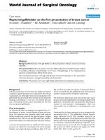
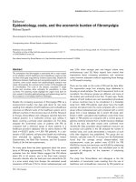
L. pneumophila (IgG titer = 512). F. tularensis was identified
by Western immunoblotting following cross-adsorption
(Figure 1). The patient retrospectively reported close con-
tact with rabbits 2 weeks prior to the beginning of the
symptoms of pericarditis.
Discussion
To study the etiological diagnosis of pericardial effusion,
we previously developed a diagnostic strategy that recom-
mends the systematic use of a combination of non-inva-
sive tests used to diagnose benign pericardial effusions
[3]. This strategy leads to a reduction in the number of
pericarditis cases classified as idiopathic compared with
an intuitive prescription of tests [4,5]. In our previous
experience of the etiological diagnosis of 204 cases of peri-
cardial effusions [3], F. tularensis was never found. Rare
cardiac complications have been reported in tularemic
infections including one case of endocarditis [6]. In 1958,
a historic description reported 28 cases of pericarditis due
to tularemia [7]. The postulate at that time was that peri-
carditis developed by direct extension from adjacent pleu-
ral effusion or from areas of pneumonia. Rare cases of
pericarditis have been described as complications of
tularemia sepsis caused by hematogenic spread during the
course of disease [2]. In our case, the pericardial effusion
was the only clinical manifestation of the disease.
Diagnosis is guided by clinical symptoms and confirmed
by serological results or culture. F. tularensis is a fastidious
organism that grows poorly on standard culture media.
Owing to achievements in technology, however,
tularemia can now be rapidly and specifically diagnosed.
Conventional polymerase chain reaction has been suc-
cessfully applied on wound specimens of patients acquir-
ing tularemia, and prospects for application on other
specimens in humans are promising [8].
Serological testing, especially the indirect immunofluo-
rescent antibody assay, remains the most commonly used
diagnostic test and is frequently the only available means
for the laboratory diagnosis of F. tularensis. Several serol-
ogy methods are available, including tube agglutination,
microagglutination, hemagglutination and enzyme-
linked immunosorbent assays [1]. Serological diagnosis
requires a four-fold or greater rise in antibody titer
between acute-phase and convalescent-phase sera. IgM,
IgA and IgG antibodies appear simultaneously after initial
infection and IgM antibodies can last for many years [9].
Initially, Evans reported that Brucella spp. and F. tularensis
Western immunoblottingFigure 1
Western immunoblotting. Legionella pneumophilia (LPNE)
and Francisella tularensis (FTUL).
Publish with BioMed Central and every
scientist can read your work free of charge
"BioMed Central will be the most significant development for
disseminating the results of biomedical research in our lifetime."
Sir Paul Nurse, Cancer Research UK
Your research papers will be:
available free of charge to the entire biomedical community
peer reviewed and published immediately upon acceptance
cited in PubMed and archived on PubMed Central
yours — you keep the copyright
Submit your manuscript here:
/>BioMedcentral
Journal of Medical Case Reports 2008, 2:206 />Page 3 of 3
(page number not for citation purposes)
contained common antigens [2]. Some serological cross-
reactions have been described, especially in IgM with Bru-
cella spp., Proteus OX19, and Yersinia pestis [10]. Serologi-
cal cross-reactions have also been encountered between
Legionella and Campylobacter, Mycoplasma, Chlamydia, Cit-
robacter freundii, Leptospira, and some mycobacteria [11]. To
the best of the authors' knowledge, there is no previous
description of serological cross-reaction between F. tula-
rensis and L. pneumophila. Western immunoblotting may
be useful in making etiological diagnoses and overcoming
confusing cross-reactivity. In our case, the specific anti-
bodies reactive to F. tularensis were detectable (FTUL, Fig-
ure 1).
Conclusion
Pericardial effusion due to F. tularensis is a rare complica-
tion. Serological cross-reactivity between Francisella and
other bacteria precludes identification of the species caus-
ing the infection when using migration inhibitory factor.
However, Western immunoblotting may help to over-
come some of these limitations in situations where sera
are the only available samples.
Competing interests
The authors declare that they have no competing interests.
Authors' contributions
CL participated in the analysis of bacterial tests and in
writing a first draft, PYL participated in collecting the data
and in following the patient's case, and contributed to the
discussion, GH participated in the diagnosis of pericardial
effusion in Marseille and generated the data, DR partici-
pated in the generation of the data, provided the results of
the bacterial tests and contributed to the discussion. All
authors read and approved the final manuscript.
Consent
Written informed consent was obtained from the patient
for publication of this case report and any accompanying
images. A copy of the written consent is available for
review by the Editor-in-Chief of this journal.
Acknowledgements
We thank Sandy Jones for reviewing the manuscript.
References
1. Ellis J, Oyston PC, Green M, Titball RW: Tularemia. Clin Microbiol
Rev 2002, 15:631-646.
2. Evans ME, Gregory DW, Schaffner W, McGee ZA: Tularemia: a 30-
year experience with 88 cases. Medicine (Baltimore) 1985,
64:251-269.
3. Levy PY, Corey R, Berger P, Habib G, Bonnet JL, Levy S, Messana T,
Djiane P, Frances Y, Botta C, DeMicco P, Dumon H, Mundler O,
Chomel JJ, Raoult D: Etiologic diagnosis of 204 pericardial effu-
sions. Medicine (Baltimore ) 2003, 82:385-391.
4. Levy PY, Khan M, Raoult D: Acute pericarditis. N Engl J Med 2005,
352:1154-1155.
5. Levy PY, Moatti JP, Gauduchon V, Vandenesch F, Habib G, Raoult D:
Comparison of intuitive versus systematic strategies for
aetiological diagnosis of pericardial effusion. Scand J Infect Dis
2005, 37:216-220.
6. Tancik CA, Dillaha JA: Francisella tularensis endocarditis. Clin
infect dis 2000, 30:399-400.
7. Adams CW: Tularemic pericarditis; reports of two cases and
reviews of literature. Dis Chest 1958, 34:632-639.
8. Tarnvik A, Chu MC: New approaches to diagnosis and therapy
of tularemia. Ann N Y Acad Sci 2007, 1105:378-404.
9. Tarnvik A, Berglund L: Tularaemia. Eur Respir J 2003, 21:361-373.
10. Behan KA, Klein GC: Reduction of Brucella species and Fran-
cisella tularensis cross-reacting agglutinins by dithiothreitol.
J Clin Microbiol 1982, 16:756-757.
11. Bornstein N, Fleurette J, Bosshard S, Bouvet C, Thouvenet D,
Aymard M: Evaluation de la fréquence des réactions
sérologiques croisées entre Legionella et Mycoplasma ou
Chlamydia. Pathol Biol 1984, 32:165-168.