Báo cáo y học: "Sweet''''s syndrome in a patient with Crohn''''s disease: a case report" pot
Bạn đang xem bản rút gọn của tài liệu. Xem và tải ngay bản đầy đủ của tài liệu tại đây (349.68 KB, 4 trang )
BioMed Central
Page 1 of 4
(page number not for citation purposes)
Journal of Medical Case Reports
Open Access
Case report
Sweet's syndrome in a patient with Crohn's disease: a case report
Nadia M Mustafa*
1
and Mark Lavizzo
†2
Address:
1
Internal Medicine Residency Program, College of Medicine, University of Illinois at Urbana-Champaign, USA and
2
Assistant professor,
College of Medicine, University of Illinois at Urbana-Champaign, USA
Email: Nadia M Mustafa* - ; Mark Lavizzo -
* Corresponding author †Equal contributors
Abstract
Background: Sweet's syndrome, also known as acute febrile neutrophilic dermatosis, has been
associated with malignancy, autoimmune disease and collagen vascular disease. The association of
Crohn's disease and Sweet's syndrome is rare. We report a case of Sweet's syndrome in a patient
with Crohn's disease.
Case presentation: A 63-year-old man with a history of Crohn's disease presented with one-
week duration of abdominal pain, diarrhea and hematochezia. He also noticed eruption of painful
skin rashes all over his body at the same time. Colonoscopy and esophagogastroduodenoscopy
(EGD) showed inflammation involving different parts of the gastrointestinal tract consistent with
Crohn's disease. Punch biopsy of the skin lesion was consistent with Sweet's syndrome, which has
a rare association with Crohn's disease.
Conclusion: Crohn's disease should be excluded in patients presenting with Sweet's syndrome
and diarrhea. Alternatively, Sweet's syndrome should be considered as a diagnosis when a patient
with Crohn's disease develops skin lesions.
Introduction
Sweet's syndrome, also known as acute febrile neu-
trophilic dermatosis, has rarely been associated with
Crohn's disease. We report a case of Sweet's syndrome in
a patient with Crohn's disease.
Case Presentation
A 63 year-old man with a history of Crohn's disease for
the past thirty years and hyperlipidemia presented with
one week of abdominal pain, diarrhea and hematochezia.
Abdominal pain was generalized, 6 by 10 in intensity on
the pain scale, and dull in character. It was worsened by
food intake and relieved by bowel movement. The
abdominal pain was associated with fever, chills, nausea
and vomiting. The patient also complained of painful
rashes all over his body that had erupted suddenly about
a week ago. The rashes were nonpruritic and had started
on the dorsum of his hands and spread to involve his face,
neck, chest and legs. He denied using any new creams,
soaps, detergents or perfumes or any change in his bed
sheets or clothing. He also denied contact with pets,
recent travel, a similar rash in any other family member,
or being bitten by an insect. He denied having had any
similar rash in the past. He had a history of Crohn's dis-
ease for the past thirty years which had been in remission
for several years, until the past few months when he began
to have episodes of diarrhea and rectal bleeding. Colonos-
copy two years ago had showed inflammatory bowel dis-
ease of segmental nature with rectal sparing and primarily
involving the ascending and sigmoid colon. His medica-
Published: 28 June 2008
Journal of Medical Case Reports 2008, 2:221 doi:10.1186/1752-1947-2-221
Received: 6 August 2007
Accepted: 28 June 2008
This article is available from: />© 2008 Mustafa and Lavizzo; licensee BioMed Central Ltd.
This is an Open Access article distributed under the terms of the Creative Commons Attribution License ( />),
which permits unrestricted use, distribution, and reproduction in any medium, provided the original work is properly cited.
Journal of Medical Case Reports 2008, 2:221 />Page 2 of 4
(page number not for citation purposes)
tions included Asacol which he had been taking for past
few months and azathioprine which was started two
weeks prior to his admission. He had previously been on
prednisone which was started two months earlier with his
last dose being four days prior to admission. His vital
signs on presentation were: Temperature 100.5°F, blood
pressure 95/58 mmHg, heart rate 120/min and respiratory
rate 21 b/min. On physical examination his abdomen was
mildly distended with tenderness to palpation in the left
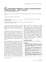
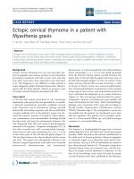
lower quadrant. He also had a papular rash and plaques,
with surrounding erythema, scattered over his face, neck,
chest and legs. (See Figure 1) These lesions were tender to
palpation. Laboratory results showed an elevated white
blood count (WBC) of 20.7 × 10
9
with 78% neutrophils
and 14% bands. Comprehensive metabolic panel was sig-
nificant for low sodium of 133 mEq/L and mildly elevated
renal function with a blood urea nitrogen of 20 mg/dL
and creatinine of 1.3 mg/dL and. His erythrocyte sedi-
mentation rate (ESR) and C reactive protein were also
high at 49 mm/hr and 161.6 mg/L respectively. Blood cul-
tures were negative. Other laboratory tests, which
included fungal serology, potassium hydroxide mount,
gram stain, acid fast bacilli smear, bacterial culture, fungal
culture and an acid fast culture of the skin rash, were all
negative. He was started empirically on intravenous van-
comycin for possible Methicillin Resistant Staphylococcus
Aureus folliculitis, pending the results of investigations.
Computed tomography (CT) scan of the abdomen on
admission showed inflammation involving the colon and
gastric and duodenal regions. Magnetic resonance angiog-
raphy (MRA) of the abdomen was negative for mesenteric
artery occlusion. Colonoscopy and esophagogastroduo-
denoscopy revealed pancolitis and gastroduodenitis con-
sistent with Crohn's disease. Biopsy specimens taken from
stomach, duodenum, ileum, ileocecal valve and colon
revealed pancolitis, duodenitis and gastritis with no evi-
dence of granuloma. The patient was diagnosed with an
exacerbation of Crohn's disease and started on intrave-
nous methylprednisolone 60 mg q 12 hrs, with continua-
tion of azathioprine and Asacol. He was also given a dose
of intravenous Infliximab. The rash showed no improve-
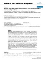
ment after three days of antibiotics. A punch biopsy of one
of the skin lesions revealed dense dermal infiltrate com-
posed predominantly of neutrophils, with no evidence of
vasculitis. This was consistent with the diagnosis of
Sweet's syndrome. (Figure 2). Antibiotic treatment was
stopped. The patient's symptoms and rash rapidly
improved with systemic corticosteroid treatment.
Discussion
Sweet's syndrome, also known as acute febrile neu-
trophilic dermatosis, was first described by Robert Doug-
las Sweet in 1964 [1]. Sweet's syndrome is characterized
by fever, neutrophilia, cutaneous eruptions consisting of
erythematous papules and plaques, and a dermal nonvas-
culitic neutrophilic infiltration on skin biopsy [2,3]. These
plaques are painful but nonpruritic [4]. Other skin mani-
festations such as pustules, vesicles, purpura, ulcers and
hemorrhagic lesions have been described [1]. Seventy-five
percent of patients have some prodromal illness, most
commonly an upper respiratory tract infection [5].
Common complications of Sweet's syndrome include
arthralgia, arthritis, conjunctivitis, iridocyclitis, and rarely
involvement of the central nervous system [4]. Sweet's
syndrome is more common in females with a female to
male ratio of 3.7:1, with the mean age of 52 years [1].
Pustular lesions with central necrosis on the patient's legFigure 1
Pustular lesions with central necrosis on the patient's
leg.
Punch biopsy of a skin lesion showing neutrophilic infiltration in the dermis, with no evidence of vasculitisFigure 2
Punch biopsy of a skin lesion showing neutrophilic
infiltration in the dermis, with no evidence of vasculi-
tis.
Journal of Medical Case Reports 2008, 2:221 />Page 3 of 4
(page number not for citation purposes)
Sweet's syndrome should be regarded as a cutaneous
marker of systemic disease. It has been associated with
malignancies in about 20 to 25% of patients [6]. Most
malignancies are hematopoietic, especially myelodysplas-
tic syndromes and acute myeloid leukemia. Fifteen per-
cent are due to solid tumors including breast,
genitourinary and gastrointestinal malignancies [7].
Other causes of Sweet's syndrome are listed in table 1.
Only a few cases of Sweet's syndrome associated with
Crohn's disease have been reported in the literature [1].
There is a higher incidence of colonic involvement and
extraintestinal features in these patients. The skin lesions
have been observed in patients with active Crohn's dis-
ease, but sometimes it can precede the onset of intestinal
symptoms. It appears that the syndrome is not initiated by
the underlying disease but rather shares with it a concur-
rent pathogenic mechanism.
The pathogenesis of Sweet's syndrome is poorly under-
stood. Cytokines, such as granulocyte colony stimulating
factor (G-CSF), interleukin (IL)-1, IL-6, or IL-8, if depos-
ited in the dermis, may be responsible for the immun-
opathologic and clinical manifestations of Sweet's
syndrome. The fact that Sweet's syndrome can occur after
G-
CSF treatment shows that IL-1, which is produced by
acute myelocytic leukemia (AML) cells and stimulates the
G-CSF gene, plays a role in the pathogenesis of Sweet's
syndrome [1].
For a definitive diagnosis of Sweet's syndrome, both
major and two minor criteria should be met. The two
major criteria are 1) abrupt onset of painful erythematous
plaques or nodules occasionally with vesicles, pustules, or
bullae and, 2) neutophilic infiltration in the dermis with-
out leukocytoclastic vasculitis. The minor criteria are 1)
skin lesions preceded by a nonspecific respiratory or gas-
trointestinal tract infection, vaccination or associated with
inflammatory diseases such as autoimmune disorders,
infections, hemoproliferative disorders, solid malignant
tumors or pregnancy, 2) accompanied by periods of gen-
eral malaise and fever (> 38°C), 3) laboratory values dur-
ing onset: ESR > 20 mm, C reactive protein positive,
segmented neutrophils >70% in peripheral blood smear,
leukocytosis > 8000 (3 of 4 of these values are necessary),
and 4) excellent response to treatment with systemic cor-
ticosteroids or potassium iodide [1,8].
Sweet's syndrome is one of the groups of neutrophilic der-
matoses that include pyoderma gangrenosum and whose
association with ulcerative colitis and Crohn's disease is
well established. Sweet's syndrome can be distinguished
from pyoderma gangrenosum by the absence of vasculitis
and lack of dermal necrosis, but histological features may
occasionally overlap. The abrupt tendency for Sweet's syn-
drome to form multiple eruptions on the upper half of the
body and the lack of ulceration also distinguishes the rash
from pyoderma gangrenosum. However, the two condi-
tions can occur in the same patient, as may other neu-
trophilic dermatosis, vesiculopapular eruptions or other
cutaneous features of inflammatory bowel disease such as
Table 1: Causes of Sweet's syndrome
Malignancies
Hematopeitic: myelodysplastic syndromes and acute myeloid leukemia, hairy cell leukemia, B and T cell lymphoma, agnogenic myeloid metaplasia
Solid tumors: breast, testicular, prostate, ovarian, vaginal squamous cell, genitourinary and gastrointestinal malignancies
Viral infections
Chronic active hepatitis, cytomegalovirus, human immunodeficiency virus
Bacterial infections
Streptococcus, mycobacterium, yersinia, typhus, salmonella
Autoimmune and collagen vascular diseases
Rheumatoid arthritis, systemic lupus erythematosus, mixed connective tissue disease, hashimoto thyroiditis, Sjogren's disease, behcet's disease
Medications
Furosemide, hydralazine, lithium, oral contraceptive pills, trimethoprim- sulfamethoxazole, minocycline and imatinib mesylate
Inflammatory bowel disease
Ulcerative Colitis, Crohn's disease
Pregnancy
Complement deficiency
Subacute necrotizing lymhadenitis
POEMS syndrome
Publish with BioMed Central and every
scientist can read your work free of charge
"BioMed Central will be the most significant development for
disseminating the results of biomedical researc h in our lifetime."
Sir Paul Nurse, Cancer Research UK
Your research papers will be:
available free of charge to the entire biomedical community
peer reviewed and published immediately upon acceptance
cited in PubMed and archived on PubMed Central
yours — you keep the copyright
Submit your manuscript here:
/>BioMedcentral
Journal of Medical Case Reports 2008, 2:221 />Page 4 of 4
(page number not for citation purposes)
erythema nodosum or polyarthritis. The simultaneous
occurrence of different rashes in the same person can be
viewed as the dermatological expression of a neutrophilic
reaction to a common stimulus [9].
Sweet's syndrome, if left untreated, usually heals within
six to eight weeks [5].
Prednisone at an initial dose of 40–60 mg per day, with
gradual tapering off over four to six weeks, is the standard
treatment for Sweet's syndrome [3,5]. Relapses are com-
mon if steroids are tapered too quickly. In recurrent dis-
ease, therapy with colchicine, potassium iodide, dapsone,
doxycycline, indomethacin, clofazimine, isotretinoin and
cyclosporine have all been described [1,5].
Potassium iodide administered orally as 300 mg enteric-
coated tablets, 3 times each day, for a daily dose of 900
mg, or as a saturated solution of potassium iodide
(Lugol's solution), beginning at a dose of 3 drops 3 times
each day (9 drops/day = 450 mg per day) and increasing
by 1 drop 3 times per day, typically to a final dose of 21
drops/day (1050 mg) to 30 drops/day (1500 mg), usually
results in resolution of fever and other symptoms within
1 to 2 days and resolution of skin lesions within 3 to 5
days of initiation of therapy. Vasculitis and hypothy-
roidism are potential adverse effects of potassium iodide
[10].
Conclusion
Sweet's syndrome should be considered an extraintestinal
manifestation of Crohn's disease, and should be differen-
tiated from other more frequent inflammatory diseases
that accompany Crohn's disease, like erythema nodusum,
pyoderma gangrenosum and leukocytoclastic vasculitis.
Awareness of this association may guide appropriate diag-
nostic procedures and therapy.
Competing interests
The authors declare that they have no competing interests.
Authors' contributions
NM was involved in the management of the patient while
in hospital, wrote the manuscript, collected all relevant
data, and finalized the manuscript for submission to the
journal, ML was involved in giving intellectual advice and
reviewing the manuscript.
Consent
Written informed consent was obtained from the patient
for publication of this case report and any accompanying
images.
Acknowledgements
Dr Niveditha Reddy MD.
References
1. Foster EN, et al.: Crohn's disease associated with Sweet's syn-
drome and Sjogren's syndrome treated with infliximab. Clin
Dev Immunol 2005, 12:145-9.
2. Kemmett D, Hunter JA: Sweet's syndrome: A clinicopathologic
review of 29 cases. J Am Acad Dermatol 1990, 23:503.
3. Driesch P Von den: Sweet's syndrome (acute febrile neu-
trophilic dermatosis). J Am Acad Dermatol 1994, 31:535.
4. Zamanian Abbas, Ameri Ahmad: Acute febrile neutrophilic der-
matosis (Sweet's syndrome): a study of 15 cases in Iran. Int J
Dermatol 2007, 46:571-4.
5. Vaz A, Kramer K, Kalish RA: Sweet's syndrome in association
with Crohn's disease. Postgrad Med J 2000, 76:713-4.
6. Cohen PR, Talpez M, Kurzrock R: Malignancy associated Sweet's
syndrome. Review of world literature. J Clin Oncology 1988,
6(12):1887-1897.
7. Cohen PR, Holder WR, Tucker SB, et al.: Sweet's syndrome
patients with solid tumors. Cancer 1993, 72:2723.
8. Su WP, Liu HN: Diagnostic criteria for Sweet's syndrome. Cutis
1986, 37:167.
9. Travis , et al.: Sweet's syndrome: an unusual cutaneous feature
of Crohn's disease or ulcerative colitis. The South West Gas-
troenterology Group. European journal of gastroenterology
and hepatology. Eur J Gastroenterol Hepatologyl 1986, 9(7):715-20.
10. Cohen Philip R: Sweet's syndrome – a comprehensive review
of an acute febrile neutrophilic dermatosis. Orphanet J Rare Dis
2007, 2:34.