Báo cáo y học: "Cervical lymphadenopathy – an unusual presentation of carcinoma of the cervix: a case report" ppt
Bạn đang xem bản rút gọn của tài liệu. Xem và tải ngay bản đầy đủ của tài liệu tại đây (299.02 KB, 5 trang )
BioMed Central
Page 1 of 5
(page number not for citation purposes)
Journal of Medical Case Reports
Open Access
Case report
Cervical lymphadenopathy – an unusual presentation of carcinoma
of the cervix: a case report
Madhavi Manoharan*
1,2
, Durga Satyanarayana
1
and Arjun R Jeyarajah
1
Address:
1
St Bartholomew's Hospital, London, UK and
2
17, Benhurst Avenue, Elm Park, Hornchurch, Essex, RM12 4QS, UK
Email: Madhavi Manoharan* - ; Durga Satyanarayana - ;
Arjun R Jeyarajah -
* Corresponding author
Abstract
Introduction: The clinical presentation of carcinoma of the cervix as cervical lymphadenopathy
has not been described before. We report a case of this unusual manifestation of cervical cancer.
Case presentation: A 51-year-old woman presented to our Head and Neck department with
cervical lymphadenopathy. A positron emission tomography scan revealed the primary tumour to
be in the cervix and a cervical biopsy confirmed carcinoma of the cervix.
Conclusion: Recurrences of carcinoma of the cervix presenting as lymphadenopathy have been
described before but this is the first time a clinical presentation of carcinoma of the cervix as
cervical lymphadenopathy has been described. Although metastasis from the cervix to the cervical
lymph nodes is rare, this can be explained by outlining the drainage of the lymphatic system from
the cervix.
Introduction
Carcinoma of the cervix commonly metastasizes by direct
extension or lymphatic dissemination within the pelvis.
Clinical presentation of carcinoma of the cervix as cervical
lymphadenopathy has not been described before. We
report a case of this unusual manifestation of cervical can-
cer.
Case presentation
A 51-year-old woman was referred to the ENT department
with a 2-week history of a lump on the right side of her
neck. There was no history of change to her voice or dys-
phagia.
She is a para 4 with all normal vaginal deliveries and has
had normal cervical smears in the past. Her periods were
regular and she gave no history of intermenstrual or post-
coital bleeding. She smoked about 20–30 cigarettes per
day.
On further questioning in the clinic, she gave a history of
increasing lethargy for the past 3 months and was also
unable to report to work due to severe back pain.
Five years before the present episode, she reported feeling
unwell with significant weight loss and heavy periods. She
was found to be anaemic and was given five units of
blood. She was investigated for a possible colon cancer
which proved to be negative. She was referred to a Men-
strual Disorder Clinic but failed to attend the clinic twice.
On examination, multiple cervical lymph nodes were pal-
pable on both sides of the neck. Ultrasound scan of the
neck revealed two large supraclavicular lymph nodes with
Published: 28 July 2008
Journal of Medical Case Reports 2008, 2:252 doi:10.1186/1752-1947-2-252
Received: 8 July 2007
Accepted: 28 July 2008
This article is available from: />© 2008 Manoharan et al; licensee BioMed Central Ltd.
This is an Open Access article distributed under the terms of the Creative Commons Attribution License ( />),
which permits unrestricted use, distribution, and reproduction in any medium, provided the original work is properly cited.
Journal of Medical Case Reports 2008, 2:252 />Page 2 of 5
(page number not for citation purposes)
several abnormal looking lymph nodes in the right
carotid chain.
An X-ray of the chest showed no abnormality. Fine needle
aspiration of the lymph nodes yielded squamous carci-
noma cells.
Metastatic squamous cell carcinoma of an unknown pri-
mary tumour was suspected and investigations were per-
formed to find a possible primary site. Clinical
examination and endoscopy of the upper digestive tract
did not yield an obvious primary tumour in the nasophar-
ynx, larynx and hypopharynx.
Computerised Tomography (CT) of the neck, chest and
abdomen revealed marked mediastinal and para-aortic
lymphadenopathy suggestive of spread of the known
squamous cell carcinoma. There was evidence of dilata-
tion of the collecting system bilaterally with dilatation of
the proximal ureters suggesting an obstruction within the
pelvis.
A Positron Emission Tomography-CT (PET-CT) scan was
performed which showed markedly increased uptake in
the right cervical lymph nodes, as well as in the right par-
atracheal, anterior mediastinal, lower para-aortic, and
bilateral iliac lymph nodes with an obturator node show-
ing a photopaenic centre. In addition, there was a focal
area of increased uptake in the pelvis, suggesting a lesion
within the rectal wall or in the vaginal vault (Figures 1 and
2).
Given the histology of squamous carcinoma, the PET scan
suggested that the uptake in the pelvis may represent a pri-
mary gynaecological problem rather than a second malig-
nancy in the rectum. But given the distribution of the
disease which was very unusual for cervical carcinoma, a
review of the histology was suggested with a differential
diagnosis of lymphoma to be considered. The histology
from fine needle aspiration of the cervical lymph node
confirmed it to be carcinoma cells of squamous origin.
Our patient was then referred to the gynae-oncology team.
On examination, the uterus was anteverted, mobile and
bulky corresponding to about 14 weeks' size with no pal-
pable adnexal masses. Her cervix appeared normal to the
naked eye and a smear was obtained which was reported
as normal.
Magnetic Resonance Imaging (MRI) of the pelvis and
abdomen was performed which revealed a highly abnor-
mal cervix, diffusely infiltrated by an intermediate to high
T2 signal intensity mass measuring approximately 3 × 4 ×
3.5 cm. The mass involved the endocervical canal and the
stroma with suspected early parametrial invasion anteri-
orly. There was no convincing evidence to suggest bladder
involvement and the rectum was clear of disease. Several
small intramural fibroids were demonstrated within the
myometrium as well as a submucosal fibroid in the ante-
rior body of the uterus (Figure 3).
There was extensive lymphadenopathy along both pelvic
side walls, common iliac regions and the para-aortic
regions but with no evidence of inguinal lymphadenopa-
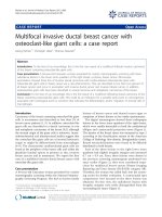
Coronal PET image of FDG uptake and excretion in the chest, abdomen and pelvisFigure 2
Coronal PET image of FDG uptake and excretion in
the chest, abdomen and pelvis.
Anterior mediastinal nodes
Right para-aortic nodes
Abnormal focal uptake in pelvis
(?vagina or rectum)
*
*
*
*
+
Normal colonic mucosal uptake(+)
Normal renal tract excretion (
*
)
Coronal PET image of FDG uptake in the head and neckFigure 1
Coronal PET image of FDG uptake in the head and
neck.
*
Right cervical nodes
Right paratracheal nodes
Normal cerebral uptake (
*)
Journal of Medical Case Reports 2008, 2:252 />Page 3 of 5
(page number not for citation purposes)
thy. Bilateral hydronephrosis was noted. No bony deposit
was seen. In conclusion, the MRI reported that the appear-
ance was consistent with a cervical carcinoma with exten-
sive lymphadenopathy and hydronephrosis, stage FIGO
3b.
Routine blood investigations before examination under
anaesthesia showed her to be anaemic with a haemo-
globin level of 6 g/dl. She was transfused with four units
of blood. Her liver function tests and renal function tests
were normal and serology showed her to be negative for
HIV.
She had an examination under anaesthesia, cervical
biopsy and an endocervical and endometrial curettage.
Examination under anaesthesia showed the cervix to be
bulky with an intact surface epithelium. There was no par-
ametrial involvement and the rectum and bladder were
free. Hysteroscopy revealed a pedunculated fibroid on the
anterior wall of the uterus. Large biopsies of the anterior
and posterior lip of the cervix were taken which identified
a poorly differentiated squamous cell carcinoma of the
anterior lip of the cervix. The endocervical curettings were
positive for squamous cell carcinoma and the endometrial
curettings showed proliferative phase endometrium.
With an impression of metastatic squamous cell carci-
noma of the cervix, she was started on palliative chemo-
therapy with carboplatin and paclitaxel. She has
responded well to the therapy with a reported decrease in
the size of the neck nodes.
Discussion
In the case of carcinoma of the cervix, metastasis to the
neck signals a grave prognosis for the patient. Although
very uncommon, spread of carcinoma from the uterine
cervix to the supraclavicular region is best understood
through a description of the lymphatic system. Carcinoma
of the cervix uteri spreads by lymphatics from the pelvis
up to the para-aortic nodes, into the mediastinum and
then into the thoracic duct. Spread can occur from the pel-
vis into the hepatic region through the diaphragm and the
thoracic duct. The thoracic duct communicates with the
central venous system in the neck at the junction of the
left subclavian and internal jugular vein. The left-sided
supraclavicular node represents the final common path of
the body's infra-diaphragmatic lymphatic drainage [1].
Small communications exist from the left side to the right
side of the neck.
On reaching the lymph nodes, the embolus of tumour
cells begins to multiply, and penetrates the subcapsular
tissue leading to local spread into the region surrounding
the lymph node. Blockage of the lymph nodes leads to ret-
rograde spread of tumour. This would account for spread
from the left side to the right side of the neck, even though
there is no direct connection to the right side.
In Henriksen's study [2], incidence of metastasis of carci-
noma of the cervix to left supraclavicular nodes was 0.1%
in untreated patients but up to 1.5% in treated patients. As
further recent studies have shown, modern radiotherapy
achieves better control of cancer in the pelvis and allows
more patients to survive longer, which, in turn, permits
distant metastases to become clinically evident. Hilar,
mediastinal [3,4] and supraclavicular lymphadenopathy
[5] have been described as the first evidence of tumour
recurrence.
But the first presentation of cancer of the cervix with dis-
tant metastases in the supraclavicular nodes with a nor-
mal looking cervix has not been described before.
The eventual diagnosis of cervical cancer in our patient
has been difficult. When she first presented to the Head
and Neck department, diagnostic work-up for cervical
metastases from an unknown primary was done. As part
of this intensive work-up, a (18)F-fluorodeoxyglucose
positron emission tomography with computed tomogra-
phy (FDG-PET-CT) was done, which surprisingly sug-
gested the possibility of a primary in the cervix.
PET is a functional diagnostic imaging technique and has
the advantage of being non-invasive and able to study the
biological function of the tumour. Increased glucose
metabolism has been observed in tumours [6] and F-18
Sagittal T2-weighted MR image through the midline of the pelvisFigure 3
Sagittal T2-weighted MR image through the midline
of the pelvis.
*
+
Submucosal fibroids
Mass in endocervix
Uterine fundus (+)
Bladder (
*)
Journal of Medical Case Reports 2008, 2:252 />Page 4 of 5
(page number not for citation purposes)
fluoro-2-deoxy-d-glucose (FDG) is a commonly used radi-
opharmaceutical and is an analogue of glucose [7].
Guntinas-Lichius et al. [8] have shown FDG-PET to have
the best sensitivity of 69% and the highest negative pre-
dictive value of 87% in detecting unknown primary
tumours.
Other studies have shown FDG-PET to have a sensitivity
of 100% and sensitivity of 94% in the detection of
unknown primary tumours. For the conventional diag-
nostic modalities (CT and/or MRI, panendoscopy), these
values were 92% and 76% [9].
On retrospective review of her past history, her admis-
sions and blood transfusions for anaemia could have
been related to underlying cancer of the cervix. But since
she did not keep her appointments with the gynaecology
clinics, that window of opportunity was lost.
In keeping with her past history, investigation by the
gynae-oncology team soon after the CT scan (which had
suggested extensive lymphadenopathy) for a possible cer-
vical cause for the lymphadenopathy, would have proba-
bly been more cost effective. Due to limited availability
and higher cost of the PET scan, it is not routinely used as
a primary tool of evaluation. A more thorough work-up
and use of other less expensive modalities would have
shown the primary to be in the cervix.
It is very unusual for squamous cell carcinoma of the cer-
vix to behave in an aggressive way with metastasis to
extrapelvic lymph nodes. Small cell cancer of the cervix is
known to be aggressive with early haematogenous and
extrapelvic lymph node metastasis [10].
The prognosis for metastatic carcinoma of the cervix is
poor. Metastases to the neck signal a grave prognosis for
the patient. Diddle [5], in his retrospective review of 18
cases of cervical cancer with metastases to supraclavicular
nodes, has quoted a survival time of between 1 and 16
months after the appearance of metastases.
If left alone, cervical nodes grow rapidly with the attend-
ant sequelae of ulceration and pain, making treatment dif-
ficult or impossible. Treatment is usually with local
irradiation [11].
Treatment of advanced cervical cancer is usually palliative.
Several chemotherapy regimes have been described. Cis-
platin has emerged as the most active single agent with
overall response rates of 19% [12]. Recent phase III trials
have documented response rates of 27% and 39% when
cisplatin was combined with either paclitaxel or topote-
can, respectively [12].
The comparison of cisplatin to cisplatin plus topotecan in
GOG-179 has shown a statistically significant impact on
the overall response rate, median progression-free sur-
vival, and median survival, with all outcome measures
favouring the two-drug regimen [13].
Our patient is presently undergoing palliative chemother-
apy with a combination of carboplatin and paclitaxel. Her
initial response has been encouraging with an anticipated
improvement in quality-of-life scores.
Conclusion
Recurrences of carcinoma of the cervix presenting as lym-
phadenopathy have been described before but this is the
first time a clinical presentation of carcinoma of the cervix
as cervical lymphadenopathy has been described.
Although metastasis to the cervical lymph nodes is rare,
this can be explained by outlining the drainage of the lym-
phatic system from the cervix. Prognosis in such patients
is usually poor and treatment is mainly palliative.
Although the management of our patient has not
changed, this case report highlights an unusual presenta-
tion of carcinoma of the cervix and the investigative
modalities which were needed to reach the final diagno-
sis.
Abbreviations
ENT: Ear, Nose and Throat; CT: Computerised tomogra-
phy; PET: Positron emission tomography; MRI: Magnetic
resonance imaging; FIGO: International Federation of
Gynaecology and Obstetrics; HIV: human immunodefi-
ciency virus; FDG: fluorodeoxyglucose.
Competing interests
The authors declare that they have no competing interests.
Authors' contributions
MM: Literature review, conceived and drafted the manu-
script, DS, Helped in collecting records and preparing the
manuscript, AJ, Department chair who provided general
support. All authors revised and approved the final draft
of the manuscript.
Consent
Written consent was obtained from the patient for publi-
cation of the case report and any accompanying images. A
copy of the written consent is available for review by the
Editor-in-Chief of this journal.
Acknowledgements
The authors would like to thank Dr Sameer Gangoli for help with the
images. They would also like to declare that no funding has been received
for the preparation of the manuscript.
Publish with BioMed Central and every
scientist can read your work free of charge
"BioMed Central will be the most significant development for
disseminating the results of biomedical research in our lifetime."
Sir Paul Nurse, Cancer Research UK
Your research papers will be:
available free of charge to the entire biomedical community
peer reviewed and published immediately upon acceptance
cited in PubMed and archived on PubMed Central
yours — you keep the copyright
Submit your manuscript here:
/>BioMedcentral
Journal of Medical Case Reports 2008, 2:252 />Page 5 of 5
(page number not for citation purposes)
References
1. Ellison E, LaPuerta P, Martin SE: Supraclavicular masses: results
of a series of 309 cases biopsied by fine needle aspiration.
Head Neck 1999, 21(3):239-246.
2. Henriksen E: Lymphatic spread of carcinoma of cervix and
body of uterus. Am J Obstet Gynecol 1949, 58:924.
3. Shin MS, Shingleton HM, Partridge EE, Nicolson VM, Ho KJ: Squa-
mous cell carcinoma of the uterine cervix. Patterns of tho-
racic metastases. Invest Radiol 1995, 30(12):724-729.
4. Scott I, Bergin CJ, Muller NL: Mediastinal and hilar lymphaden-
opathy as the only manifestation of metastatic carcinoma of
the cervix. Can Assoc Radiol J 1986, 37(1):52-53.
5. Diddle AW: Carcinoma of the cervix uteri with metastases to
the neck. Cancer 1972, 29(2):453-455.
6. Warburg O: The Metabolism of Tumours New York, NY: Richard R.
Smith Inc; 1931:129-169.
7. Kumar R, Chauhan A, Jana S, Dadparvar S: Positron emission tom-
ography in gynecological malignancies. Expert Rev Anticancer
Ther 2006, 6(7):1033-1044.
8. Guntinas-Lichius O, Klissmann JP, Dinh S, Dinh M, Schmidt M, Semrau
R, Mueller RP: Diagnostic work up and outcome of cervical
metastases from an unknown primary. Acta Otolaryngol 2006,
126(5):536-544.
9. Regelink G, Brouwer J, de Bree R, Pruim J, Laan BF van der, Vaalburg
W, Hoekstra OS, Comans EF, Vissink A, Leemans CR, Roodenburg
JL: Detection of unknown primary tumours and distant
metastases in patients with cervical metastases: value of
FDG-PET versus conventional modalities. Eur J Nucl Med Mol
Imaging 2002, 29(8):1024-1030.
10. Trinh XB, Bogers JJ, Van Marck EA, Tjalma WA: Treatment policy
of neuroendocrine small cell cancer of the cervix. Eur J Gynae-
col Oncol 2004, 25(1):40-44.
11. Carlson V, Delclos L, Fletcher GH: Distant metastases in squa-
mous cell carcinoma of the uterine cervix. Radiology 1967,
88:961-966.
12. Moore DH, Blessing JA, McQuellon RP, Thaler HT, Cella D, Benda J,
Miller DS, Olt G, King S, Boggess JF, Rocereto TF: Phase III study
of cisplatin with or without paclitaxel in stage IVB, recur-
rent, or persistent squamous cell carcinoma of the cervix: a
gynaecologic oncology group study. J Clin Oncol 2004,
22(15):3113-3119.
13. Tewari KS, Monk BJ: Gynecologic oncology group trials of
chemotherapy for metastatic and recurrent cervical cancer.
Curr Oncol Rep 2005, 7(6):419-434.