Báo cáo y học: " Isolated left ventricular non-compaction as an unusual cause of heart failure: a case report" pdf
Bạn đang xem bản rút gọn của tài liệu. Xem và tải ngay bản đầy đủ của tài liệu tại đây (244.11 KB, 3 trang )
BioMed Central
Page 1 of 3
(page number not for citation purposes)
Journal of Medical Case Reports
Open Access
Case report
Isolated left ventricular non-compaction as an unusual cause of
heart failure: a case report
Nicholas LM Cruden and Martin A Denvir*
Address: Department of Cardiology, Royal Infirmary of Edinburgh, Little France Crescent, Little France, Edinburgh, EH16 5SA, UK
Email: Nicholas LM Cruden - ; Martin A Denvir* -
* Corresponding author
Abstract
Introduction: Isolated left ventricular non-compaction is a recently described form of
cardiomyopathy that is associated with a significant risk of life-threatening arrhythmia and
thromboembolic complications.
Case presentation: We report the presentation, diagnosis and management of isolated left
ventricular non-compaction in a 54-year-old Caucasian woman presenting with progressive
symptoms of heart failure.
Conclusion: Advances in diagnostic imaging have undoubtedly led to an increase in the detection
of isolated left ventricular non-compaction. Diagnosing and differentiating this uncommon
condition from other forms of cardiomyopathy are important as treatment and prognosis may
differ significantly. Our current understanding of isolated left ventricular non-compaction, including
diagnostic criteria, management and prognosis, is discussed.
Introduction
During normal foetal development, myocardial compac-
tion usually occurs by day 70 in utero. Where this process
fails to take place, prominent left ventricular trabecula-
tions may remain that persist into adult life. In the
absence of significant cardiac outflow tract obstruction,
the presence of extensive left ventricular trabeculation is
associated with the development of left ventricular systo-
lic impairment, cardiac arrhythmias and systemic throm-
boembolism. Recent advances in diagnostic imaging
techniques have led to an increase in the detection of this
previously rare form of cardiomyopathy, known as iso-
lated left ventricular non-compaction (IVNC). It is impor-
tant that clinicians recognise and differentiate this
condition from other forms of cardiomyopathy as treat-
ment and prognosis may differ significantly.
Case presentation
A 54 old-year-old Caucasian woman was admitted with a
3-month history of progressive exertional breathlessness,
orthopnoea and chest tightness. On examination she was
in sinus rhythm with a rate of 66 beats/minute and a
blood pressure of 90/60 mmHg. Auscultation revealed a
first and second heart sound with no added sounds and
no murmurs, reduced air entry at both lung bases and
coarse crepitations at the left lung base.
Serum urea, electrolytes, thyroid function, ferritin and full
blood count were all within normal limits. A chest X-ray
demonstrated cardiomegaly with small bilateral pleural
effusions. The electrocardiogram confirmed sinus rhythm
with left atrial enlargement, low voltage QRS complexes
and lateral T wave inversion. Transthoracic echocardiogra-
Published: 13 August 2008
Journal of Medical Case Reports 2008, 2:269 doi:10.1186/1752-1947-2-269
Received: 20 February 2007
Accepted: 13 August 2008
This article is available from: />© 2008 Cruden and Denvir; licensee BioMed Central Ltd.
This is an Open Access article distributed under the terms of the Creative Commons Attribution License ( />),
which permits unrestricted use, distribution, and reproduction in any medium, provided the original work is properly cited.
Journal of Medical Case Reports 2008, 2:269 />Page 2 of 3
(page number not for citation purposes)
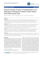
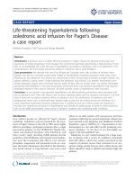
phy demonstrated a dilated left ventricle (end systolic
diameter 5.5 cm; end diastolic diameter 5.9 cm) with
severe systolic impairment and hypertrabeculation of the
left ventricular apex (Fig. 1) in the absence of significant
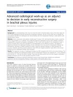
valvular heart disease. Doppler colour flow mapping con-
firmed colour flow between the trabeculations (Fig. 2).
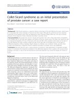
Intravenous injection of ultrasound contrast agent con-
firmed an area of non-compacted myocardium subtend-
ing a thinner walled area of compaction and a diagnosis
of IVNC was made (Fig. 3).
Discussion
Isolated left ventricular non-compaction is a recently
described cardiomyopathy [1], the true prevalence of
which remains unknown. Advances in diagnostic imaging
modalities have undoubtedly led to an increase in detec-
tion of this rare condition and it is likely that earlier cases
have been misdiagnosed as phenotypically similar cardio-
myopathies, such as apical hypertrophic cardiomyopathy
[2], where prognosis and treatment may differ signifi-
cantly. The purpose of this case report is to highlight the
diagnosis of IVNC and briefly review our current under-
standing of the condition.
The presence of marked left ventricular trabeculation in
patients with IVNC is believed to arise as a result of intra-
uterine arrest of left ventricular myocardial compaction,
although the trigger for this phenomenon is not yet
known. Both familial and sporadic forms of IVNC have
been described and although no causative gene has yet
been identified, familial screening is recommended [3,4].
Echocardiography remains the reference standard for the
diagnosis of IVNC [5]. Jenni and colleagues identified
four criteria for the diagnosis of IVNC by echocardiogra-
phy [5]. A thick, inner layer of non-compacted myocar-
dium is present subtending an outer, thin compacted
layer of myocardium with ratio of non-compacted to
Transthoracic echocardiographyFigure 1
Transthoracic echocardiography. Apical four chamber
view demonstrating marked trabeculation of the left ven-
tricular apex (arrow). RA, right atrium; LA, left atrium; LV,
left ventricle; RV, right ventricle; PE, pleural effusion.
Transthoracic echocardiographyFigure 2
Transthoracic echocardiography. Doppler colour flow
mapping suggesting blood flow present between the ventricu-
lar trabeculations (arrow). RA, right atrium; LA, left atrium;
LV, left ventricle; RV, right ventricle.
Transthoracic echocardiographyFigure 3
Transthoracic echocardiography. Following intravenous
injection, contrast agent is visualised between the ventricular
trabeculations (arrow). LV, left ventricle.
Publish with BioMed Central and every
scientist can read your work free of charge
"BioMed Central will be the most significant development for
disseminating the results of biomedical research in our lifetime."
Sir Paul Nurse, Cancer Research UK
Your research papers will be:
available free of charge to the entire biomedical community
peer reviewed and published immediately upon acceptance
cited in PubMed and archived on PubMed Central
yours — you keep the copyright
Submit your manuscript here:
/>BioMedcentral
Journal of Medical Case Reports 2008, 2:269 />Page 3 of 3
(page number not for citation purposes)
compacted myocardium during systole being greater than
2:1. When the left ventricle is divided into nine segments,
non-compacted myocardium is present predominantly
(more than 80%) on the apical and mid-ventricular
aspects of the inferior and lateral walls. Deeply perfused
intertrabecular recesses that do not communicate with the
coronary circulation can be identified using colour Dop-
pler (Fig. 2). The use of an intravenous ultrasound con-
trast agent (Fig. 3) or Doppler tissue imaging may
improve visualisation of the left ventricular intertrabecu-
lar recesses. These features should be present in the
absence of any other significant cardiac abnormality.
Cardiac magnetic resonance imaging may also be of use in
the diagnosis of IVNC, in particular in individuals where
the image quality at echocardiography is limited [6]. In
contrast to echocardiography, with cardiac magnetic reso-
nance imaging the ratio of non-compacted to compacted
myocardium should be measured during diastole, a ratio
of greater than 2.3:1 confirming pathological trabecula-
tion of the left ventricle [6].
Treatment for patients with IVNC should be directed at
the management of left ventricular systolic impairment
where present; the detection, treatment and prevention of
arrhythmias; and the prevention of systemic embolic
events [3,4]. In addition to treatment with angiotensin-
converting enzyme inhibitors, β-blockers and, where
appropriate, diuretics and/or digoxin, all patients with
IVNC should be screened annually with 24-hour electro-
cardiogram recordings and considered for long-term pro-
phylactic anticoagulation with warfarin. The high
incidence of sudden death reported in patients with IVNC
has prompted some authors to advocate a strategy of
"early" automated implantable cardiodefibrillator
implantation [3]. The role of biventricular pacemakers in
this population remains unclear. Finally, where pharma-
cological therapy fails to halt the progression to cardiac
failure, heart transplantation should be considered [4].
Initial data from Europe and America reported a 4- to 6-
year combined mortality or transplantation rate of ~50%
to 60% [3,4] although recent UK data indicate the prog-
nosis may be more favourable [7]. Our patient responded
well to the introduction of angiotensin-converting
enzyme inhibition, beta-blockade and warfarin anticoag-
ulation but is currently being considered for cardiac trans-
plantation.
Conclusion
Isolated left ventricular non-compaction is a rare but
important form of cardiomyopathy that should not be
overlooked in patients presenting with cardiac failure.
This case report emphasises the importance of differenti-
ating this condition from alternative diagnoses where
treatment and prognosis may vary significantly.
Abbreviations
IVNC: Isolated left ventricular non-compaction.
Competing interests
The authors declare that they have no competing interests.
Authors' contributions
NLMC and MAD made the diagnosis and were involved in
subsequent management. MAD was the Consultant
responsible for the patient. Both authors have drafted,
read and approved the manuscript.
Consent
Written informed consent was obtained from the patient
for publication of this case report and accompanying
images. A copy of the written consent is available for
review by the Editor-in-Chief of this journal.
References
1. Chin TK, Perloff JK, Williams RG, Jue K, Mohrmann R: Isolated non-
compaction of left ventricular myocardium. A study of eight
cases. Circulation 1990, 82:507-513.
2. McCulloch C, Jadhav S, Glen S, Bridges A: Isolated left ventricular
non-compaction, not hypertrophic cardiomyopathy. QJM
2004, 97:827-828.
3. Oechslin EN, Attenhofer Jost CH, Rojas JR, Kaufmann PA, Jenni R:
Long-term follow-up of 34 adults with isolated left ventricu-
lar noncompaction: a distinct cardiomyopathy with poor
prognosis. J Am Coll Cardiol 2000, 36:493-500.
4. Weiford BC, Subbarao VD, Mulhern KM: Noncompaction of the
ventricular myocardium. Circulation 2004, 109:2965-2971.
5. Jenni R, Oechslin EN, Loo B van der: Isolated ventricular non-
compaction of the myocardium in adults. Heart 2007,
93:11-15.
6. Petersen SE, Selvanayagam JB, Wiesmann F, Robson MD, Francis JM,
Anderson RH, Watkins H, Neubauer S: Left ventricular non-com-
paction: insights from cardiovascular magnetic resonance
imaging. J Am Coll Cardiol 2005, 46:101-105.
7. Murphy RT, Thaman R, Blanes JG, Ward D, Sevdalis E, Papra E, Kiot-
sekolglou A, Tome MT, Pellerin D, McKenna WJ, Elliott PM: Natural
history and familial characteristics of isolated left ventricular
non-compaction. Eur Heart J 2005, 26:187-192.