Báo cáo y học: "Bezoar in gastro-jejunostomy presenting with symptoms of gastric outlet obstruction: a case report and review of the literature" pdf
Bạn đang xem bản rút gọn của tài liệu. Xem và tải ngay bản đầy đủ của tài liệu tại đây (772.09 KB, 5 trang )
BioMed Central
Page 1 of 5
(page number not for citation purposes)
Journal of Medical Case Reports
Open Access
Case report
Bezoar in gastro-jejunostomy presenting with symptoms of gastric
outlet obstruction: a case report and review of the literature
Edmund Leung*, Ruth Barnes and Ling Wong
Address: Department of Surgery, University Hospitals Coventry and Warwickshire, Coventry, CV2 2DX, UK
Email: Edmund Leung* - ; Ruth Barnes - ; Ling Wong -
* Corresponding author
Abstract
Introduction: Gastric outlet obstruction usually presents with non-bilious vomiting, colicky
epigastric pain, loss of appetite and occasionally, upper gastrointestinal bleeding. Causes can be
classified as benign or malignant, or as extra- or intraluminal. Gastrojejunostomy is a well-
recognised surgical procedure performed to bypass gastric outlet obstruction. A bezoar occurs
most commonly in patients with impaired gastrointestinal motility or with a history of gastric
surgery. It is an intestinal concretion, which fails to pass along the alimentary canal.
Case presentation: A 62-year-old Asian woman with a history of gastrojejunostomy for peptic
ulcer disease was admitted to hospital with epigastric pain, vomiting and dehydration. All
investigations concluded gastric outlet obstruction secondary to a "stricture" at the site of
gastrojejunostomy. Subsequent laparotomy revealed that the cause of the obstruction was a
bezoar.
Conclusion: Many bezoars can be removed endoscopically, but some will require operative
intervention. Once removed, emphasis must be placed upon prevention of recurrence. Surgeons
must learn to recognise and classify bezoars in order to provide the most effective therapy.
Introduction
Gastric outlet obstruction (GOO) in adults is not a single
entity; it is the pathophysiological consequence of any
disease process that produces a mechanical impediment
to gastric emptying. There are benign and malignant
causes. In the past, peptic ulcer disease was more preva-
lent than malignant causes, currently, it only accounts for
5% of all cases of GOO [1]. With the advent of proton
pump inhibitors and Helicobacter pylori eradication ther-
apy, this benign cause has become less common. Anders-
son and Bergdahl reported [2] that 67% of patients have
GOO secondary to malignancy. Other benign intralumi-
nal causes in adults include gastric polyps, caustic inges-
tion, gallstone obstruction (Bouveret syndrome), and
bezoars.
Bezoars, concretions of indigestible material in the gas-
trointestinal tract, have been known to occur in animals
for centuries. The incidence of bezoars in adult patients
has increased as a result of operative manipulation of the
gastrointestinal tract. Although bezoars are often recog-
nised radiologically, endoscopy provides the most accu-
rate means of identification. Many bezoars can be
removed endoscopically, but some will require operative
intervention. Once removed, emphasis must be placed
upon prevention of recurrence. Surgeons must learn to
Published: 2 October 2008
Journal of Medical Case Reports 2008, 2:323 doi:10.1186/1752-1947-2-323
Received: 5 March 2008
Accepted: 2 October 2008
This article is available from: />© 2008 Leung et al; licensee BioMed Central Ltd.
This is an Open Access article distributed under the terms of the Creative Commons Attribution License ( />),
which permits unrestricted use, distribution, and reproduction in any medium, provided the original work is properly cited.
Journal of Medical Case Reports 2008, 2:323 />Page 2 of 5
(page number not for citation purposes)
recognise and classify bezoars in order to provide the most
effective therapy.
We report a case of a 62-year-old Asian woman with a his-
tory of gastrojejunostomy, who was admitted to hospital
with GOO secondary to a bezoar. We present the case, dis-
cuss management and review the literature.
Case presentation
A 62-year-old Asian woman presented acutely to the
emergency department with a 1-day history of colicky epi-
gastric pain and postprandial vomiting. She had been tol-
erating only liquids rather than solid food for 2 months.
There was no history of weight loss, but she did report
early satiety and loss of appetite.
This woman had a history of peptic ulcer disease over 20
years ago in Kenya. It had led to GOO requiring truncal
vagotomy and gastrojejunostomy. In order to investigate
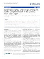
the cause of her dysphagia and loss of appetite, she had
undergone an upper gastrointestinal endoscopy 3 weeks
before this admission. This showed inflammation and
oedema at the anastomotic site of the gastrojejunostomy,
but no evidence of obstruction or stricture (Figure 1). She
was then prescribed daily omeprazole, which was the only
medication she was taking on admission.
The patient was clinically dehydrated on examination.
She had a very thin body habitus. Her abdomen was soft,
but mildly tender over her epigastrium. Succussion splash
was demonstrated and a 10 cm × 8 cm mass was palpable
just right of the umbilicus. Bowel sounds were scanty.
There were no clinical signs for upper gastrointestinal
bleeding.
Her admission blood profiles were essentially unremark-
able. There was no biochemical evidence of fluid shifts or
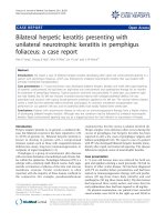
dehydration. Plain abdominal radiograph did not show
any diagnostic features. However, her erect chest radio-
graph showed an air-fluid level within a dilated stomach
(Figure 2a).
In view of the examination and chest radiograph findings,
she had a nasogastric tube and urinary catheter inserted
for gastric decompression and urine output monitoring,
respectively. An urgent contrasted computed tomography
of the abdomen was arranged. Meanwhile, the nasogastric
tube successfully prevented further vomiting, and there
was little drainage from it. She was commenced on intra-
venous omeprazole and fluid therapy.
The abdominal computed tomography (Figure 2b)
showed a fluid filled, non-dilated stomach. The anasto-
mosis between the proximal jejunum and body of the
stomach was shown to be patent. The afferent loop was
not dilated but the efferent loop was dilated. Just past the
midline, approximately 20 cm from the anastomotic site,
there was a change in calibre of the bowel with the jeju-
num becoming significantly narrowed. The bowel distal
to this site was collapsed. The proposed diagnosis was a
stricture at the site of the gastrojejunostomy, but the exact
cause was uncertain.
The patient provided consent for expedited laparotomy
and relief of obstruction. Intra-operatively, the jejunum
was found to be dilated from the duodenojejunal flexure
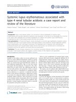
to a large bolus obstruction. A conical mass suspicious of
a bezoar was found measuring 10 cm in length, situated
20 cm beyond the gastrojejunostomy. The small bowel
distal to this site was collapsed. Attempts to break up this
hard bolus mass externally were unsuccessful. The bezoar
eventually had to be removed in whole via an enterotomy.
Careful examination confirmed that it was indeed a phy-
tobezoar (Figure 3).
Image taken during upper endoscopyFigure 1
Image taken during upper endoscopy. a) Oedema
present at the anastomotic site of the gastrojejunostomy. b)
No evidence of obstruction beyond the anastomosis.
b
a
Journal of Medical Case Reports 2008, 2:323 />Page 3 of 5
(page number not for citation purposes)
The patient had an uneventful recovery and was dis-
charged home 1 week after surgery. Before discharge, she
was seen by the dietician with regard to different types of
fibre diet. She was also advised on the importance of
longer mastication of food.
Discussion
A bezoar is also known as an enterolith, a concretion of
foreign or indigestible matter found in the alimentary
canal. There are two main types of bezoars: trichobezoar –
a bezoar formed from hair and phytobezoar – formed by
indigestible cellulose. Rarely, pharmacobezoars from
masses of tablets are found.
This was an unusual presentation of symptoms and signs
of GOO secondary to a phytobezoar, in that this woman
had already had a gastrojejunostomy to bypass previous
GOO caused by peptic ulcer disease. The oedema seen in
her upper endoscopy 3 weeks before admission may have
been the result of a distal subacute obstruction. Postpran-
dial non-bilious vomiting is the cardinal symptom of
GOO, which may lead to electrolyte abnormalities. The
frequency of vomiting puts patients at risk of aspiration
pneumonia. Early satiety and better tolerance to liquid
than solid food may represent gastric dilatation, which
may be appreciated by succussion splash. Management
includes identification of the cause and reversal of any
complications of GOO such as metabolic alkalosis, elec-
trolyte abnormalities, and aspiration pneumonia. Diag-
nosis can result from upper endoscopy or imaging studies.
Regardless of the cause, 75% of all cases of GOO require
surgical intervention [3]. Definitive treatment consists of
laparotomy with milking of the contents to the caecum, or
enterotomy. Medical treatment is usually inadequate.
Recently, the laparoscopic approach has become increas-
ingly popular. A recent study compared laparoscopic ver-
sus open treatment for bezoar-induced small bowel
obstruction [4]. The report concluded that laparoscopy is
safe and effective and is associated with a better postoper-
ative outcome and a shorter hospital stay. One author
describes how a jejunal bezoar in a 59-year-old man was
laparoscopically milked into the caecum through the ile-
ocaecal valve [5].
ImagingFigure 2
Imaging. a) Erect chest radiograph showing an air-fluid level within a dilated stomach. Lung fields were clear. There is no air
under the diaphragm. b) Contrasted abdominal computed tomography showed possible stricture at the site of the gastrojeju-
nostomy.
b
a
Journal of Medical Case Reports 2008, 2:323 />Page 4 of 5
(page number not for citation purposes)
Bezoars tend to be rare, except in patients with previous
gastric surgery [6] or gastrointestinal dysmotility. In a 10-
year retrospective review of all patients with small bowel
obstruction in a hospital in Hong Kong, the incidence of
bezoar was reported as approximately 2% [7]. A 4-year
study in an Italian unit confirmed a similar incidence with
nine of 369 patients having bowel obstruction secondary
to bezoars [8]. It appears that geographical or dietary var-
iation does not participate in the risk of developing bez-
oar obstruction.
Delayed gastric emptying and abnormal gastric motility
patterns were prominent in one series of patients with
bezoars, suggesting that these events were the underlying
factors [9]. There was another series of patients with bez-
oar obstruction, who had had pyloroplasty for peptic
ulcer disease. These patients did not demonstrate delayed
gastric emptying when assessed by technetium-99m-
labelled studies [10]. However, Cifuentes et al. [11]
reported that 84% of cases of bezoar obstruction occurred
in those who had had a bilateral truncal vagotomy and
pyloroplasty. The authors proposed that, in this acid
reducing procedure, there is hypochlorhydria, which
reduces gastric antral motility and gives poor degradation
of food. This predisposes to the formation of a ball of
sticky concretions, which pass into the duodenum and
jejunum unfragmented.
More evidence has since emerged supporting this theory.
Another study [12] involving 117 patients with gastroin-
testinal bezoars revealed that 87% occurred in the small
bowel and 30% in the stomach. Furthermore, 70% of
patients had previous surgery for peptic ulcer disease, and
80% of these patients had a bilateral truncal vagotomy
with pyloroplasty. Of the 87 patients presenting with
intestinal bezoars, excessive intake of dietary fibre
occurred in 40%, and 24% had alterations of mastication
and dentition. There are other risk factors for bezoar
obstruction. Children themselves are at higher risk than
adults in that they have smaller gastrointestinal lumens,
especially with trichobezoar obstruction. There is also an
association between bezoar obstruction and mentally
retarded patients [13].
As discussed, patients with bezoars often present with
symptoms and clinical or radiological signs of bowel
obstruction. Dilated small bowel loops may be seen in
plain abdominal radiographs. In one retrospective study,
the abdominal computed tomography scan was declared
to be the most useful imaging modality for detecting bez-
oars [14]. The study advocated that abdominal computed
tomography should be performed early in patients at
higher risk of developing bezoars. The classical appear-
ance of a bezoar on computed tomography is a well-
defined ovoid intraluminal mass with mottled gas pattern
at the site of obstruction.
Besides obstruction and its associated complications,
other complications of bezoars include ulceration, intus-
susception, and bowel perforation. Intraluminal bezoar is
a serious condition, with a mortality rate as high as 30%
being reported in a retrospective analysis of 34 cases [15].
Early diagnosis and aggressive treatment is the key to suc-
cessful management of the condition, which is curable.
Conclusion
Bezoar induced bowel obstruction is uncommon and
remains a diagnostic challenge. It should be suspected in
patients with an increased risk, such as those with previ-
ous gastric surgery, poor dentition, mental retardation
and a suggestive history of increased fibre intake. Com-
puted tomography of the abdomen should be performed
early in these at-risk patients presenting with symptoms of
GOO or small bowel obstruction in order to reduce
unnecessary delays before appropriate surgical interven-
tion. Bezoar is a curable condition but can potentially
cause significant morbidity and mortality.
Consent
Written informed consent was obtained from the patient
for publication of this case report and any accompanying
images. A copy of the written consent is available for
review by the Editor-in-Chief of this journal.
Competing interests
The authors declare that they have no competing interests.
A 10 cm conical phytobezoar was found 20 cm distal to the gastrojejunostomyFigure 3
A 10 cm conical phytobezoar was found 20 cm distal
to the gastrojejunostomy. It was removed by an enterot-
omy.
Publish with BioMed Central and every
scientist can read your work free of charge
"BioMed Central will be the most significant development for
disseminating the results of biomedical research in our lifetime."
Sir Paul Nurse, Cancer Research UK
Your research papers will be:
available free of charge to the entire biomedical community
peer reviewed and published immediately upon acceptance
cited in PubMed and archived on PubMed Central
yours — you keep the copyright
Submit your manuscript here:
/>BioMedcentral
Journal of Medical Case Reports 2008, 2:323 />Page 5 of 5
(page number not for citation purposes)
Authors' contributions
EL revised the manuscript and researched the case while
LW supervised the ethical approval, patient consent,
patient management and amendments to the manuscript.
RB acquired patient information, designed and drafted
the manuscript, sorted patient consent, carried out day-to-
day management of patient and provided intellectual con-
tent to the discussion of this case report.
References
1. Gibson JB, Behrman SW, Fabian TC: Gastric outlet obstruction
resulting from peptic ulcer disease requiring surgical inter-
vention is infrequently associated with Helicobacter pylori
infection. J Am Coll Surg 2000, 191(1):32-37.
2. Andersson A, Bergdahl L: Carcinoid tumours of the appendix in
children. A report of 25 cases. Acta Chir Scand 1977,
143(3):173-175.
3. Doberneck RC, Berndt GA: Delayed gastric emptying after pal-
liative gastrojejunostomy for carcinoma of the pancreas.
Arch Surg 1987, 122(7):827-829.
4. Yau KK, Siu WT, Law BK, Cheung HY, Ha JP, Li MK: Laparoscopic
approach compared with conventional open approach for
bezoar-induced small-bowel obstruction. Arch Surg 2005,
140(10):972-975.
5. Yol S, Bostanci B, Akoglu M: Laparoscopic treatment of small
bowel phytobezoar obstruction. J Laparoendosc Adv Surg Tech A
2003, 13(5):325-326.
6. Moshe R, Mordechai S, Franklin G, Zeev R, Shlomo L: Phytobezoar:
A rare cause of intestinal obstruction. Dig Surg 1998, 15:52-54.
7. Lo CY, Lau PW: Small bowel phytobezoars: an uncommon
cause of small bowel obstruction. Aust N Z J Surg 1994,
64:187-189.
8. Dervisoglou A, Condilis N, Liveranou S, Pinis S: A causal factors
and treatment of obstructive ileus in 369 patients. Ann Ital Chir
2005, 76(5):477-480.
9. Brady PG: Gastric phytobezoars consequent to delayed gas-
tric emptying. Gastrointest Endosc 1978, 24:159-161.
10. Calabuig R, Navarro S, Carrio I, Artigas V, Mones J, Puig LaCalle J:
Gastric emptying and bezoars. Am J Surg 1989, 157:287-290.
11. Cifuentes J, Robles C, Parrilla P, Lujan M, Escamilla C, Liron R, Perl-
licer F: Gastric surgery and bezoars. Dig Dis Sci 1992,
37(11):1694-1696.
12. Robles R, Parrilla P, Escamilla C, Lujan JA, Torralba JA, Liron R,
Moreno A: Gastrointestinal bezoars. Br J Surg 1994,
81:1000-1001.
13. Van Goor H, Van Rooyen W: Bezoars: a special cause of ileal
obstruction in mentally retarded patients. Neth J Surg 1991,
43:43-44.
14. Ho TW, Koh DC: Small-bowel obstruction secondary to bez-
oar impaction: a diagnostic dilemma. World J Surg 2007,
31(5):1073-1079.
15. Erzurumlu K, Malazgirt Z, Bektas A, Dervisoglu A, Polat C, Senyurek
G, Yetim I, Ozkan K: Gastrointestinal bezoars: a retrospective
analysis of 34 cases. World J Gastroenterol 2005, 11(12):1813-1817.