Cephalometry A Color Atlas and Manual - part 7 doc
Bạn đang xem bản rút gọn của tài liệu. Xem và tải ngay bản đầy đủ của tài liệu tại đây (2.47 MB, 37 trang )
CHAPTER 5
215
5.1 Definition of 3-D Cephalometric Soft Tissue Landmarks
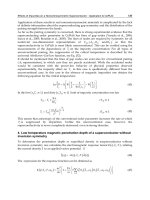
Cheilion: ch
r
,ch
l
Definition of the cheilion Landmarks
Cheilion (ch) is the point located at each labial com-
missure.
Virtual Definition of the cheilion Landmarks
Step 1: Define the cheilion
r
and cheilion
l
landmarks
on the frontal view of the 3-D soft tissue sur-
face representation (Fig. 5.56).
Fig. 5.56. Cheilion
r
and cheilion
l
.Frontal view (3-D CT, patient K.C.)
CHAPTER 5
216
3-D Cephalometric Soft Tissue Landmarks
Labiale (or labrale) inferius: li
Definition of the labiale inferius Landmark
Labiale inferius (li) is the midpoint of the vermilion
line of the lower lip.
Virtual Definition of the labiale inferius Landmark
Step 1: Define labiale inferius on the right profile
view of the 3-D soft tissue surface representa-
tion (Fig. 5.57) and verify its position on the
left profile view (Fig. 5.58).The position of the
labiale inferius landmark can also be verified
on the virtual lateral cephalogram (Figs. 5.76,
5.77).
Step 2: Verify the midline position of the labiale
inferius landmark on the submental view
of the 3-D soft tissue surface representation
(Fig. 5.59).
Fig. 5.57. Labiale inferius. Profile view right (3-D CT,patient K.C.) Fig. 5.58. Labiale inferius. Profile view left (3-D CT,patient K.C.)
Fig. 5.59. Labiale inferius. Submental view (3-D CT, patient K.C.)
CHAPTER 5
217
5.1 Definition of 3-D Cephalometric Soft Tissue Landmarks
Soft tissue gonion: go
r
,go
l
Definition of the soft tissue gonion Landmarks
Soft tissue gonion (go) is the most lateral point on the
soft tissue contour of each mandibular angle,located at
the same level as the 3-D hard tissue cephalometric
Gonion landmark (Chap. 4).
Virtual Definition of the soft tissue gonion Landmarks
Step 1: Define soft tissue gonion
r
and soft tissue
gonion
l
on the right (Fig. 5.60) and left (Fig.
5.61) profile views of the 3-D transparent soft
tissue surface representation.
Step 2: Verify the position of both soft tissue gonion
landmarks on the frontal view of the 3-D
transparent soft tissue surface representation
(Fig. 5.62).
Step 3: Visualize both soft tissue gonion landmarks
on the frontal view of the 3-D soft tissue sur-
face representation (Fig. 5.63).
Fig. 5.60. Soft tissue gonion
r
. Profile view right (3-D CT, transparent soft
tissues,patient K.C.)
Fig. 5.61. Soft tissue gonion
l
.Profile view left (3-D CT,transparent soft tissues,
patient K.C.)
CHAPTER 5
218
3-D Cephalometric Soft Tissue Landmarks
Fig. 5.62. Soft tissue gonion
r
and soft tissue gonion
l
. Frontal view.The trans-
parent soft tissue representation visualizes the underlying bony structures and
allows accurate definition of the soft tissue gonion landmarks (3-D CT, trans-
parent soft tissues,patient K.C.)
Fig. 5.63. Soft tissue gonion
r
and soft tissue gonion
l
. Frontal view (3-D CT,
patient K.C.)
CHAPTER 5
219
5.1 Definition of 3-D Cephalometric Soft Tissue Landmarks
Sublabiale: sl
Definition of the sublabiale Landmark
Sublabiale (sl) is the most posterior midpoint on the
labiomental soft tissue contour that defines the border
between the lower lip and the chin.
Virtual Definition of the sublabiale Landmark
Step 1: Define sublabiale on the right profile view of
the 3-D soft tissue surface representation
(Fig. 5.64) and verify its position on the left
profile view (Fig. 5.65). The position of the
sublabiale landmark can also be verified on
the virtual lateral cephalogram (Figs. 5.76,
5.77).
Step 2: Verify the midline position of the sublabiale
landmark on the submental view of the 3-D
soft tissue surface representation (Fig. 5.66).
Fig. 5.64. Sublabiale. Profile view right (3-D CT,patient K.C.) Fig. 5.65. Sublabiale. Profile view left (3-D CT,patient K.C.)
Fig. 5.66. Sublabiale. Submental view (3-D CT, patient K.C.)
CHAPTER 5
220
3-D Cephalometric Soft Tissue Landmarks
Soft tissue pogonion: pg
Definition of the soft tissue pogonion Landmark
Soft tissue pogonion (pg) is the most anterior mid-
point of the chin.
Virtual Definition of the soft tissue pogonion Landmark
Step 1: Define soft tissue pogonion on the right pro-
file view of the 3-D soft tissue surface repre-
sentation (Fig. 5.67) and verify its position on
the left profile view (Fig. 5.68).The position of
the soft tissue pogonion landmark can also be
verified on the virtual lateral cephalogram
(Figs. 5.76, 5.77).
Step 2: Verify the midline position of the soft tissue
pogonion landmark on the submental view of
the 3-D soft tissue surface representation
(Figs. 5.69, 5.70).
Fig. 5.67. Soft tissue pogonion.Profile view right (3-D CT,patient K.C.) Fig. 5.68. Soft tissue pogonion.Profile view left (3-D CT, patient K.C.)
CHAPTER 5
221
5.1 Definition of 3-D Cephalometric Soft Tissue Landmarks
Fig. 5.69. Soft tissue pogonion.Submental view left (3-D CT,patient K.C.) Fig. 5.70. Soft tissue pogonion. Profile view right. Note that the soft tissue
pogonion landmark is usually more superiorly located than the bony Pogonion
landmark (3-D CT,transparent soft tissues,patient K.C.)
CHAPTER 5
222
3-D Cephalometric Soft Tissue Landmarks
Soft tissue gnathion (or menton): gn
Definition of the soft tissue gnathion Landmark
Soft tissue gnathion (gn) is the most inferior midpoint
on the soft tissue contour of the chin located at the
level of the 3-D cephalometric hard tissue Menton
landmark (Chap. 4). In 3-D cephalometry, soft tissue
gnathion is a well-defined soft tissue landmark and is
therefore not the same as the anthropometric gnathion
landmark according to L.G. Farkas, which is identical
to the bony Gnathion.
Virtual Definition of the soft tissue gnathion Landmark
Step 1: Define soft tissue gnathion on the right profile
view of the transparent 3-D soft tissue sur-
face representation (Fig. 5.71) and verify its
position on the left profile view of the trans-
parent 3-D soft tissue surface representation
(Fig. 5.72). The position of the soft tissue
gnathion landmark can also be verified on the
virtual lateral cephalogram (Figs. 5.76, 5.77).
Step 2: Verify the midline position of the soft tissue
gnathion landmark on the base view of
the 3-D soft tissue surface representation
(Fig. 5.73).
Fig. 5.71. Soft tissue gnathion. Profile view right (3-D CT, transparent soft
tissues,patient K.C.)
Fig. 5.72. Soft tissue gnathion. Profile view left (3-D CT, transparent soft
tissues,patient K.C.)
Fig. 5.73. Soft tissue gnathion.Base view (3-D CT,patient K.C.)
CHAPTER 5
223
5.2 Set-up of 3-D Cephalometric Soft Tissue Landmarks
5.2
Set-up of 3-D Cephalometric Soft Tissue Landmarks
Fig. 5.74 a–d. Set-up of 3-D cephalometric soft tissue landmarks.(3-D CT,patient K.C.)
a
cd
b
CHAPTER 5
224
3-D Cephalometric Soft Tissue Landmarks
Fig. 5.75. Set-up of 3-D cephalometric soft tis-
sue landmarks.Virtual lateral and frontal cephalo-
grams linked to the 3-D soft tissue surface repre-
sentation (3-D CT,patient K.C.)
CHAPTER 5
225
5.2 Set-up of 3-D Cephalometric Soft Tissue Landmarks
Fig. 5.77 a,b. Virtual lateral cephalograms linked to the 3-D hard and transparent soft tissue surface representations, illustrating 3-D soft tissue cephalometric
landmarks located in the midplane (3-D CT,patient K.C.)
Fig. 5.76. Virtual lateral cephalogram linked to the 3-D hard tissue surface
representation, illustrating 3-D soft tissue cephalometric landmarks located in
the midplane (3-D CT,patient K.C.)
ab
CHAPTER 5
226
3-D Cephalometric Soft Tissue Landmarks
5.3
Additional 3-D Cephalometric Soft Tissue Landmarks
The following list shows some additional anthropo-
metric landmarks described by L.G. Farkas that can be
used in 3-D cephalometry, once these are validated.
▬
Eurion: Landmark defined as the most lateral point
of the parieto-temporal region of the skull
▬
Opisthocranion: Landmark defined as the most pos-
terior point of the occipital region of the head and
the most distant from glabella
▬
Otobasion inferius: Landmark defined as the point
of attachment of the ear lobe to the cheek, which
determines the lower border of the ear insertion
▬
Otobasion superius: Landmark defined as the point
of attachment of the helix in the temporal region,
which determines the upper border of the ear inser-
tion
▬
Porion (soft): Landmark defined as the highest point
on the upper margin of the cutaneous auditory mea-
tus
▬
Postaurale: Landmark defined as the most posterior
point on the free margin of the ear
▬
Preaurale: Landmark defined as the most anterior
point of the ear, located at the level of the helix
attachment to the head
▬
Subaurale: Landmark defined as the lowest point on
the free margin of the ear lobe
▬
Superaurale: Landmark defined as the highest point
on the free margin of the auricle
▬
Ve r t e x : Landmark defined as the highest point of
the head when the head is oriented to the Frankfort
horizontal
Fig. 5.78 a–c. Ear related 3-D cephalometric soft tissue landmarks. Right profile (a), frontal (b), dorsal (c) view (3-D CT,patient K.C.)
b
a
c
3-D Cephalometric Planes
Gwen R. J. Swennen
6.1 3-D Cephalometric Planes
230
6.2 Set-up of 3-D Cephalometric Planes 240
CHAPTER 6
CHAPTER 6
227
CHAPTER 6
229
Once virtual definition of the 3-D cephalometric hard
and soft tissue landmarks has been accomplished,
3-D cephalometric planes can be automatically com-
puted. 3-D cephalometric hard and soft tissue planes
can be used for both qualitative and quantitative assess-
ment of craniofacial morphology.
The 3-D virtual scene approach allows the genera-
tion of several types of 3-D cephalometric planes that
are automatically computed based on one or more
3-D cephalometric hard tissue (Chap. 4) or soft
tissue (Chap. 5) landmarks, with regard to the virtual
cephalograms or the 3-D cephalometric reference
planes (Chap. 3):
▬
A 3-D cephalometric plane computed from one
3-D cephalometric landmark is defined by a plane
that passes one landmark and that is parallel to one
of the 3-D cephalometric reference planes
▬
A 3-D cephalometric plane computed from two
3-D cephalometric landmarks is defined by a plane
that passes two landmarks and that is perpendicular
to one of the 3-D cephalometric reference planes
▬
A 3-D cephalometric plane computed from three
3-D cephalometric landmarks is defined by a plane
that passes three landmarks (e.g. maxillary plane,
mandibular plane, facial midplane)
▬
A 3-D cephalometric plane computed from four
3-D cephalometric landmarks is defined by a plane
that passes two landmarks and the mean of two
other landmarks (e.g. Frankfort horizontal plane)
▬
A 3-D cephalometric plane computed from more
than four 3-D cephalometric landmarks is defined
by a plane that passes the means of different pairs of
landmarks (e.g. occlusal plane)
In this chapter important craniofacial 3-D cephalo-
metric planes are described whose accuracy and relia-
bility has been tested (Chap. 7).Other 3-D cephalomet-
ric hard and soft tissue planes can easily be computed
depending on the clinical or research purpose.
CHAPTER 6
230
3-D Cephalometric Planes
6.1
3-D Cephalometric Planes
Frankfort horizontal Plane: FH-Pl
Virtual Definition of the Frankfort horizontal Plane
The Frankfort horizontal plane is defined by a plane
that passes both Orbita (Orbita
r
and Orbita
l
) land-
marks and the mean of the two Porion (Porion
r
and
Porion
l
) landmarks.
Fig. 6.1. Orbita
r
,Orbita
l
and the mean of Porion
r
and Porion
l
define the Frank-
fort horizontal plane (linked virtual lateral and frontal cephalograms, patient
K.C.)
Figs. 6.2, 6.3. Orbita
r
,Orbita
l
and the mean of Porion
r
and Porion
l
define the Frankfort horizontal plane [linked virtual lateral and frontal cephalograms to
3-D hard (6.2) and soft (6.3) tissue representations, patient K.C.]
CHAPTER 6
231
6.1 3-D Cephalometric Planes
Figs. 6.4, 6.5. Frankfort horizontal plane [linked virtual lateral cephalogram to 3-D hard (6.4) and soft (6.5) tissue representations,patient K.C.]
Fig. 6.6. Frankfort horizontal plane (3-D hard tissue representation, patient
K.C.)
Fig. 6.7. Frankfort horizontal plane (3-D soft tissue representation, patient
K.C.)
CHAPTER 6
232
3-D Cephalometric Planes
Maxillary Plane: Mx-Pl Virtual Definition of the Maxillary Plane
The maxillary plane is defined by a plane that passes
the Anterior Nasal Spine and both Posterior Maxillary
Point (PMP
r
–PMP
l
) landmarks.
Fig. 6.8. Anterior Nasal Spine and both Posterior Maxillary Point (PMP
r
– PMP
l
)
landmarks define the maxillary plane (linked virtual lateral and frontal cephalo-
grams,patient K.C.)
Figs. 6.9, 6.10. Anterior Nasal Spine and both Posterior Maxillary Point (PMP
r
-PMP
l
) landmarks define the maxillary plane [linked virtual lateral and frontal
cephalograms to 3-D hard (6.9) and soft (6.10) tissue representations, patient K.C.]
CHAPTER 6
233
6.1 3-D Cephalometric Planes
Figs. 6.11, 6.12. Maxillary plane [linked virtual lateral cephalogram to 3-D hard (6.11) and soft (6.12) tissue representations, patient K.C.]
Fig. 6.13. Maxillary plane (3-D hard tissue representation, patient K.C.) Fig. 6.14. Maxillary plane (3-D soft tissue representation,patient K.C.)
CHAPTER 6
234
3-D Cephalometric Planes
Occlusal Plane: Occ-Pl Virtual Definition of the Occlusal Plane
The occlusal plane is defined by a plane that passes (1)
the mean of Upper Incisor
r
– Upper Incisor
l
and Lower
Incisor
l
– Lower Incisor
l
landmarks, (2) the mean of
Upper Molar Cusp
r
and Lower Molar Cusp
r
landmarks
and (3) the mean of Upper Molar Cusp
r
and Lower
Molar Cusp
r
landmarks.
Fig. 6.15. Tooth-related landmarks that define the occlusal plane (linked vir-
tual lateral and frontal cephalograms,patient K.C.)
Figs. 6.16,6.17. Tooth-related landmarks that define the occlusal plane [linked virtual lateral and frontal cephalograms to 3-D hard (6.16) and soft (6.17) tissue
representations, patient K.C.]
CHAPTER 6
235
6.1 3-D Cephalometric Planes
Figs. 6.18, 6.19. Occlusal plane [linked virtual lateral cephalogram to 3-D hard (6.18) and soft (6.19) tissue representations,patient K.C.]
Fig. 6.20. Occlusal plane (3-D hard tissue representation,patient K.C.) Fig. 6.21. Occlusal plane (3-D soft tissue representation, patient K.C.)
CHAPTER 6
236
3-D Cephalometric Planes
Mandibular Plane: Md-Pl Virtual Definition of the Mandibular Plane
The mandibular plane is defined by a plane that passes
the Menton and both Gonion (Go
r
– Go
l
) landmarks.
Fig. 6.22. Menton and both Gonion (Go
r
– Go
l
) landmarks define the
mandibular plane (linked virtual lateral and frontal cephalograms, patient K.C.)
Figs. 6.23,6.24. Menton and both Gonion (Go
r
– Go
l
) landmarks define the mandibular plane [linked virtual lateral and frontal cephalograms to 3-D hard (6.23)
and soft (6.24) tissue representations, patient K.C.]
CHAPTER 6
237
6.1 3-D Cephalometric Planes
Figs. 6.25, 6.26. Mandibular plane [linked virtual lateral cephalogram to 3-D hard (6.25) and soft (6.26) tissue representations,patient K.C.]
Fig. 6.27. Mandibular plane (3-D hard tissue representation,patient K.C.) Fig. 6.28. Mandibular plane (3-D soft tissue representation, patient K.C.)
CHAPTER 6
238
3-D Cephalometric Planes
Facial Midplane Virtual Definition of the Facial Midplane
The facial midplane is defined by a plane that passes
the Sella, Nasion and Menton landmarks.
Fig. 6.29. Sella, Nasion and Menton landmarks define the facial midplane
(linked virtual lateral and frontal cephalograms, patient K.C.)
Figs. 6.30, 6.31. Sella, Nasion and Menton landmarks define the facial midplane [linked virtual lateral and frontal cephalograms to 3-D hard (6.30) and soft
(6.31) tissue representations, patient K.C.]
CHAPTER 6
239
6.1 3-D Cephalometric Planes
Figs. 6.32, 6.33. Facial midplane [linked virtual lateral cephalogram to 3-D hard (6.32) and soft (6.33) tissue representations,patient K.C.]
Fig. 6.34. Facial midplane (3-D hard tissue representation, patient K.C.) Fig. 6.35. Facial midplane (3-D soft tissue representation, patient K.C.)
CHAPTER 6
240
3-D Cephalometric Planes
6.2
Set-up of 3-D Cephalometric Planes
Fig. 6.36. Set-up of 3-D cephalometric reference system in conjunction with
the Frankfort horizontal plane, maxillary plane, occlusal plane and mandibular
plane (3-D hard tissue and transparent soft tissue surface representation,
patient K.C.)
Fig. 6.37. Set-up of 3-D cephalometric reference system in conjunction with
the Frankfort horizontal plane, maxillary plane, occlusal plane and mandibular
plane (3-D soft tissue representation,patient K.C.)