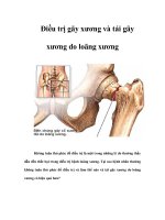
Gãy xương do loãng xương pot
Bạn đang xem bản rút gọn của tài liệu. Xem và tải ngay bản đầy đủ của tài liệu tại đây (724.17 KB, 11 trang )
Vol 11, No 2, March/April 2003
109
Osteoporosis is a systemic disease
characterized by decreased bone
mass and deteriorated bone microar-
chitecture. In the elderly (≥65 years),
it is a contributing factor in 75% of
fractures caused by low-energy
falls.
1
Fractures resulting from os-
teoporosis generally involve the
metaphyseal regions of the skeleton.
These regions are affected earlier
and more profoundly during the de-
velopment of osteoporosis because
they are composed mostly of cancel-
lous bone, which has a greater sur-
face area for bone turnover com-
pared with the compact cortical bone
of the diaphysis.
In the United States, 1.5 million
fractures are reported annually,
most from low-energy falls, includ-
ing 300,000 proximal femur frac-
tures, 250,000 distal radius fractures,
and 300,000 fractures in other bones
affected by osteoporosis. Of the 28
million Americans with osteoporo-
sis, 80% are women. Fifty percent
of women and 18% of men older
than 50 years will sustain an osteo-
porotic fracture.
1
Although $13.8
billion is spent annually to manage
these fractures, <50% of hip fracture
patients recover fully after treat-
ment.
2,3
These statistics emphasize
the need for skilled fracture care for
osteoporotic patients. Reasonable
return of function in the elderly
requires solid internal fixation and
rapid initiation of rehabilitation.
Conversely, inadequate fixation or
prolonged immobilization with
nonsurgical care increases the risk
of thromboembolic disease, pul-
monary complications, decubitus
ulceration, and generalized muscu-
loskeletal deterioration from which
complete recovery is unlikely.
Achieving stable internal fixation
for fractures in osteoporotic bone
can be problematic but is central to
effective care.
Fracture Management in
Osteoporotic Patients
The goal of definitive fracture care
in elderly patients is early restora-
tion of function. Treatment should
be timely; generally, these patients
are in the best condition to undergo
surgery within the first 48 hours
after injury.
4,5
Nevertheless, the
presence of concurrent illness re-
quires thorough evaluation before
surgery. Preoperative management
to optimize the patient’s condition
or correct any decompensation
resulting from the injury can benefit
survival.
5
Procedures should be
kept as simple as possible to mini-
mize surgical time, blood loss, and
physiologic stress. Early weight
bearing is possible only after suc-
cessful stable fracture fixation in the
lower extremity. Although anatom-
ic restoration is important for intra-
articular fractures, metaphyseal
Dr. Cornell is Associate Attending Ortho-
paedic Surgeon, Hospital for Special Surgery,
New York, NY.
Reprint requests: Dr. Cornell, 535 East 70th
Street, New York, NY 10021.
Copyright 2003 by the American Academy of
Orthopaedic Surgeons.
Abstract
Because of the decreased holding power of plate-and-screw fixation in osteoporotic
bone fractures, internal fixation can have a high failure rate, ranging from 10% to
25%. Screws placed into cortical bone have better resistance to pullout than do
those placed into adjacent trabecular bone. Plates should not be used to bridge
unstable regions of bony comminution in osteoporotic patients. Fixation stability
is optimized by securing stable bone contact across the fracture site and by plac-
ing screws both as close to and as far from the fracture as possible. Intentional
shortening can improve stability and load sharing of the fracture construct.
Structural bone graft or other types of fillers can be used to fill voids when com-
minution prevents stable contact. Load-sharing fixation devices such as the slid-
ing hip screw, intramedullary nail, antiglide plate, and tension band constructs
are better alternatives for osteoporotic metaphyseal locations. Proper planning is
essential for improved fracture fixation in this high-risk patient group.
J Am Acad Orthop Surg 2003;11:109-119
Internal Fracture Fixation in
Patients With Osteoporosis
Charles N. Cornell, MD
and diaphyseal fractures are best
managed by attempts to primarily
achieve stability rather than ana-
tomic reduction.
Appropriate treatment of frac-
tures secondary to osteoporosis re-
quires understanding the effect of
the disease on the material and
structural properties of bone, as well
as any effect on the process of frac-
ture healing. Decline in the capacity
for fracture repair is age related.
6
Disturbance of the development of
strength within fracture callus in the
elderly has been shown in ex-
perimental rat models,
7
but little is
known about the causes of osteo-
porosis and its effect on the fracture
repair process in humans.
8
None-
theless, impaired fracture healing in
osteoporotic patients is assumed.
The principles of biologic fracture
repair should be applied whenever
possible.
9
Careful handling of the
surrounding soft tissues and avoid-
ing unnecessary stripping of fracture
fragments preserve blood supply to
the fracture site. Minimizing ex-
posure of the fracture with preserva-
tion of the fracture hematoma may
speed development of callus.
Bone failure, not implant break-
age, is the primary mode of failure
of internal fixation in osteoporotic
bone. Because bone mineral density
correlates with the holding power of
screws, osteoporotic bone often
lacks the strength to hold plates and
screws securely.
10-12
Furthermore,
comminution can be severe in osteo-
porotic fractures. Surgical treatment
of fractures of the proximal humerus,
proximal and distal femur, and
proximal tibia has resulted in an
increased incidence of poor results
in elderly, osteoporotic patients. In
proximal humerus fractures in
elderly patients, more than 50%
have fair or poor results because of
screw loosening and pullout from
the humeral head.
13
Internal fixa-
tion of intertrochanteric fractures
fails in 10% of cases because of
cutout of the lag screw from the
cancellous bone of the femoral
head.
14
Although open reduction
and internal fixation yields results
superior to those of nonsurgical
management for supracondylar
femur fractures, 25% of patients
treated with the angled blade plate
have fair to poor results because of
loss of reduction caused by loosen-
ing of the implant in the osteoporotic
bone of the femoral condyles.
15,16
Traditional internal fixation tech-
niques must be modified to achieve
satisfactory results in osteoporotic
bone. Internal fixation devices that
allow load sharing with host bone
should be used to minimize stress at
the bone-implant interface. Sliding
nail plate devices, intramedullary
nails, antiglide plates, and tension
band constructs are better than
more rigid techniques to treat osteo-
porotic bone fracture.
Implant Fixation in
Osteoporotic Bone
Screws
Resistance to pullout of a screw
placed in bone depends on the
length of the screw purchase, thread
diameter, and quality of the bone
into which it is inserted. Recent
studies also have indicated that the
trabecular orientation within the
bone is important. Bone is highly
anisotropic. Screws placed parallel
to the trabecular pattern have greater
pullout strength than do those
placed across the trabeculae.
17
The
variable of bone quality becomes the
prime determinant of screw holding
power in osteoporotic bone.
17,18
When bone mineral content falls
below 0.4 gm/cm
2
, the effect of
varying thread diameter is lost.
18
Therefore, a plan to place screws
into osteoporotic bone should be
designed to place them as parallel as
possible to the cancellous trabeculae.
Also, the screws should have the
largest thread diameter compatible
with the scale of the fracture being
repaired. Most importantly, if possi-
ble, screws should be placed to
secure fixation into cortical bone.
Cortical bone has greater mineral
density and, therefore, greater resis-
tance to screw pullout than does the
adjacent trabecular bone. Thus, in
poor quality bone, a smaller diame-
ter cortical screw may be better than
a larger diameter cancellous screw
that does not secure cortical pur-
chase.
In cases of severe osteoporosis,
screw fixation may be augmented
with polymethylmethacrylate
(PMMA).
16,19,20
Although PMMA
has relatively poor adhesion to
bone, its intrusion into the cancel-
lous structure results in a much
stronger composite after the cement
polymerizes. One screw fixation
augmentation technique
21
begins
with removal of any screws that
have inadequate purchase or have
stripped with tightening. The
PMMA powder and liquid should
be cooled to slow polymerization.
Once the components are mixed,
the liquid cement is placed into a
10-mL syringe with the tip widened
by drilling it out with a 3.5-mm
drill. The cement then can be in-
jected into the stripped screw holes,
and the screws replaced but incom-
pletely tightened. The screws are
fully tightened once the cement has
set. (Manipulation of the screw
while the cement is setting loosens
the bond between the cement, bone,
and screw, lowering the pullout
strength.) Struhl et al
19
described an
alternative method similar to ce-
ment techniques used for fixation of
intramedullary prostheses. The
medullary canal is blocked proxi-
mal and/or distal to the fracture
site, and the entire medullary cavity
is filled with cement. After the frac-
ture is reduced and the cement has
cured, the screws are inserted by
drilling and tapping. Screws placed
during the curing process are tight-
ened once the cement has cured.
This technique is very useful when
Internal Fracture Fixation in Patients With Osteoporosis
Journal of the American Academy of Orthopaedic Surgeons
110
poor screw fixation is combined
with significant bone loss.
Plates
The strength of plate fixation is
directly affected by the degree of
comminution and the resulting size
of any gap at the fracture site. In
addition, the pattern of screw place-
ment influences the strain experi-
enced within the plate and its
screws.
22,23
The most important fac-
tor that reduces strain in plated frac-
tures is the degree to which cortical
contact can be achieved at the frac-
ture site. Experimental fractures
stabilized by plates spanning a gap
had three times the strain of frac-
tures stabilized with secure cortical
contact.
23
Additionally, for a given
fracture pattern, the screw spacing
is more important than the number
of screws used for fixation.
22
Strain
within a plated construct is least
when screws are placed both as
close to and as far from the fracture
site as possible. In two-part frac-
tures or those with solid cortical
contact, the farther the screws are
placed from the fracture, the less the
strain experienced within the plate.
Thus, in longer plates with screws
placed as close to and as far as pos-
sible from the fracture site, interven-
ing screws add little to overall fixa-
tion strength. In comminuted frac-
tures or those with a gap, the longer
plate retains its advantage, but an
increased number of screws adja-
cent to the fracture site reduces the
strain within the plate. Ellis et al
23
concluded that three screws should
be placed in the holes adjacent to
either side of the fracture gap as
well as the most distant hole of the
plate, since additional intervening
screws add little to the load experi-
enced by the plate.
Longer plates with widely spaced
screws should be used in osteoporotic
bone. Cortical contact at the fracture
site is paramount; if moderate areas
of comminution exist, the fracture
should be shortened to achieve con-
tact, especially in the cortex opposite
the plate.
15,16
Plates should not be
used to bridge gaps in osteoporotic
bone but should be used as tension
bands, which require an intact, load-
sharing cortex opposite the plate.
When comminution is extensive and
prevents stable contact opposite the
plate, double-plating should be con-
sidered to secure stability. In addi-
tion, the plates should be placed to
act as antiglide plates whenever pos-
sible, especially in short oblique or
spiral oblique fracture patterns. In
such situations, the plate can be posi-
tioned to create an axilla with the cor-
tex at the apex of the oblique tongue
of the fracture (Fig. 1). The plate
position acts to prevent fracture dis-
placement, placing less importance
on screw fixation within the weak,
adjacent metaphyseal bone. It also
positions the plate for insertion of lag
screws, which are important to treat
the oblique fracture pattern.
Intramedullary Nails
Intramedullary nail fixation is
well suited for diaphyseal fractures
in osteoporotic bone and is the treat-
ment of choice for diaphyseal frac-
tures of the femur and tibia.
24
The
nails provide broad areas of pur-
chase, allow load sharing, and offer
sufficiently secure fixation to allow
immediate weight bearing in many
circumstances.
25
The development
of interlocking nails has extended
the indications for intramedullary
nailing to include metaphyseal frac-
tures. Intramedullary nails are posi-
tioned closer to the mechanical axis
Charles N. Cornell, MD
Vol 11, No 2, March/April 2003
111
Figure 1 The antiglide fixation method. A, Direction of the fracture displacement (arrow).
B, The plate is positioned to create an axilla at the apex of the fracture, and the distal frag-
ment reduces into the axilla. The arrows indicate the corrective force exerted by the plate.
C, The plate minimizes the tendency for displacement and achieves compression along the
fracture line (arrows). Strong screw fixation in the diaphysis of the proximal fragment
holds the reduction (arrows) and makes the distal screws unnecessary. Placement of the
plate in the plane of the fracture obliquity makes it easy to place a lag screw through the
plate. (Adapted with permission from Carr JB, Trafton PG: Malleolar fractures and soft
tissue injuries of the ankle, in Browner BB, Jupiter JB, Levine AM, Trafton PG [eds]:
Skeletal Trauma, ed 2. Philadelphia, PA: WB Saunders, 1998, vol 2, pp 2327-2404.)
A B C
of the bone and, as a result, are sub-
ject to smaller bending forces than
are plated constructs placed on the
external surface of the bone. Fa-
tigue failure is less likely with
intramedullary nails than with plate
constructs. Mechanically locked in-
tramedullary nails provide greater
strength in axial loading than do
condylar blade plates but are mark-
edly less stable during bending and
torsion when used in the distal
femur.
26,27
Thus, although locked
nails provide less stability than do
condylar blade plates in simple,
metaphyseal fractures, they are bet-
ter suited for fixation of severely
comminuted osteoporotic bone frac-
tures with no reconstructable medial
buttress.
The major weakness of locked
intramedullary nails is the security
of the locking screws, which may
loosen in osteoporotic metaphyseal
bone. This is particularly likely in
the bone of the distal femur and can
lead to loss of control of the distal
fragment, which often results in ro-
tational and varus/valgus malalign-
ment. Locking screw fixation can be
improved by using different planes
of screw orientation (eg, anteropos-
terior and transverse placement),
28
by using osteoporotic nuts and
washers on the medial side of the
femur where the locking bolts emerge,
or by using cement to improve fixa-
tion.
16
Tension Band Wiring
Tension band wiring is usually
applied to transverse fractures,
which are distracted by the pull of
attached tendons and ligaments.
This technique provides strong and
secure fixation, which allows im-
mediate mobilization of involved
joints. Fractures of the olecranon and
patella can be successfully treated
with this method. The tension band
wire has additional advantages in
osteoporotic bone. In metaphyseal
locations, such as the proximal
humerus or medial malleolus, ten-
don and ligament insertions to bone
can provide better strength for fixa-
tion than does the bone itself. In
these areas, placement of tension
band wires within the soft-tissue
attachments can provide excellent
anchorage. Hawkins et al
29
report-
ed better clinical results with ten-
sion band wiring than with plate-
and-screw fixation in proximal
humerus fractures. A similar tech-
nique can be used to secure ex-
tremely osteoporotic or comminuted
medial malleolar fractures (Fig. 2).
The fracture is reduced and main-
tained with small Kirschner wires.
The tension band wire is passed
within the fibers of the deltoid liga-
ment and proximally secured to
bone by passing it around a screw
placed through the tibia. The wire is
placed in figure-of-8 fashion and can
be tightened by opposing twists.
Tension band wires also can supple-
ment plate-and-screw fixation in
fractures that may be subjected to
tensile loading. After securing the
fracture with plate and screws, a
tension band wire is passed within
adjacent tendinous attachments and
beneath the plate to help neutralize
tensile forces across the construct
(Fig. 3).
Augmentation
Bone grafting plays several
important roles in the treatment of
osteoporotic fractures. Cancellous
bone graft can be used to augment
or encourage rapid fracture healing.
Cancellous bone is osteoinductive,
osteoconductive, and osteogenic,
30
and it can stimulate new bone for-
mation periosteally in fracture gaps
created by comminution. There is
no evidence that osteoporotic bone
is an inferior graft material.
Corticocancellous bone graft can
be used in osteoporotic fractures
to replace regions of skeletal loss
caused by comminution or crush,
thus enhancing fracture construct
stability. This is especially true for
metaphyseal and joint depression
fractures, such as split-depression tib-
ial plateau fractures, intra-articular
fractures of the distal radius, distal
humerus fractures, and tibial plafond
fractures. Surgical repair requires
elevation of the articular surface to
restore joint congruity with the use
of structural bone graft to fill in the
metaphyseal void and provide sup-
port to the subchondral region.
The iliac crest is the most com-
mon donor site for autogenous bone
graft. The morbidity associated
with the harvest of autogenous
bone is a concern,
31
especially in the
Internal Fracture Fixation in Patients With Osteoporosis
Journal of the American Academy of Orthopaedic Surgeons
112
Figure 2 Tension band wiring. Kirschner
wires are placed to hold the medial malleo-
lus fracture reduced. A figure-of-8 wire is
then passed distal to the wires through the
substance of the deltoid ligament and
anchored to a screw placed proximal to the
fracture. (Adapted with permission from
Carr JB, Trafton PG, Simpson LA:
Fractures and soft tissue injuries of the
ankle, in Browner BD, Jupiter JB, Levine
AM, Trafton PG [eds]: Skeletal Trauma, ed
2. Philadelphia, PA: WB Saunders, 1998,
vol 2, pp 1871-1957.)
elderly. In older osteoporotic indi-
viduals, the quantity and quality of
bone available at the iliac crest is
often insufficient, requiring a larger
exposure, which increases the risk
of donor site complications. Bone
graft substitutes can provide an at-
tractive alternative to autograft in
osteoporotic patients.
32
Bone graft
substitutes include allograft bone,
demineralized allograft bone prod-
ucts, and synthetic osteoconductive
materials, which can be used as
bone void fillers. Several of these
products have been shown to be
essentially equivalent to autogenous
graft for treatment of acute frac-
tures.
33,34
Replacement of severely com-
minuted areas with PMMA to re-
gain stablility is sometimes required
after severe skeletal loss. It has been
used successfully, especially in
supracondylar fractures of the
femur
16,19
and intertrochanteric frac-
tures. However, PMMA is not an
ideal material for this purpose
because it is a permanent implant
and a foreign body within bone. It
also generates considerable heat
with polymerization, which may be
harmful to bone and surrounding
soft tissue. Cements made from cal-
cium phosphate adhere better to
bone and have the advantage of
being resorbed and replaced by host
bone. These cements are not used
to augment screw fixation but to fill
voids caused by comminution or
severe osteoporosis. Calcium phos-
phate cements have been useful in
intertrochanteric and distal radius
fractures.
35,36
These new cements
can provide enough support to
allow earlier load bearing and
decrease the dependency on inter-
nal fixation devices.
PMMA can be used to augment
screw purchase in the severely
osteoporotic diaphysis, but another
useful approach is to place an aug-
mentation device into the medullary
canal to incorporate as bone or be
resorbed. Fibular allograft struts
are used for this purpose (Fig. 4).
The fibular strut improves local
bone stock for screw purchase and
can be incorporated to provide a
span across regions of diaphyseal
deficiency. Creative strategies such
as these can be extremely successful
for treating osteoporotic diaphyseal
fracture and nonunion.
Fracture Types
Intertrochanteric Fractures
The sliding hip screw (SHS) has
markedly advanced the treatment of
intertrochanteric fractures.
37,38
The
success of the SHS is based on its
design, and in many ways it is the
ideal device for this typically osteo-
porotic fracture. The SHS has a lag
screw that gains broad purchase in
the highest quality bone in the fem-
oral head. The dynamic slide of the
lag screw and side plate allows
impaction at the fracture site with
load sharing along the plane of the
fracture. The success of load shar-
ing is evident in that the length of
the side plate makes little difference
in the stability of an SHS con-
struct.
39
When the SHS is inserted
correctly, in all but the most unsta-
ble fractures the failure rate of fixa-
tion is <5%, even in extremely os-
teoporotic patients.
14,40
The most
important aspect of SHS insertion is
to ensure that the lag screw is
placed in the center of the femoral
head (within 10 mm of the femoral
head apex in both the anteroposte-
rior and lateral radiographic views).
Charles N. Cornell, MD
Vol 11, No 2, March/April 2003
113
Figure 3 Anteroposterior radiograph of a
tension band wire augmenting plate fixa-
tion in the proximal humerus. The wire is
passed in figure-of-8 fashion beneath the
supraspinatus tendon and distally beneath
the plate (arrow). The wire provides a
strong purchase and acts to neutralize the
deforming pull of the rotator cuff.
Figure 4 Anteroposterior radiograph of a
repair of a humeral nonunion with seg-
mental bone loss. A fibular strut allograft
was fashioned into an intramedullary peg.
It spanned the defect and improved the
screw purchase proximal and distal to the
fracture site. The loose distal screw was
hidden within soft tissues left from a prior
attempt at reconstruction.
Additionally, the lag screw must be
able to slide within the side plate
barrel to allow stable impaction at
the fracture site. To this end, the
side plate angle should be ≥135°.
With a short lag screw, a short-bar-
rel plate should be used to assure
adequate slide. Generally, a short-
barrel side plate should be used
when the lag screw is <85 mm. The
surgeon should ensure that 10 mm
of slide can occur.
Unfortunately, similar success
does not occur with unstable four-
part fractures, reverse obliquity
fractures, or fractures with sub-
trochanteric extension because the
lack of a lateral buttress and the loss
of the posteromedial bone for load
sharing prevents them from dynam-
ically achieving stability.
41
As a
result, the SHS allows maximum
medial displacement of the shaft,
leading to either unacceptable short-
ening at the fracture site or cutout of
the lag screw from the femoral head
because the lag screw threads have
come to rest on the barrel of the side
plate (Fig. 5). In these unstable frac-
ture patterns, devices are needed to
recreate a lateral buttress or allow
vertical, dynamic impaction. Fixed-
angle devices such as the 95° condy-
lar screw can be used.
42
This type of
device provides a lateral buttress,
but because it has a fixed angle, it
cannot load-share unless an intact
medial buttress can be reconstituted.
Failure is almost guaranteed with-
out this medial support opposite the
plate (Fig. 6). Additionally, these
devices do not allow weight bearing
immediately after surgery.
Alternative devices for treating
unstable peritrochanteric fracture
include the intramedullary hip
screw (Gamma nail) and vertically
sliding plate. The intramedullary
SHS provides the advantages of an
intramedullary nail combined with a
dynamic hip screw that allows
impaction of the peritrochanteric
fracture.
25,43
The intramedullary
position decreases the lever-arm on
the device and creates its own lateral
buttress that prevents excessive lat-
eral migration of the proximal frag-
ment. The strength of the device
allows immediate weight bearing.
Use of the long intramedullary nail
helps avoid fracture of the femoral
shaft that can occur when the short
Gamma nail is used. The insertion
of intramedullary hip screws can be
technically demanding because the
fracture must be reduced before
reaming and nail insertion to avoid
comminution of the fracture site and
adjacent cortex. Open reduction be-
fore nailing is recommended unless
a nearly perfect closed reduction can
be achieved.
The vertical SHS allows fractures
to impact without excessive lateral
displacement (Fig. 7). Although
clinical experience with these de-
vices is preliminary, initial results
are encouraging.
44
The primary
advantages of this device are the
ease with which it can be inserted as
well as its use to salvage an SHS
that has been complicated by lateral
cortex comminution during inser-
tion.
Supracondylar Fractures
of the Distal Femur
Osteoporosis weakens the supra-
condylar region of the distal femur in
the elderly, allowing even low-energy
injuries to result in complex fractures.
Intra-articular involvement and com-
minution of the metaphysis is com-
mon in this population. The chal-
Internal Fracture Fixation in Patients With Osteoporosis
Journal of the American Academy of Orthopaedic Surgeons
114
Figure 5 Anteroposterior radiograph of a
reverse obliquity fracture pattern. A slid-
ing hip screw was used to stabilize the frac-
ture, which lacks a stable lateral buttress.
Without a lateral buttress, the dynamic
slide of the plate could not achieve stability.
The proximal fragment displaced until no
further slide could occur, so the dynamic
hip screw became a fixed-angle, load-bear-
ing device. As a result, the lag screw cut
out of the femoral head.
Figure 6 Anteroposterior radiograph of a
subtrochanteric fracture treated with a 95°
dynamic condylar screw plate. A defect in
the medial cortex persisted after recon-
struction. The plate acted as a bridge plate
rather than a tension band, which resulted
in loosening of the screws of the side plate,
with loss of reduction of the fracture.
lenge of treating these fractures led
to the development of closed treat-
ment methods using traction and
cast bracing, but those techniques
resulted in loss of range of motion.
The development of internal fixa-
tion methods for these fractures has
allowed restoration of anatomy and
early knee rehabilitation.
Many features make the 95°
blade plate designed for supra-
condylar fractures ideal for fixation
in osteoporotic bone. Retrograde
intramedullary nails do not provide
the same stability as do the 95°
devices
26,27,45
and should be re-
served for fractures around total
knee replacements,
46
those with se-
vere comminution into the diaphy-
sis, or those with severe skin com-
promise around the knee. The 95°
condylar screw was designed to
make insertion of the device easier
compared with the blade plate.
However, it sacrifices more bone
from the distal femur with insertion
and cannot be as easily revised. It is
also slightly larger and can impinge
on the lateral soft tissues of the
knee. In contrast, the blade plate is
low profile and requires very little
bone sacrifice; also, the position of
the blade can be revised without
compromising fixation. However,
the blade plate is more technically
demanding to insert.
Although the blade plate can be
used successfully as a bridging plate
in younger individuals with good
bone stock, it should be used only
as a tension band in the elderly.
This requires reconstitution of a
load-sharing medial femoral cortex
opposite the blade plate. For many
fractures, this can be accomplished
by shortening the fracture into a
position of stability by impacting
the fracture surfaces. Shortening of
as much as 1 to 2 cm can be done
without notable loss of function.
15
Healing is usually rapid after short-
ening because the comminuted
medial bone functions as bone
graft.
16
In severely comminuted
fractures in which sufficient stability
cannot be accomplished by shorten-
ing, the surgeon can resort to dou-
ble plating the distal femur
47
or
replacing the region of bone loss
with cement.
Lateral Tibial Plateau Fractures
The split-depression or Schatzker
type II (AO classifications B2 and
B3) is the most common lateral tibial
plateau fracture in the osteoporotic
patient. Fractures with <5 mm of
joint surface depression in a patient
with a knee that is stable to varus/
valgus stress can be managed non-
surgically. Surgical reconstruction
is required if the degree of joint
depression is >5 mm and if there is
>5° of varus/valgus instability. The
surgical technique for repair of the
split-depression lateral tibial pla-
teau fracture has been reported
extensively.
47,48
Benirschke et al
49
suggested modifications of this
technique that call for use of a small
fragment plate specifically designed
for the lateral tibial plateau. The
small fragment plate is low profile
and allows placement of proximal
screws very close to the subchon-
dral plate of the reduced tibial pla-
teau. As many as four screws can
be inserted, providing extensive
support of the reduced joint surface.
The small screws can be placed into
opposing cortex to secure cortical
purchase without the risk of soft-tis-
sue irritation associated with pro-
truding large fragment cancellous
screws. Because the screws placed
under the subchondral bone are
reminiscent of rafters supporting a
roof or a floor, this has been referred
to as the rafter plate technique (Fig.
8). This modification of the stan-
dard technique is useful for the os-
teoporotic patient.
49
Insertion of a bone graft to fill the
metaphyseal defect created after
elevation of the joint surface is stan-
dard, but substitution of other osteo-
conductive materials, such as calcium
phosphate cements and hydroxy-
Charles N. Cornell, MD
Vol 11, No 2, March/April 2003
115
Figure 7 Anteroposterior radiograph of a
sliding hip screw with vertical slide capa-
bility. This plate was chosen after attempt-
ed insertion of a standard side plate result-
ed in comminution of the lateral cortex
around the lag screw insertion site. The
vertical slide allows axial settling in this
unstable fracture pattern. Stable bone con-
tact is achieved without excessive lateral
displacement of the head and neck.
Figure 8 The rafter plate technique. Four
3.5-mm screws are inserted through a cus-
tom plate and placed close to the subchon-
dral plate, providing a broad area of sup-
port. (Adapted with permission from
Benirschke SK, Swiontkowski MF: Knee,
in Hansen ST Jr, Swiontkowski MF [eds]:
Orthopaedic Trauma Protocols. New York,
NY: Raven Press, 1993, pp 291-329.)
apatite implants, has proved to be
successful.
33
Ankle Fractures and
the Distal Fibula
Ankle and foot fractures are
among the most common fractures
sustained by women, and most ankle
fractures occur in women aged 75 to
84 years. The prognosis for ankle
fractures is worse in the elderly than
in younger individuals, but the treat-
ment principles are identical.
50
Even
small amounts of residual displace-
ment in the mortise markedly alter
the load-bearing distribution on the
talus, leading to poor clinical out-
comes.
51,52
As in younger patients,
ankle fractures must be treated with
anatomic reduction until healing.
Isolated fractures of the lateral
malleolus without injury to the
medial malleolus or deltoid liga-
ment can be treated nonsurgically,
whereas unstable ankle fractures
require open reduction and internal
fixation. Surgical fixation of the
ankle in the elderly can be made
more difficult by poor skin integ-
rity, swelling, diabetes, and vascular
disease. In addition, bone loss from
an osteoporotic lateral malleolus
can compromise the ability to se-
cure internal fixation of the lateral
aspect of the ankle. Most lateral
malleolar fractures are short oblique
or spiral oblique patterns, with the
apex of the fracture posterior and
proximal. The most accepted fixa-
tion technique is placement of a
plate on the lateral surface of the
malleolus through a lateral incision.
Modifying this technique by placing
the plate on the posterior surface of
the fibula puts it in the antiglide
position, which results in superior
biomechanical performance
53
(Fig. 9).
Additionally, with the plate placed
posteriorly, the skin incision can be
more posterior, allowing for better
coverage of the fibula if wound-
healing problems occur. The best
advantage of the antiglide position
is that the purchase of the distal
screws is relatively unimportant; the
important screw fixation is in the
more proximal cortical region of the
fibula. The antiglide principle can
be applied to any oblique fracture,
especially medial tibial metaphyseal
fractures and spiral oblique fractures
of the distal tibial metaphysis.
Proximal Humerus Fractures
Proximal humerus fractures com-
monly occur in elderly women; for-
tunately, 80% of these fractures are
impacted or minimally displaced
and heal with a brief period of im-
mobilization. Unstable fractures
displace because of the pull of the
musculature attached to the upper
humerus. If not reduced, they will
result in malunion, with loss of
range of motion, strength, and func-
tion of the shoulder girdle. Loss of
shoulder motion can diminish the
ability to dress and attend to per-
sonal hygiene. Poor functional out-
come after a proximal humerus frac-
ture in the elderly can markedly
reduce social independence. Open
reduction and internal fixation of
unstable two- and three- part frac-
tures of the humerus leading to im-
proved functional outcome is ad-
vantageous in the elderly.
Repair of proximal humerus frac-
tures is extremely challenging, and
results are often disappointing even
with experienced surgeons. Pros-
thetic replacement of the humeral
head is indicated in three- or four-
part fractures in which the head is
only a deficient shell of subchondral
bone. However, functional results
of two- and three-part fractures are
better if open reduction and preser-
vation of the humeral head can be
done.
29
Plating of the proximal hu-
merus is often unsatisfactory be-
cause of poor screw purchase and
acromial impingement caused by
the bulkiness of the hardware.
13,54
Hawkins et al
29
first described the
advantage of tension band fixation
for such fractures after recognizing
that the tendinous attachment of the
rotator cuff to the tuberosities pro-
vides excellent purchase for figure-
of-8 wires. The tendon provides
better anchorage than does the soft
bone of the humeral head, and the
reduced bulkiness of wire con-
structs makes tension band fixation
ideal for this region. Excellent clini-
cal results are possible with modifi-
cations of Hawkins’ original tech-
nique, which involves exposure of
the fracture site through an extend-
ed deltopectoral approach.
54-56
The
fracture is mobilized and the head
and shaft are impacted to achieve
stability along the fracture site.
Intramedullary nails or a simple lag
screw can be placed to provide ini-
tial stability while the tension band
wires are positioned. One is placed
under the rotator cuff tendons, and
a second can be used to wire the
tuberosities together. The wires are
attached to the shaft through a drill
hole placed lateral through the shaft
(Fig. 10). The stability of this con-
struct allows immediate shoulder
rehabilitation, thereby optimizing
outcome.
Extensive metaphyseal com-
minution, which would lead to
excessive shortening with impaction
at the fracture site, precludes use of
Internal Fracture Fixation in Patients With Osteoporosis
Journal of the American Academy of Orthopaedic Surgeons
116
Figure 9 Lateral radiograph of a lateral
malleolar fracture stabilized with a plate
placed in the posterior or antiglide position.
this technique alone. Such shorten-
ing can cause subluxation of the
shoulder because of laxity in the
deltoid muscle. In these cases, a
plate should be used to restore and
maintain proper height. A modified
cloverleaf plate works well because
it is small, can be used to place mul-
tiple screws into the head, and can
be supplemented with a tension
band wire for added support. Re-
cently, interlocking proximal hu-
meral nails and blade plates have
been advocated for these fractures.
Proximal humeral plates with fixed-
angle screws also have been intro-
duced. Although these devices are
promising, clinical experience has
yet to be reported.
Postoperative Care
Postoperative care of patients with
osteoporotic fracture should include
both physical rehabilitation and psy-
chosocial treatment. Many elderly
patients have marked preinjury
functional compromise, and the
additional disability associated with
recovery makes short-term rehabili-
tation necessary for most before
eventual return home. Depression
and hopelessness are common in the
elderly after injury and must be
addressed by the health care team.
These patients are best treated by a
multidisciplinary service in which
their medical, psychological, and
social concerns are addressed. Many
elderly patients enter the hospital in
a malnourished state and therefore
have a high mortality rate. Malnu-
trition results in immunocompro-
mise and is associated with higher
complication rates for fracture sur-
gery.
57
Clinical evaluation of nutri-
tional status can easily be done by
assessing the patient’s dietary habits
and measuring the serum albumin.
A serum albumin <3.5 mg/dL indi-
cates chronic protein malnutrition.
Elderly patients may have diffi-
culty complying with restricted
weight bearing after surgery on the
lower extremity and should be
allowed to bear weight as tolerated
with a walker. Because load sharing
is the most important principle of
osteoporotic fracture surgery, weight
bearing as tolerated is not contraindi-
cated postoperatively. Most elderly
patients will not adhere to partial
weight-bearing protocols; therefore,
no weight bearing is recommended
if there is uncertainty about the sta-
bility of the fracture construct.
Finally, it should be assumed that
any patient past middle age with a
low-energy metaphyseal fracture
has osteoporosis. These patients
should undergo bone mineral den-
sity testing and be placed on a regi-
men to combat further bone loss.
They should be encouraged to take
calcium 1,000 to 1,500 mg/d with a
multivitamin to ensure adequate vi-
tamin D intake. Elderly fracture pa-
Charles N. Cornell, MD
Vol 11, No 2, March/April 2003
117
A C
Figure 10 Anterior (A) and lateral (B) views and anteroposterior radiograph (C) of the tension band technique used to treat proximal
humerus fractures. A preliminary lag screw impacts the fracture and maintains reduction, then two tension band wires are placed. The
first wire is passed beneath the supraspinatus tendon, and the second through the tuberosities. The figure-of-8 wires are passed through a
drill hole in the shaft. This construct takes advantage of the strong rotator cuff tendinous insertion and permits immediate postoperative
rehabilitation of the shoulder.
Wire 1
Wire 1
Wire 2
Wire 2
B
tients also should be encouraged to
start bisphosphonate therapy be-
cause alendronate has been proved
to reduce the risk of additional frac-
tures after hip fracture.
58
Treatment
of the underlying osteoporosis is
part of the fracture treatment.
Summary
Fracture care techniques often
require modification to be useful to
treat osteoporotic bone. Screws
should be placed into the best-quali-
ty-bone available, which is usually
an opposing cortex. Screw fixation
can be augmented by using PMMA.
With plate fixation, stable bone con-
tact at the fracture site is the most
important factor for reducing strain
in the plate. Shortening of the af-
fected bone can achieve this contact
in comminuted fractures. Plates
should not be used to bridge areas of
comminution in osteoporotic bone
and should be as long as possible,
with screws placed close to and far
from the fracture site. Locked
intramedullary nails can be used for
diaphyseal fractures or fractures with
metaphyseal-diaphyseal comminu-
tion. Angled blade plates are very
applicable to osteoporotic metaph-
yseal fractures but should be used as
tension band plates that require sta-
ble, load-sharing contact opposite
the plate. Antiglide plating and use
of tension band wires also are effec-
tive strategies for osteoporotic frac-
tures. Use of bone graft substitutes is
particularly applicable to reduce the
morbidity of bone graft harvest and
ensure adequate volumes of graft in
the elderly. Patients with evidence
of osteoporosis should be started on
a medical regimen that includes cal-
cium supplementation with a pre-
scription for bisphosphonates or
other antiresorptive regimes to com-
bat further bone loss.
Internal Fracture Fixation in Patients With Osteoporosis
Journal of the American Academy of Orthopaedic Surgeons
118
References
1. Lucas TS, Einhorn TA: Osteoporosis:
The role of the orthopaedist. J Am
Acad Orthop Surg 1993;1:48-56.
2. Egol KA, Koval KJ, Zuckerman JD:
Functional recovery following hip frac-
ture in the elderly. J Orthop Trauma
1997;11:594-599.
3. Tinetti ME, Baker DI, Gottschalk M, et
al: Home-based multicomponent reha-
bilitation program for older persons
after hip fracture: A randomized trial.
Arch Phys Med Rehabil 1999;80:916-922.
4. Aharonoff GB, Koval KJ, Skovron ML,
Zuckerman JD: Hip fractures in the
elderly: Predictors of one year mortality.
J Orthop Trauma 1997;11:162-165.
5. Kenzora JE, McCarthy RE, Lowell JD,
Sledge CB: Hip fracture mortality:
Relation to age, treatment, preoperative
illness, time of surgery, and complica-
tions. Clin Orthop 1984;186:45-56.
6. Silver JJ, Einhorn TA: Osteoporosis
and aging: Current update. Clin Orthop
1995;316:10-20.
7. Ekeland A, Engesoeter LB, Langeland
N: Influence of age on mechanical
properties of healing fractures and
intact bones in rats. Acta Orthop Scand
1982;53:527-534.
8. Lill CA, Fluegel AK, Schneider E:
Sheep model for fracture treatment in
osteoporotic bone: A pilot study about
different induction regimens. J Orthop
Trauma 2000;14:559-566.
9. Mast J, Jakob R, Ganz R (eds): Planning
and Reduction Technique in Fracture
Surgery. Berlin, Germany: Springer-
Verlag, 1989.
10. Sjostedt A, Zetterberg C, Hansson T,
Hult E, Ekstrom L: Bone mineral con-
tent and fixation strength of femoral
neck fractures: A cadaver study. Acta
Orthop Scand 1994;65:161-165.
11. Alho A: Mineral and mechanics of
bone fragility fractures: A review of
fixation methods. Acta Orthop Scand
1993;64:227-232.
12. Strømsøe K, Kok WL, Høiseth A, Alho
A: Holding power of the 4.5 mm
AO/ASIF cortex screw in cortical bone
in relation to bone mineral. Injury
1993;24:656-659.
13. Kristiansen B, Christensen SW: Plate
fixation of proximal humeral fractures.
Acta Orthop Scand 1986;57:320-323.
14. Baumgaertner MR, Curtin SL, Lindskog
DM, Keggi JM: The value of the tip-
apex distance in predicting failure of
fixation of peritrochanteric fractures of
the hip. J Bone Joint Surg Am 1995;77:
1058-1064.
15. Blatter G, König H, Janssen M, Magerl F:
Primary femoral shortening osteosynthe-
sis in the management of comminuted
supracondylar femoral fractures. Arch
Orthop Trauma Surg 1994;113:134-137.
16. Schatzker J: Fractures of the distal fe-
mur revisited. Clin Orthop 1998;347:
43-56.
17. An YH, Young FA, Kang Q, Williams
KR: Effects of cancellous bone struc-
ture on screw pullout strength. Medical
University of South Carolina Orthopedic
Journal 2000;3:22-26.
18. Turner IG, Rice GN: Comparison of
bone screw holding strength in healthy
bovine and osteoporotic human cancel-
lous bone. Clin Mater 1992;9:105-107.
19. Struhl S, Szporn MN, Cobelli NJ, Sadler
AH: Cemented internal fixation for
supracondylar femur fractures in osteo-
porotic patients. J Orthop Trauma 1990;
4:151-157.
20. Motzkin NE, Chao EYS, An K-N,
Wikenheiser MA, Lewallen DG: Pull-
out strength of screws from poly-
methylmethacrylate cement. J Bone
Joint Surg Br 1994;76:320-323.
21. Helfet DL: Fractures of the distal
femur, in Browner BD, Jupiter JB,
Levine AM, Trafton PG (eds): Skeletal
Trauma: Fractures, Dislocations, Ligamen-
tous Injuries. Philadelphia, PA: WB
Saunders, 1992, vol 2, pp 1643-1683.
22. Törnkvist H, Hearn TC, Schatzker J: The
strength of plate fixation in relation to
the number and spacing of bone screws.
J Orthop Trauma 1996;10:204-208.
23. Ellis T, Bourgeault CA, Kyle RF: Screw
position affects dynamic compression
plate strain in an in vitro fracture
model. J Orthop Trauma 2001;15:333-337.
24. McConnell T, Court-Brown C, Sar-
miento A: Isolated tibial shaft frac-
ture. J Orthop Trauma 2000;14:306-308.
25. Rodriguez Alvarez J, Casteleiro Gon-
zolez R, Laguna Aranda R, Ferrer
Blanco M, Cuervo Dehesa M: Indica-
tions for use of the long Gamma nail.
Clin Orthop 1998;350:62-66.
26. Ito K, Grass R, Zwipp H: Internal fixa-
tion of supracondylar femoral fractures:
Comparative biomechanical perfor-
mance of the 95-degree blade plate and
two retrograde nails. J Orthop Trauma
1998;12:259-266.
27. Firoozbakhsh K, Behzadi K, DeCoster
TA, Moneim MS, Naraghi FF: Mechan-
ics of retrograde nail versus plate fixa-
tion for supracondylar femur fractures.
J Orthop Trauma 1995;9:152-157.
28. Kummer FJ, Koval KJ, Kauffman JI:
Improving the distal fixation of intra-
medullary nails in osteoporotic bone.
Bull Hosp Jt Dis 1997;56:88-90.
29. Hawkins RJ, Bell RH, Gurr K: The
three-part fracture of the proximal
part of the humerus: Operative treat-
ment. J Bone Joint Surg Am 1986;68:
1410-1414.
30. Aronson J, Cornell CN: Bone healing
and grafting, in Beaty JH (ed): Ortho-
paedic Knowledge Update 6: Home Study
Syllabus. Rosemont, IL: American
Academy of Orthopaedic Surgeons,
1999, pp 25-35.
31. Younger EM, Chapman MW: Morbid-
ity at bone graft donor sites. J Orthop
Trauma 1989;3:192-195.
32. Gazdag AR, Lane JM, Glaser D, Forster
RA: Alternatives to autogenous bone
graft: Efficacy and indications. J Am
Acad Orthop Surg 1995;3:1-8.
33. Bucholz RW, Carlton A, Holmes R:
Interporous hydroxyapatite as a bone
graft substitute in tibial plateau frac-
tures. Clin Orthop 1989;240:53-62.
34. Chapman MW, Bucholz R, Cornell C:
Treatment of acute fractures with a
collagen-calcium phosphate graft
material: A randomized clinical trial.
J Bone Joint Surg Am 1997;79:495-502.
35. Goodman SB, Bauer TW, Carter D, et
al: Norian SRS cement augmentation
in hip fracture treatment: Laboratory
and initial clinical results. Clin Orthop
1998;348:42-50.
36. Jupiter JB, Winters S, Sigman S, et al:
Repair of five distal radius fractures
with an investigational cancellous
bone cement: A preliminary report.
J Orthop Trauma 1997;11:110-116.
37. Koval KJ, Zuckerman JD: Hip frac-
tures: II. Evaluation and treatment of
intertrochanteric fractures. J Am Acad
Orthop Surg 1994;2:150-156.
38. Kyle RF, Gustilo RB, Premer RF:
Analysis of six hundred and twenty-
two intertrochanteric hip fractures.
J Bone Joint Surg Am 1979;61:216-221.
39. McLoughlin SW, Wheeler DL, Rider J,
Bolhofner B: Biomechanical evalua-
tion of the dynamic hip screw with
two- and four-hole side plates. J Orthop
Trauma 2000;14:318-323.
40. Larsson S, Friberg S, Hansson LI:
Trochanteric fractures: Influence of
reduction and implant position on
impaction and complications. Clin
Orthop 1990;259:130-139.
41. Haidukewych GJ, Israel TA, Berry DJ:
Reverse obliquity fractures of the
intertrochanteric region of the femur.
J Bone Joint Surg Am 2001;83:643-650.
42. Kinast C, Bolhofner BR, Mast JW,
Ganz R: Subtrochanteric fractures of
the femur: Results of treatment with
the 95 degrees condylar blade-plate.
Clin Orthop 1989;238:122-130.
43. Kummer FJ, Olsson O, Pearlman CA,
Ceder L, Larsson S, Koval KJ: Intramed-
ullary versus extramedullary fixation of
subtrochanteric fractures: A biome-
chanical study. Acta Orthop Scand
1998;69:580-584.
44. Lunsjo K, Ceder L, Stigsson L, Haug-
gaard A: Two-way compression along
the shaft and the neck of the femur
with the Medoff sliding plate: One-
year follow-up of 108 intertrochanteric
fractures. J Bone Joint Surg Br 1996;78:
387-390.
45. Koval KJ, Kummer FJ, Bharam S,
Chen D, Halder S: Distal femoral fix-
ation: A laboratory comparison of the
95 degrees plate, antegrade and retro-
grade inserted reamed intramedul-
lary nails. J Orthop Trauma 1996;10:
378-382.
46. Moran MC, Brick GW, Sledge CB,
Dysart SH, Chien EP: Supracondylar
femoral fracture following total knee
arthroplasty. Clin Orthop 1996;324:
196-209.
47. Jazrawi LM, Kummer FJ, Simon JA, et
al: New technique for treatment of
unstable distal femur fractures by
locked double-plating: Case report and
biomechanical evaluation. J Trauma
2000;48:87-92.
48. Tscherne H, Lobenhoffer P: Tibial
plateau fractures: Management and
expected results. Clin Orthop 1993;292:
87-100.
49. Benirschke SK, Swiontkowski MF:
Knee, in Hansen ST Jr, Swiontkowski
MF (eds): Orthopaedic Trauma Protocols.
New York, NY: Raven Press, 1993, pp
291-329.
50. Litchfield JC: The treatment of unsta-
ble fractures of the ankle in the elder-
ly. Injury 1987;18:128-132.
51. Ramsey PL, Hamilton W: Changes in
tibiotalar area of contact caused by lat-
eral talar shift. J Bone Joint Surg Am
1976;58:356-357.
52. Yablon IG, Heller FG, Shouse L: The
key role of the lateral malleolus in dis-
placed fractures of the ankle. J Bone
Joint Surg Am 1977;59:169-173.
53. Winkler B, Weber BG, Simpson LA:
The dorsal antiglide plate in the treat-
ment of Danis-Weber type-B fractures
of the distal fibula. Clin Orthop 1990;
259:204-209.
54. Szyszkowitz R, Seggl W, Schleifer P,
Cundy PJ: Proximal humeral fractures:
Management techniques and expected
results. Clin Orthop 1993;292:13-25.
55. Cornell CN, Levine D, Pagnani MJ:
Internal fixation of proximal humerus
fractures using the screw-tension band
technique. J Orthop Trauma 1994;8:23-27.
56. Cornell CN: Proximal humeral frac-
tures: Open reduction internal fixation,
in Wiss DA (ed): Master Techniques in
Orthopaedic Surgery: Fractures. Philadel-
phia, PA: Lippincott-Raven, 1998, pp
35-45.
57. Patterson BM, Cornell CN, Carbone B,
Levine B, Chapman D: Protein deple-
tion and metabolic stress in elderly
patients who have a fracture of the hip.
J Bone Joint Surg Am 1992;74:251-260.
58. Ensrud KE, Black DM, Palermo L, et al:
Treatment with alendronate prevents
fractures in women at highest risk:
Results from the Fracture Interven-
tion Trial. Arch Intern Med 1997;157:
2617-2624.
Charles N. Cornell, MD
Vol 11, No 2, March/April 2003
119