Báo cáo y học: "The effect of voluntary fasting and dehydration on flicker-induced retinal vascular dilation in a healthy individual: a case report" doc
Bạn đang xem bản rút gọn của tài liệu. Xem và tải ngay bản đầy đủ của tài liệu tại đây (275.4 KB, 7 trang )
BioMed Central
Page 1 of 7
(page number not for citation purposes)
Journal of Medical Case Reports
Open Access
Case report
The effect of voluntary fasting and dehydration on flicker-induced
retinal vascular dilation in a healthy individual: a case report
Rebekka Heitmar, Doina Gherghel*, Richard Armstrong, Robert Cubbidge
and Sarah Hosking
Address: School of Life and Health Sciences, Aston University, Birmingham, UK
Email: Rebekka Heitmar - ; Doina Gherghel* - ; Richard Armstrong - ;
Robert Cubbidge - ; Sarah Hosking -
* Corresponding author
Abstract
Introduction: Dynamic retinal vessel analysis represents a well-established method for the
assessment of vascular reactivity during both normal conditions and after various provocations. We
present a case where the subject showed abnormal retinal vessel reactivity after fasting voluntarily
for 20 hours.
Case presentation: A healthy, 21-year-old man who fasted voluntarily for 20 hours exhibited
abnormal retinal vascular reactivity (dilation and constriction) after flicker provocation as measured
using the Dynamic Retinal Vessel Analyser (Imedos, Jena, Germany).
Conclusion: The abnormal vascular reactivity induced by fasting was significant; abnormal levels
of important nutrients due to fasting and dehydration could play a role through altering the
concentration of vasoactive substances such as nitric oxide. This hypothesis needs further
investigation.
Introduction
The assessment of retinal vessel diameters during both
normal conditions and after various provocations could
represent an important tool in investigating ocular dis-
eases as well as for the initial diagnosis and subsequent
followup of systemic disorders such as hypertension, car-
diovascular disease and diabetes [1]. Since retinal vessel
size is a major determinant of vascular resistance and
hence of blood flow, any alterations in the diameter of
this vascular bed could signal perfusion-related pathology
occurring either locally or even systemically. Indeed,
abnormalities of retinal vascular dynamics have been
found in both ocular [2,3] and systemic vascular diseases
[1,4].
Case presentation
A 21-year-old healthy man presented voluntarily for a
routine research appointment involving the assessment of
retinal vessel reactivity using the Dynamic Retinal Vessel
Analyser (DVA) at the Aston Academy of Life Sciences,
Aston University, Birmingham. This device consists of a
digital fundus camera combined with a CCD camera for
electronic online image acquisition coupled to a video
recorder for archiving [5]. During a routine experiment, a
chosen vessel segment of approximately 500 µm is
scanned at a rate of 25 Hz. After baseline measurements,
flicker light is generated optoelectronically by chopping
the fundus illumination at a frequency of 12.5 Hz.
Published: 13 May 2008
Journal of Medical Case Reports 2008, 2:153 doi:10.1186/1752-1947-2-153
Received: 23 August 2007
Accepted: 13 May 2008
This article is available from: />© 2008 Heitmar et al; licensee BioMed Central Ltd.
This is an Open Access article distributed under the terms of the Creative Commons Attribution License ( />),
which permits unrestricted use, distribution, and reproduction in any medium, provided the original work is properly cited.
Journal of Medical Case Reports 2008, 2:153 />Page 2 of 7
(page number not for citation purposes)
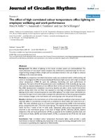
(A) Initial measurement of retinal arteriole diameter under fasting and normal conditions (blue and red, respectively), (black lines showing normal range in healthy subjects)Figure 1
(A) Initial measurement of retinal arteriole diameter under fasting and normal conditions (blue and red, respectively), (black
lines showing normal range in healthy subjects). (B) Average curve of all four observations under fasting and normal conditions
(blue and red, respectively).
Journal of Medical Case Reports 2008, 2:153 />Page 3 of 7
(page number not for citation purposes)
After 20 minutes of room acclimatization to obtain stabile
haemodynamic conditions, systemic blood pressure (BP)
was measured three times (1 minute apart) using a man-
ual sphygmomanometer. Systolic BP (SBP) and diastolic
BP (DBP) values were obtained; the average readings for
SBP and DBP were then used to calculate the mean arterial
BP (MABP) using the formula: MABP = 2/3 × DBP+1/3 ×
SBP. After instilling one drop of oxybuprocaine hydro-
chloride 0.4% in the right eye, intraocular pressure (IOP)
was also measured by means of a handheld contact
tonometer. The IOP and MBP measurements were used to
calculate the mean ocular perfusion pressure (MOPP)
according to the formula: MOPP = 2/3 × MABP-IOP.
After full pupil dilation was reached by using tropicamide
1.0% retinal diameters of the superior temporal retinal
artery and vein, measured approximately 1.5 disc diame-
ters away from the optic nerve head, were assessed contin-
uously over 350 seconds by using the DVA machine
according to a previously established protocol [6]. Briefly,
the measurement steps were: 50 seconds of still illumina-
tion (baseline recording) followed by three cycles of 20
seconds of flicker stimulation interspersed with 80 sec-
onds still illumination (representing recovery time). The
main measured outcome is the vessel width expressed in
units of measurement; for the stimulation with flicker
light, the outcome was defined as percent changed to
baseline. In addition to this standard measure, the time
needed to reach maximum dilation in both the artery and
the vein during fasting and normal conditions (called the
'reaction time') was calculated.
The results of the initial measurements under fasting con-
ditions and after a meal are shown in Table 1, and Figure
1A and Figure 2A. As compared with what is expected in a
healthy subject under normal conditions [1,6] our results
have shown a subnormal retinal vessels response in both
the arteriole (mean dilation = 2.59 ± 1.98%) and venule
(mean dilation = 5.19 ± 2.53%). This observation has trig-
gered a more detailed history of the patient's background
and dietary habits; this later step revealed that the subject
had been voluntarily fasting for 20 hours prior to the
research appointment. In order to enable us to see if this
particular aspect has had any influence on the measure-
ments, the subject was sent to have a meal and all meas-
urements were repeated immediately afterwards.
The results of the second set of measurements are shown
in Table 2. The difference between the measurements per-
formed before and after the meal were statistically signifi-
cant for the retinal venous dilation (p = 0.03) but not for
the retinal artery dilation (p = 0.08).
With the subject's consent, the experiment was repeated
on three additional occasions, each one month apart.
During these subsequent tests, the subject was in a similar
physiological condition to the initial experiment. Results
of all observations are shown in Table 3, and Figures 1B,
2B and 3. Retinal vein dilation and reaction time, but not
artery dilation and reaction time, were statistically signifi-
cantly lower in fasting versus non-fasting conditions (dila-
tion: p = 0.02 and p = 0.08; reaction time: p = 0.03 and p
> 0.05, respectively). These results were comparable with
the first observation that triggered the subsequent tests (a
blunted vascular response during fasting and a normal
response during after a meal).
Although SBP was statistically different when measured in
the fasting versus non-fasting state (111.5 ± 1.17; 113.5 ±
1.17; p = 0.01) the MABP, IOP and OPP were not statisti-
cally different in the fasting versus non-fasting state (p =
0.24, p = 0.11 and p = 0.64, respectively).
Discussion
This case demonstrates for the first time that despite hav-
ing comparable intraocular and systemic 'pressure' condi-
tions, the retinal vascular reactivity was blunted during
voluntary fasting in a healthy, young individual. This
observation was repeatable on four separate occasions.
The ocular circulation has a very complex regulating sys-
tem able to maintain constant blood flow and oxygen
supply to the tissue despite variations in systemic BP [1,7].
Autonomic nervous systems, as well as endothelial, meta-
bolic, myogenic and neurogenic factors play important
roles at different levels in this complex process. Molecules
such as nitric oxide (NO), arginine and glucose also inter-
vene by regulating smooth muscle cell relaxation, and
hence vascular dilation [1,8,9].
The vascular response to flickering light has been widely
studied in both animals and humans [1,6]. This provoca-
tion results in increased metabolic demand and, there-
fore, activation of NO synthase with subsequent
vasodilation occurs [8,10]. It is therefore possible that the
abnormal vascular dilation reported here, may be the
result of low NO availability; however, the mechanism
resulting in NO depletion can only be hypothesized at
this stage. Changes in the levels of active substances such
as lactate and glucose could also play role [8,10-12] in the
vascular response to flicker provocation. Therefore, beside
modifications in the NO dynamics, practically any change
in the quantity and composition of the circulating fluid
due to dehydration and/or fasting is also capable of alter-
ing vascular dynamics and BP [13]. Indeed, Alghadyan et
al. [14] found a significant increase in onset of retinal vein
occlusion (RVO) during fasting, suggesting a possible
relationship between fasting and dehydration in the
pathogenesis of RVO. Moreover, Lapeyraque et al.
reported about five cases of chronic hypovolaemia occur-
Journal of Medical Case Reports 2008, 2:153 />Page 4 of 7
(page number not for citation purposes)
(A) Initial measurement of retinal venule diameter under fasting and normal conditions (light blue and dark blue, respectively), (black lines showing upper and lower confidence limits in healthy subjects)Figure 2
(A) Initial measurement of retinal venule diameter under fasting and normal conditions (light blue and dark blue, respectively),
(black lines showing upper and lower confidence limits in healthy subjects). (B) Average curve of all four observations under
fasting and normal conditions (light blue and dark blue, respectively).
Journal of Medical Case Reports 2008, 2:153 />Page 5 of 7
(page number not for citation purposes)
Table 1: Showing retinal venule and arteriole dilation after each cycle of flickering light stimulation, as change in percentage,
compared to baseline diameter as measured under fasting conditions.
CONDITION: FASTING
FLICKER STIMULATION VENOUS DILATION (in % to baseline value)
F1 3.90
F2 8.29
F3 4.47
MEAN +/- STD 5.59 +/- 2.37
ARTERIAL DILATION (in % to baseline value)
F1 5.20
F2 3.96
F3 4.72
MEAN +/- STD 4.63 +/- 0.63
Table 2: Showing retinal venule and arteriole dilation after each cycle of flickering light stimulation, as change in percentage,
compared to baseline diameter as measured immediately after a meal.
CONDITION: AFTER MEAL
FLICKER STIMULATION VENOUS DILATION (in % to baseline value)
F1 11.91
F2 5.26
F3 8.16
MEAN +/- STD 8.45 +/- 3.34
ARTERIAL DILATION (in % to baseline value)
F1 9.18
F2 8.76
F3 11.69
MEAN +/- STD 9.88 +/- 1.58
Retinal venule reaction time under normal conditions (TX-V-norm) and under fasting conditions (TX-V-fast)Figure 3
Retinal venule reaction time under normal conditions (TX-V-norm) and under fasting conditions (TX-V-fast).
Publish with BioMed Central and every
scientist can read your work free of charge
"BioMed Central will be the most significant development for
disseminating the results of biomedical research in our lifetime."
Sir Paul Nurse, Cancer Research UK
Your research papers will be:
available free of charge to the entire biomedical community
peer reviewed and published immediately upon acceptance
cited in PubMed and archived on PubMed Central
yours — you keep the copyright
Submit your manuscript here:
/>BioMedcentral
Journal of Medical Case Reports 2008, 2:153 />Page 6 of 7
(page number not for citation purposes)
ring during continuous peritoneal dialysis which could
result in blindness due to ocular ischaemia [15].
Although the data obtained in this case study is limited
and needs further investigation to demonstrate the exact
mechanism behind our observation, it clearly shows the
clinical significance of detailed medical history before
conducting and interpreting any test results. Knowledge of
medical and/or family history and ethnic background at
the time of examination is of great importance. In addi-
tion, since fluid and/or nutrient intake plays an important
role in the physiology of important variables such as BP,
vessel dynamics and blood composition, which are com-
monly used in diagnosing various systemic vascular disor-
ders, fasting and dehydration could act as confounding
factors and the clinical and/or laboratory results could be
misleading.
Conclusion
We present a case of decreased retinal vascular reactivity
due to fasting and dehydration in a young and healthy
man. This scenario could represent a potential case of
vasoactive substance imbalance resulting in vasodilatory
inhibition at the level of retinal vessels. The importance of
this finding should be investigated in further studies and
additional tests should be included to verify our hypothe-
sis.
Abbreviations
BP: blood pressure; DBP: diastolic blood pressure; DVA:
dynamic retinal vessel analyser; IOP: intraocular pressure;
MABP: mean arterial blood pressure; MOPP: mean ocular
perfusion pressure; NO: nitric oxide; RVO: retinal vein
occlusion; SBP: systolic blood pressure.
Competing interests
The authors declare that they have no competing interests.
Authors' contributions
RH collected the data, was involved in data analysis and
interpretation and drafting of the manuscript. DG revised
the manuscript and was involved in the data analysis and
interpretation. RA was involved in the statistical analysis
and interpretation. RC made substantial contributions to
the design and collection. SH made substantial contribu-
tions to the design and collection. All authors read and
approved the final manuscript.
Consent
Written informed consent was obtained from the patient
for publication of this case report and any accompanying
images. A copy of the written consent is available for
review by the Editor-in-Chief of this journal.
References
1. Nagel E, Vilser W, Lanzl I: Age, blood pressure, and vessel diam-
eter as factors influencing the arterial retinal flicker
response. Invest Ophthalmol Vis Sci 2004, 45:1486-1492.
2. Frederiksen CA, Jeppesen P, Knudsen ST, Poulsen PL, Mogensen CE,
Bek T: The blood pressure-induced diameter response of ret-
inal arterioles decreases with increasing diabetic maculopa-
thy. Graefes Arch Clin Exp Ophthalmol 2006.
3. Garhofer G, Zawinka C, Resch H, Huemer KH, Schmetterer L,
Dorner GT: Response of retinal vessel diameters to flicker
stimulation in patients with early open angle glaucoma. J
Glaucoma 2004, 13:340-344.
4. Garhofer G, Zawinka C, Resch H, Kothy P, Schmetterer L, Dorner
GT: Reduced response of retinal vessel diameters to flicker
stimulation in patients with diabetes. Br J Ophthalmol 2004,
88:887-891.
5. Vilser W, Nagel E, Lanzl I: Retinal Vessel Analysis new possibil-
ities. Biomed Tech (Berl) 2002, 47 Suppl 1 Pt 2:682-685.
6. Nagel E, Vilser W: Flicker observation light induces diameter
response in retinal arterioles: a clinical methodological
study. Br J Ophthalmol 2004, 88:54-56.
7. Nagel E, Vilser W: Autoregulative behavior of retinal arteries
and veins during changes of perfusion pressure: a clinical
study. Graefes Arch Clin Exp Ophthalmol 2004, 242:13-17.
8. Dallinger S, Sieder A, Strametz J, Bayerle-Eder M, Wolzt M, Schmet-
terer L: Vasodilator effects of L-arginine are stereospecific
and augmented by insulin in humans. Am J Physiol Endocrinol
Metab 2003, 284:E1106-E1111.
9. Garhofer G, Resch H, Lung S, Weigert G, Schmetterer L: Intrave-
nous administration of L-arginine increases retinal and
choroidal blood flow. Am J Ophthalmol 2005, 140:69-76.
10. Dorner GT, Garhofer G, Kiss B, Polska E, Polak K, Riva CE, Schmet-
terer L: Nitric oxide regulates retinal vascular tone in
humans. Am J Physiol Heart Circ Physiol 2003, 285:H631-H636.
11. Dorner GT, Garhofer G, Huemer KH, Riva CE, Wolzt M, Schmet-
terer L: Hyperglycemia affects flicker-induced vasodilation in
the retina of healthy subjects. Vision Res 2003, 43:1495-1500.
Table 3: Showing results of all 4 observations along with p-values, as obtained from the statistical analysis.
CONDITION: FASTING CONDITION: AFTER MEAL P-VALUE
VENOUS DILATION [% change to baseline] 5.19 +/- 2.53 9.01 +/- 2.69 0.02
ARTERIAL DILATION [% change to baseline] 2.59 +/- 1.98 3.91 +/- 2.41 0.08
REACTION TIME [sec] 20 +/- 4.88 15.41 +/- 3.72 0.03
Publish with BioMed Central and every
scientist can read your work free of charge
"BioMed Central will be the most significant development for
disseminating the results of biomedical research in our lifetime."
Sir Paul Nurse, Cancer Research UK
Your research papers will be:
available free of charge to the entire biomedical community
peer reviewed and published immediately upon acceptance
cited in PubMed and archived on PubMed Central
yours — you keep the copyright
Submit your manuscript here:
/>BioMedcentral
Journal of Medical Case Reports 2008, 2:153 />Page 7 of 7
(page number not for citation purposes)
12. Koifman B, Topilski I, Megidish R, Zelmanovich L, Chernihovsky T,
Bykhovsy E, Keren G: Effects of losartan + L-arginine on nitric
oxide production, endothelial cell function, and hemody-
namic variables in patients with heart failure secondary to
coronary heart disease. Am J Cardiol 2006, 98:172-177.
13. Inan UU, Yucel A, Ermis SS, Ozturk F: The effect of dehydration
and fasting on ocular blood flow. J Glaucoma 2002, 11:411-415.
14. Alghadyan AA: Retinal vein occlusion in Saudi Arabia: possible
role of dehydration. Ann Ophthalmol 1993, 25:394-398.
15. Lapeyraque AL, Haddad E, Andre JL, Bremond-Gignac D, Taylor CM,
Rianthavorn P, Salusky IB, Loirat C: Sudden blindness caused by
anterior ischemic optic neuropathy in 5 children on continu-
ous peritoneal dialysis. Am J Kidney Dis 2003, 42:E3-E9.