Báo cáo y học: "Upregulation of a novel eukaryotic translation initiation factor 5A (eIF5A) in dengue 2 virus-infected mosquito cells" pot
Bạn đang xem bản rút gọn của tài liệu. Xem và tải ngay bản đầy đủ của tài liệu tại đây (1.72 MB, 9 trang )
RESEA R C H Open Access
Upregulation of a novel eukaryotic translation
initiation factor 5A (eIF5A) in dengue 2
virus-infected mosquito cells
Yu-Tzu Shih
1
, Chao-Fu Yang
1
, Wei-June Chen
1,2*
Abstract
Background: Dengue virus, a mosquito-borne flavivirus, is the etiological agent of dengue fever, dengue
hemorrhagic fever, and dengue shock syndrome. It generally induces apoptosis in mammalian cells, but frequently
results in persistent infection in mosquito cells. That mechanism remains to be explored. In turn, a genomic survey
through subtractive hybridization (PCR-select cDNA subtraction) was conducted in order to find gene(s) that may
play a role in in teractions between the virus and its host cells.
Results: Through this technique, we identified a novel eukaryotic translation initiation factor 5A (eIF5A) which is
upregulated in Aedes albopictus-derived C6/36 cells infected by the type 2 dengue (Den-2) virus. The full-length of
the identified eIF5A gene consisted of 1498 bp of nucleotides with a 41.39% G+C content, and it possessed a
higher similarity and shorter evolutionary distance with insects than with other organisms. Upregulation of eIF5A in
response to Den-2 virus infection was validated at both the RNA and protein levels. This phenomenon was also
observed by confo cal microscopy. In addition, cell death obviously occurred when eIF5A activity was inhibited in
C6/36 cells even when they were infected by the virus. However, viral multiplication was not obviously affected in
infected C6/36 cells when eIF5A activity was reduced.
Conclusions: Taken together, we postulated that eIF5A plays a role in preventing mosquito cells from death in
response to Den-2 viral infection, thus facilitating continued viral growth and potential persistent infection in
mosquito cells. It would be worthwhile to further investigate how its downstream factors or cofactors contribute to
this phenomenon of dengue infection.
Background
The dengue virus, one of the flaviviruses, contains ~11
kilobase (kb) single-stranded, positive-sense genomic
RNA [1]. Within host cells, viral RNA directly translates
into a single p olyprotein that is subsequently cleaved
into three structural proteins and seven nonstructural
proteins [2]. The process is carried out by the combined
action of host proteases and a trypsin-like viral NS2B/
NS3 serine protease [3].
The dengue virus is transmitted between humans by
mosquitoes, imp lying that both mammalian and mos-
quito cells are susceptible to the virus [4]. Mammalian
cells with dengue virus infection usually end up
undergoing apoptosis due to shutdown of protein synth-
esis in the host cell [5]. However, dengue and other arbo-
viruses frequently occur in mosquito cells without
causing obvious del eterious effects [6,7], implying that
specific host factors are critically involved in such
regulation.
Hypothetically, viruses invading a host cell redirect
cellular processes to meet the needs of viral propagation
[8], leading to the induction of novel changes in gene
expressions; this was reported in human umbilical vein
endothelial cells infected with dengue virus [9]. The
change in a host cell’ s protein-making machinery was
also confirmed after infection by the dengue virus [10].
In turn, the path to maturation for the dengue virus
may depend o n the cell type, leading to unique charac-
teristics of the virus.
* Correspondence:
1
Graduate Institute of Biomedical Sciences, College of Medicine, Chang
Gung University, Kwei-San, Tao-Yuan 33332, Taiwan
Full list of author information is available at the end of the article
Shih et al. Virology Journal 2010, 7:214
/>© 2010 Shih et al; licensee BioMed Cent ral Ltd. This is an Open Access article distributed under the terms of the Creative Commons
Attribution License (http://creativecom mons.org/licenses/by/ 2.0), which permits unrestricted use, distribution, and reproduction in
any medium, provided the original work is properly cited.
Through the method of polymerase chain reaction
(PCR)-select complementary (c)DNA subtraction, eukar-
yotic translation initiation factor 5A (eIF5A) was
demonstrated to be upregulated at both the messenger
(m)RNA and protein levels in C6/36 cells following den-
gue 2 (Den-2) virus infection [11]. eIF5A, formerly
called eIF-4D, was first isolated from imma ture red
blood cells [12], is an acidic pr otein with a molecular
mass of 17~21 kDa, and is relatively conserved from
yeast to humans [13]. It is the only protein in nature
known to contain the unusual amino acid, hypusi ne
[N
ε
-(4-amino-2-hydroxybutyl) lysine], derived from a
modification of lysine by spermidine [14].
The eIF5A protein was originally considered to be a
translation initiation factor based on its in vitro activity of
stimulating the formation of methionyl-puromycin, a
dipeptide analogue, used in a model system to study the
formation of the first peptide bond and to transiently
attach to the ribosome in the course of initiation of eukar-
yotic cellular protein synthesis [15]. However, its role in
translation seems controv ersial since its deletion in yeas t
leads to only a slight decrease in total protein s ynthesis
[16]. Further, eIF5A was suggested to function as a nucleo-
cytoplasmic shuttle for specific subsets of mRNAs
involved in cell division [17], and its posttranslational
modification is important for cell survival as well as prolif-
eration [18]. These functions were observed via stimula-
tion of polyamines (putrescine, spermidine, and spermine),
which are transformed to active eIF5A [19]. Herein, eIF5A
was demonstrated to be upregulated in response to Den-2
virus infection in C6/36 cells, and its role in a ssociation
with the survival of infected cells is discussed.
Results
Full-length sequence and phylogenetic analysis of eIF5A
derived from Ae. albopictus
Full-length eIF5A derived from Ae. albopictus consists
of 1498 bp of nucleotides with a 41.39% G+C content
and possesses an 85.8% similarity with that from
Ae. aegypti (AY433334). The sequence was submitted to
GenBank (accession no. EU910137). This genome
enco ded 160 amino acids, with only a single amino acid
difference (S®A) compared to that from Ae. aegypti
(ABF18091) (Figure 1).
In a compariso n of 12 eIF5A proteins, the one from
Ae. albopictus shared 99% similarity with that from Ae.
aegypti, 89% with that from Bombyx mori (AAZ15 319),
57% with that from Caenorhabditis elegans (CAA90247),
69% with that from Danio rerio (AAH67190), 80% with
that from Drosophila melanogaster (AAG17032), 67%
with that from Gallus gallus (CAG31407), 68% with that
from Homo sapiens (NP001961), 68% with that from
Rattus norvegicus (NP001028853), 62% with that from
Saccharomyces cerevisiae (BAA11826), 90% with that
from Spodoptera frugiperda (AAF13316), and 68% with
that from Xenopus tropicalis (CAJ83651). In the phylo-
genetic tree constructed using the NJ method (Figure 2),
at the protein level, the first branch that emerged from
the insect group included vertebrates as mentioned
above. The bootstrap support for the insect group was
98%; it w as as high as 80% for other organisms in the
NJ tree. In contrast, eIF5A derived from Ae. albopictus
was genetically distant from those of fungi ( S. cerevisiae)
and nematodes (C. elegans).
Elevated expression of eIF5A in C6/36 cells infected by
the Den-2 virus
Expression of eIF5A in C6/36 cells was analyzed follow-
ing Den-2 virus and UV-i nactivated Den-2 virus infec-
tion. C6/36 cells were infected with either the Den-2
virus or a UV-inac tivated Den-2 virus at an MOI of 1. At
24 h, cells were collected for RNA extraction. Significant
upregulation of eIF5A was only observed in C6/36 cells
after infection b y intact Den-2 vir us as dete cted by a
quantitative real-time PCR. Den-2 virus infection induced
a 3-fold (3.60 ± 0.30) increase in eIF5A (for comparison
with the mock; Student’ s t-test; p > 0.05), whereas the
inactivated Den-2 virus infection only in duced a 1.63-
fold (1.63 ± 0.44) increase (Student’ s t-test; p <0.05)
(Figure 3). Enhanced expression of eIF5A at the protein
level was measured by Western blottin g (Figure 4A).
Using double-staining with specific antibodies to com-
pare images under laser scanning confocal microscopy,
the expression of eIF5A was obviously enhanced in virus-
infected C6/36 cells at 24 hpi compared to mock-infected
cells in which lighter expression of the endogenous pro-
tein was shown (Figure 4B). Co-localization of eIF5A and
dengue proteins was shown in certain areas of infected
C6/36 cells (Figure 4B).
Association of eIF5A with the survival of infected
C6/36 cells
Cell death was measured at 24 and 48 hpi using the
method of PI staining. With mock infection (without
CPO treatment) in C6/36 cells, the cell death rates were
2.15% and 2.12%, respectively; the rates did not evi-
dently change even when cells were treated with CPO
(1.14% and 9.85%, respectively). When cells were
infected with the Den-2 virus (without CPO treatment),
the cell death rate slightly increased to 4.71% and 8.10 %
at 24 and 48 hpi, respectively. In the g roup with Den-2
virus infection plus CPO treatment, the cell death rate
slightly increased to 5.85% at 24 hpi, but rapidly to
28.04% at 48 hpi (Figure 5).
Effects of the eIF5A on propagation of the dengue virus
After treatment of C6/36 cells with CPO for 24 h, both
viral RNA and proteins were examined to evaluate the
Shih et al. Virology Journal 2010, 7:214
/>Page 2 of 9
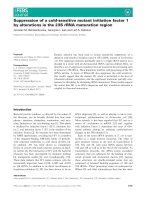
Figure 1 Alignment of the eIF5A amino acid sequence derived from C6/36 cells with 11 homologous proteins from other organisms.
The black background denotes amino acid residues identical to those in the first line, and gaps are indicated by a dash (-). Accession numbers
of listed species: Aedes albopictus (EU910137); Ae. aegypti (ABF18091); Bombyx mori (AAZ15319); Caenorhabditis elegans (CAA90247); Danio rerio
(AAH67190); Drosophila melanogaster (AAG17032); Gallus gallus (CAG31407); Homo sapiens (NP_001961); Rattus norvegicus (NP_001028853);
Saccharomyces cerevisiae (BAA11826); Spodoptera frugiperda (AAF13316); and Xenopus tropicalis (CAJ83651).
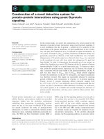
Figure 2 Neighbor-joining tree of eIF5A identified from 12
species of organisms using protein databases from GenBank.
Numbers on the branches are bootstrap proportions (1000
replicates). See the text for accession numbers.
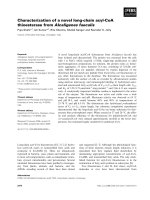
Figure 3 Validation of the eIF5A gene expression level in C6/
36 cells with Den-2 virus infection. RNAs extracted from C6/36
cells with mock infection (Mock), UV-inactivated Den-2 virus (UV), or
intact Den-2 virus (Den-2) at a multiplicity of infection (MOI) of
1 were evaluated by a quantitative real-time RT-PCR assay. The
quantitative real-time PCR analysis of eIF5A was monitored and
normalized to the expression of 18S, which was used as an internal
control. Ratios of the normalized expressions of eIF5A of Den-2-
infected cells were relative to that of mock-infected cells. The results
showed that the expression of eIF5A was significantly higher in the
group with Den-2 virus infection (p < 0.05), but not in those
inoculated with UV-inactivated Den-2 virus (p > 0.05).
Shih et al. Virology Journal 2010, 7:214
/>Page 3 of 9
effect of eIF5A on the growth of Den-2 virus. Total
RNA harvested from C6/36 cells was evaluated with the
primer pair (D2EL and D2ER) to detect the amplifica-
tion of positive- and negative-sense viral RNAs. The
results showed that viral RNA was detected in infected
cells with and without treatment with CPO although
they did not quantitatively differ (Figure 6A). At the
protein level, they did not show a quantitative difference
either (Figure 6B). In addition, virus production was not
obviously affected by CPO treatment before 24 hpi, but
slightly decreased in the period between 24 and 48 hpi
(Figure 6C).
Discussion
The dengue virus is transmitted by mosquitoes between
vertebrate hosts in nature [4], reflecting its ability to
grow in both humans and mosquitoes. This way of
transmission reveals an important event: the vector
Figure 4 Upregulation of eIF5A in C6/36 cells infected by Den-2 virus for 24 h. eIF5A was shown to have increased in expression,
according to the results of Western blotting, in response to Den-2 virus infection at 24 h post-infection (hpi) (A). With confocal microscopy,
expression of eIF5A (green) was shown to be upregulated in Den-2 virus (red)-infected cells compared to that of the mock infection (B).
Shih et al. Virology Journal 2010, 7:214
/>Page 4 of 9
must be compatible fo r virus amplification or can even
become persistently infected without causing tissue or
cell damage; which is hypothetically regulated by specific
genes upregulated by viral stimulation [11]. We recently
foundthateIF5AisupregulatedinC6/36cellswith
Den-2 infection, suggesting that this gene may have spe-
cific functions in mosquito cells infected with Den-2
virus. This implies that eIF5A may play a role different
from that in mammalian cells. Because C6/36 cells are
upregulated to express eIF5A only by intact, not by UV-
inactivated, Den-2 virus, its overexpression is supposedly
regulated w hen endocytosis is completed. Levels of the
virus in infected cells treated with CPO compared to
non-treated cells did not significantly differ. This indi-
cated that induct ion of eIF5A may just prolong the sur-
vival of infected cells, providing an environment
beneficial for viral growth.
Phylogenetic analyses using the amino acid frequen-
cies of conserved proteins are free from the drastic bias
of the genomic G+C content and may provide a robust
estimation of early divergences in the evolution of
eukaryotes [20]. As shown in the phylogenetic tree co n-
structed using the NJ method, eIF5A was suggested to
have a closer evolutionary relat ionship and common
functions due to the short genetic distance between spe-
cies compared. The high sequence conservation of
eIF5A across species suggests that t he protein has an
important common physiological role.
Unlike mammalian cells, mo squito cells are usually
susceptible to Den-2, but infection does not result in
cell death. Based on a cell-cycle analysis, both mock-
and Den-2-infected C6/36 cell s tended to remain in the
S phase, a point at which mosquito cells are g ood at
replication, protein synthesis, and assembly of the virus
[21]. It would be interesting to see if inhibiting eIF5A in
infected mammalian cells also induces G
1
arrest as
opposed to uninfected cells. This experiment could be
crucial in revealing the significance of data in this
report. In fact, reduction of eIF5A was previously
described to induce G
1
arrest in mammalian HeLa cells
[22]. In C6/36 cells not treated with CPO, the death
rate slightly increased in infected cells, indicating that
the Den-2 virus may naturally cause a low-level death
rate in mosquito cells. On the other hand, eIF5A upre-
gulation induced by the virus actually helped mosquito
cells survive the lethal effects of the virus by allowing
successful progression through the cell cycle. Although
the functions of eIF5A are still being debated, it was
reported to play roles in cell proliferation, cell viability,
and cell-cycle progression. In addition to proliferation
and cellu lar protein synthesis [15], genetic and pharma-
cological studies provided evidence that eIF-5A is
Figure 5 Effects of eIF5A on the survival of C6/36 cells with Den-2 virus infection. C6/36 cells were incubated with the Den-2 virus for 1 h
at a multiplicity of infection (MOI) of 1, and then treated with ciclopirox olamine (CPO, 10 μM). At 24 and 48 h, cells were fixed and stained with
propidium iodide for a flow cytometric analysis. Cells in the sub-G
0
/G
1
phase are marked as M1, and the rate of cell death is shown in
parentheses on each graph. Representative data of the experiments are shown.
Shih et al. Virology Journal 2010, 7:214
/>Page 5 of 9
essential for cell survival [23]. Although the expression
of the eIF5A protein i s normally low [24], the Den-2
virus likely induces eIF5A overexpression in C6/36 cells,
which is advantageous for cells’ adaptation to viral infec-
tion without deleterious effects.
Taken together, we postulate that the Den-2 virus may
stimulate the overexpression of eIF5A, which facilitates
a reduction in cell death in infected C6/36 cells. This
actually produces an advantage of continuing replication
by the virus in mosquito cells although it might not be
involved in directly promoting virus replication.
Methods
Virus and cell culture
The Den-2 virus (New Guine a C strain) was propagated
in Aedes albopictus-derived C6/36 cells, which were cul-
tured in minimal essential medium ( MEM; GIBCO™,
Invitrogen, Carlsbad, CA, U SA) supplemented with 10%
fet al bovine serum (FBS), 2% non-essential amino acids,
2 g/ml Hepes (Sigma, St. Louis, MO, USA), 2.2 g/ml
sodium bicarbonate (NaHCO
3
), and 0.4% antibiotic-anti-
mycotic at 28°C in a closed system. The virus was
titrated as described below in baby hamster kidney
(BHK)-21 cells, which were maintained in MEM con-
taining 10% FBS, 2% non-essenti al amino acids, 2.2 g/ml
sodium bicarbonate (NaHCO
3
), and 0.4% antibiotic-anti-
mycotic (GIBCO™ , Invitrogen) at 37°C in a 5% CO
2
atmosphere. Viruses produced in cultured cells w ere
titrated by a plaque assay as described previously [11].
Cell infection
C6/36 cells (~1 × 10
7
cells/tube) were harvested and
centrifuged at 3000 rpm and 4°C for 10 min. After
removing the medium, the Den-2 viral suspension or
medium (mock infection as the control) was added to
the tubes at a multiplicity of infection (MOI) of 1 for
incubation at 25°C for 1 h with gentle agitation every 15
min. Then the viral suspension was removed by centri-
fugation, and pelleted cells were seeded and incubated
at 25°C.
Figure 6 Inhibition of eIF5A via treatment with CPO showed no effect on propagation of C6/36 cells inoculated with Den-2 virus for
24 h. (A) Demonstration of viral replication using RT-PCR to amplify fragments of the E gene from extracted positive- (+) or negative (-)-strand
viral RNA of infected cells. (B) Detection of dengue E protein with an anti-E monoclonal antibody via Western blot analysis in infected cells.
(C) Growth of the Den-2 virus in C6/36 cells with or without inhibition at different times after infection.
Shih et al. Virology Journal 2010, 7:214
/>Page 6 of 9
RNA extraction and reverse-transcription polymerase
chain reaction (RT-PCR)
The procedures of RNA extraction and RT-PCR were
performed as described previously [11]. In brief, total
RNA was isolated from both mock- and Den-2 virus-
infected C6/36 cells using the Trizol reagent (Invitro-
gen). Compl ementary (c)DNA was prepared from
extracted total RNA following instructions provided by
the SMART™ PCR cDNA synthesis kit (Clontech,
Mountain View, CA, USA).
Real-time PCR
cDNA from infected (with active or UV-inactivated vir-
ions) or uninfected (mock) cells was used to validate the
expression of eIF5A using the primers GCCCATC-
CACTCACAACATG (forward) and TCGATGTCAGT-
GAGCTGGTAGTC (reverse), designed from the
sequence of the cloned eIF5A described above. The
thermal cycling conditions and presentation of results
followed a previous description [11].
Determination of the full-length sequence of eIF5A
Determination of the full-len gth sequence of eIF5A fol-
lowed an approach described elsewhere [25]. Extracted
total RNA was used to synthesize a fragment of eIF5A
with Oligo dT and the primer derived from selected
clones of eIF5A (eIF5AL: 5’-TATTTGCCC ATCCACT-
CACA). The products were then cloned into the
pGEM-T vector to subsequently sequence the 3’-end o f
thegene.The5’ -end of Ae. albopictus eIF5A was
obtained using a 5’RACE system (Invitrogen) according
to the manufacturer’ s protocol. In brief, the extracted
total RNA was first treated with 1 U/μlDNase(Pro-
mega, Madison, WI, USA) to remove t he genomic
DNA, from which 5’ -end cDNA was generated with
gene-specific primer (GSP)-1, 5’-CGATGCCAACCA-
GATGTACC-3’ ,andSuperscriptII™ RT (Invitrogen).
dCTP was added to the tail of the 5’ -end cDNA using
terminal deoxynucleotidy l transferase, and then the
dCTP-tailed cDNA was amplified by a PCR with GSP-2,
5’ -GTGTTTACCGGTCTTGGAGG-3’,anduniversal
primers provided by the manufacturer of the kit. The
resultant PCR products were then cloned into the
pGEM-T vector (Promega) for nucleotide sequencing.
The obtained sequence was used to compare ESTs
derived from both Ae. aegypti and Armigeres subalbatus
[26].
Phylogenetic analysis
The similarity of the eIF5A coding sequence derived
from Ae. albopictus-derived C6/36 c ells was compared,
using the basic local alignment search tool [27] in the
BLAST network servi ce (National Ce nter for B iotech-
nology Information, Bethesda, MD, USA), against those
from selecte d species (see “Results”) in the database. All
sequences were aligned using the default parameters of
CLUSTAL X [28] and edited by Genedoc software [29];
from this, a phylogenetic analysis using an unrooted tree
constructed with the distance-based Neighbor-joining
(NJ) method was carried out with MEGA4 [30]. One
thousand bootstrap replications were performed. Other
parameters used the default option.
UV inactivation of the Den-2 virus
The method followed a previous description [11].
Briefly, a viral suspension was exposed to a UV lamp
(254 nm; 120 mJ/cm2) for 30 min. The efficacy of viral
inactivation was examined by a real-time RT-PCR and
plaque assay. Expression of the eIF5A gene in UV-i nac-
tivated Den-2 viral-infected C6/36 cells was assayed by a
real-time RT-PCR as described above.
Detection of viral RNA synthesis
Synthesis of viral RNA including positive and negative
strands was detected by an RT-PCR as described before
[31]. Viral RNA was extracted from C6/36 cells inocu-
lated wi th a combination of Den-2 virus (at an MOI of
1) and CPO. Inoculated cells were harvested at 24 h
post-infection (hpi) to detect RNA synthesis through
amplification of a gene fragment by RT-PCR. The pri-
mer pair (D2EL: TAACACCACAGAGTTCC ATC and
D2ER: TAAACTTTCCTGTGCACATA) was used to
detect ne wly synthesized positive-strand RNA. The pri-
mers used to detect negative-strand RNA was the com-
plementary counterparts of the a bove primer pair. The
PCR product was identified as 429 bp of an amplified
cDNA fragment by running on a 2% (w/v) agarose gel.
Confocal microscopy
About 2 × 10
6
C6/36 cells were plated in 6-well culture
plates for 24 h. A Den-2 virus suspension was added to
each well and allowed to be adsorbed for 1 h, and then
the cells were was incubated for another 24 h. Cells
were fixed with 4% paraformaldehyde and subsequently
treated with 0.1% Triton X-100 for 2 min to increase
the permeability. Primary antibodies including a rabbit
anti-eIF5A antibody (1: 8000 in dilution) and a mouse
anti-Den-2 antibody (1: 100 in dilution), followed by
secondary antibodies of Alexa Fluor® 488-conjugated
goat anti-rabbit IgG (Invitrogen) and rhodamine-conju-
gated goat anti-mouse immunoglobulin G (IgG) (Chemi-
con International, Billerica, MA, USA), were used to
respectively detect eIF5A (in green) and the Den-2 (in
red) virus. 4’-6-Diamidino-2-phenylindole (DAPI) which
presented as blue was used as an indicator of cell nuclei.
Prepared specimens were observed under a laser scan-
ning confocal microscope (Zeiss LSM 510, Vertrieb,
Germany).
Shih et al. Virology Journal 2010, 7:214
/>Page 7 of 9
Effects of eIF5A on cell death measured with propidium
iodide (PI) nucleic acid staining
C6/36 cells (~2 × 10
6
cells/tube) were collected and
infected with the Den-2 virus at an MOI of 1. After 1 h
of absorption, cells were treated with 10 μM ciclopirox
olamine (CPO, Sigma) to inhibit the function of eIF5A,
while treatment with dimethyl sulfoxide (DMSO; the
solventusedwithCPO)wasusedasthecontrol.At48
hpi, cells were harvested and cen trifuged at 10 00 rpm
and 4°C for 5 min. After the suspension was removed,
the cell pellet was fixed with ice-cold 70% ethanol in a
-20°C freezer for at least 1 h. Cells were centrifuged
again at 1500 rpm and 4°C for 5 min, and washed with
PBS after the fixative solution had been discarded.
These cells were treated with 0.5% Triton X-100 and
0.05% RNase A (Si gma) in PBS for 1 h at 37°C. A fter a
final centrifugation, pelleted cells were stained with
50 μg/ml PI (Sigma) in PBS at 37°C for 20 min and
stored at 4°C in the dark. The cellular DNA content was
measured using ModFit LT software vers. 3.0 (Verity
Software House, Topsham, ME, USA) with a FASCAN
flow cytometer (BD Biosciences, San Jose, CA, USA).
Western blotting
To detect viral proteins, C6/36 cells were infected with
the Den-2 virus at an MOI of 1. At 24 hpi, cells were
harvested and washed with PBS three times. Approxi-
mately 2 × 10
6
cells were pelleted and ly sed with 100 μl
RIPA lysis buffer (50 mM Tris Cl (pH 7.4), 150 mM
NaCl, 1% NP-40, 1 mM EDTA, and a protease inhibitor
cocktail) at -80°C overnight. After centrifuga tion at
14,000 rpm for 10 min at 4°C, supernatants were boiled
in 2× sample buffer (8% sodium dodecylsulfate (SDS),
1 M Tris (pH 6.8), 40% glycerol, and 0.001 bromophenol
blue) for 10 min; these were subsequently resolved by
SDS-polyacrylamide gel electrophoresis (PAGE) and
transferred to an Immobilon™ -P transfer membrane
(Millipore, Billerica, MA, USA). Membranes were
soaked in 5% skim milk in a TBS-T solution (0.242%
Tris-base, 2.924% NaCl, and 0.1% Tween 20; pH 7.5) at
room temperature for 1 h. Membranes were then
washed with the TBS-T solution three times. For viral
protein detection, membranes were probed with an anti-
Den -2 viral E protein antibody at room temperature for
1 h; f or eIF5A detection, membranes were probed with
an anti-eIF5A antibody (both of which were prepared by
our lab). A goat anti-rabbit IgG-horseradish peroxidase
(HRP)-conjugated antibody (Perkin-Elmer™ Life
Sciences, Boston, MA, USA) was subsequently added to
the membranes and incubated for 1 h at room tempera-
ture after the membranes had been washed with TBS-T.
After the membranes were washedagain,bandprofiles
were visualized by a reaction after application of Wes-
tern Lighting® Chemiluminescence Reagent Plus (Perkin-
Elmer™ Life Science, Waltham, MA, USA) and exposure
to K odak BioMax XAR film (Eastman Kodak, Rochester,
NY, USA).
Statistical analysis
Comparisons between two means were analyzed by Stu-
dent’s t-test at a significance level of 5%.
Acknowledgements
We thank Dr. Chih-Yu Wu for technical assistance. This work was supported
by a grant from the National Science Council of Taiwan (NSC 96-2628-B-182-
003-MY3) and partially by Chang Gung Memorial Hospital (CMRPD 160163).
Author details
1
Graduate Institute of Biomedical Sciences, College of Medicine, Chang
Gung University, Kwei-San, Tao-Yuan 33332, Taiwan.
2
Department of Public
Health and Parasitology, College of Medicine, Chang Gung University, Kwei-
San, Tao-Yuan 33332, Taiwan.
Authors’ contributions
YTS carried out all the experiments and analyzed results. CFY helped to
perform confocal microscopy. WJC designed the study and wrote the
manuscript. All authors read and approved the final manuscript.
Competing interests
The authors declare that they have no competing interests.
Received: 27 March 2010 Accepted: 7 September 2010
Published: 7 September 2010
References
1. Lindenbach D, Rice CM: Molecular biology of flaviviruses. Adv Virus Res
2003, 59:23-61.
2. Rey FA, Heinz F, Mandl C, Kunz C, Harrison S: The envelope glycoprotein
from tick-borne encephalitis virus at 2 Å resolution. Nature 1995,
375:291-298.
3. Murthy HM, Judge K, DeLucas L, Padmanabhan R: Crystal structure of
dengue virus NS3 protease in complex with a Bowman-Birk inhibitor:
implications for flaviviral polyprotein processing and drug design. J Mol
Biol 2000, 301:759-767.
4. Gubler DJ: The global emergence/resurgence of arboviral diseases as
public health problems. Arch Med Res 2002, 33:330-342.
5. Courageot M, Catteau A, Despres P: Mechanisms of dengue virus-induced
cell death. Adv Virus Res 2003, 60:157-186.
6. Newton SE, Short NJ, Dalgarno L: Bunyamwera virus replication in
cultured Aedes albopictus (mosquito) cells: establishment of a persistent
viral infection. J Virol 1981, 38:1015-1024.
7. Chen WJ, Chen SL, Fang AH: Phenotypic characteristics of dengue
persistently infected in C6/36 cell clone of Aedes albopictus cells.
Intervirology 1994, 37:25-30.
8. Frolova EI, Fayzulin RZ, Cook SH, Griffin DE, Rice CM, Frolov I: Roles of
nonstructural protein nsP2 and alpha/beta interferons in determining
the outcome of Sindbis virus infection. J Virol 2002, 76:11254-11264.
9. Warke RV, Xhaja K, Martin KJ, Fournier MF, Shaw SK, Brizuela N, de Bosch N,
Lapointe D, Ennis FA, Rothman AL, Bosch I: Dengue virus induces novel
changes in gene expression of human umbilical vein endothelial cells. J
Virol 2003, 77:11822-11832.
10. Conceição TM, El-Bacha T, Villas-Bôas CS, Coello G, Ramírez J, Montero-
Lomeli M, Da Poian AT: Gene expression analysis during dengue virus
infection in HepG2 cells reveals virus control of innate immune
response. J Infect 2010, 60:65-75.
11. Lin CC, Yang CF, Tu CH, Huang CG, Shih YT, Chuang CK, Chen WJ: A novel
tetraspanin C189 upregulated in C6/36 mosquito cells following dengue
2 virus infection. Virus Res 2007, 124:176-183.
12. Smit-McBride Z, Schnier J, Kaufman RJ, Hershey JW: Protein synthesis
initiation factor eIF-4D: functional comparison of native and
unhypusinated forms of the protein. J Biol Chem 1989, 264:18527-18530.
Shih et al. Virology Journal 2010, 7:214
/>Page 8 of 9
13. Gordon ED, Mora R, Meredith SC, Lee C, Lindquist SL: Eukaryotic initiation
factor 4D, the hypusine-containing protein, is conserved among
eukaryotes. J Biol Chem 1987, 262:16585-16589.
14. Chattopadhyay MK, Park MH, Tabor H: Hypusine modification for growth
is the major function of spermidine in Saccharomyces cerevisiae
polyamine auxotrophs grown in limiting spermidine. Proc Nat Acad Sci
USA 2007, 105:6554-6559.
15. Hershey JW, Smit-McBride Z, Schnier J: The role of mammalian initiation
factor eIF-4D and its hypusine modification in translation. Biochim
Biophys Acta 1990, 1050:160-162.
16. Kang HA, Hershey JW: Effect of initiation factor eIF-5A depletion on
protein synthesis and proliferation of Saccharomyces cerevisiae. J Biol
Chem 1994, 269:3934-3940.
17. Xu A, Chen KY: Hypusine is required for a sequence-specific interaction
of eukaryotic initiation factor 5A with postsystematic evolution of
ligands by exponential enrichment RNA. J Biol Chem 2001, 276:2555-2561.
18. Park MH, Wolff EC, Folk JE: Is hypusine essential for eukaryotic cell
proliferation? Trends in Biochem Sci 1993, 18:475-479.
19. Igarashi K, Kashiwagi K: Polyamines: mysterious modulators of cellular
functions. Biochem Biophys Res Comm 2000, 271:559-564.
20. Hashimoto T, Nakamura Y, Nakamura F, Shirakura T, Adachi J, Goto N,
Okamoto K, Hasegawa M: Protein phylogeny gives a robust estimation
for early divergences of eukaryotes: phylogenetic place of a
mitochondria-lacking protozoan, Giardia lamblia. Mol Biol Evol 1994,
11:65-71.
21. Helt AM, Harris E: S-phase-dependent enhancement of dengue virus 2
replication in mosquito cells, but not in human cells. J Virol 2005,
79:13218-13230.
22. Urbani L, Sherwood SW, Schimke RT: Dissociation of nuclear and
cytoplasmic cell cycle progression by drugs employed in cell
synchronization. Exp Cell Res 1995, 219:159-168.
23. Xu A, Jao DL, Chen KY: Identification of mRNA that binds to eukaryotic
initiation factor 5A by affinity co-purification and differential display.
Biochem J 2004, 384:585-590.
24. Bevec D, Klier H, Holter W, Tschachler E, Valent P, Lottspeich F,
Baumruker T, Hauber J: Induced gene expression of the hypusine-
containing protein eukaryotic initiation factor 5A in activated human T
lymphocytes. Proc Nat Acad Sci USA 1994, 91:10829-10833.
25. Huang CG, Tsai KH, Wu WJ, Chen WJ: Intestinal expression of H
+
V-ATPase
in the mosquito Aedes albopictus is tightly associated with gregarine
infection. J Eukaryot Microbiol 2006, 53:127-135.
26. Bartholomay LC, Cho WL, Rocheleau TA, Boyle JP, Beck ET, Fuchs JF, Liss P,
Rusch M, Butle KM, Wu RC, Lin SP, Kuo HY, Tsao IY, Huang CY, Liu TT,
Hsiao KJ, Tsai SF, Yang UC, Nappi AJ, Perna NT, Chen CC, Christensen BM:
Description of the transcriptomes of immune response-activated
hemocytes from the mosquito vectors Aedes aegypti and Armigeres
subalbatus. Infect Immun 2004, 72:4114-4126.
27. ltschul SF, Gish W, Miller WE, Myers W, Lipman DJ: Basic local alignment
search tool. J Mol Biol 1990, 215:403-410.
28. Jeanmougin F, Thompson JD, Gouy M, Higgins DG, Gibson TJ: Multiple
sequence alignment with Clustal X. Trends in Biochem Sci 1998,
23:403-405.
29. Nicholas KB, Nicholas HBJ, Deerfield DW: GeneDoc: analysis and
visualization of genetic variation. Eur. Manag. Busin. News 1998, 14:30.
30. Tamura K, Dudley J, Nei M, Kumar S, MEGA4: Molecular evolutionary
genetics analysis (MEGA) software version 4.0. Mol. Biol. Evol 2007,
24:1596-1599.
31. Chiou SS, Chen WJ: Mutations in the NS3 gene and 3’-NCR of Japanese
encephalitis virus isolated from an unconventional ecosystem and
implications for natural attenuation of the virus. Virology 2001,
289:129-136.
doi:10.1186/1743-422X-7-214
Cite this article as: Shih et al.: Upregulation of a novel eukaryotic
translation initiation factor 5A (eIF5A) in dengue 2 virus-infected
mosquito cells. Virology Journal 2010 7:214.
Submit your next manuscript to BioMed Central
and take full advantage of:
• Convenient online submission
• Thorough peer review
• No space constraints or color figure charges
• Immediate publication on acceptance
• Inclusion in PubMed, CAS, Scopus and Google Scholar
• Research which is freely available for redistribution
Submit your manuscript at
www.biomedcentral.com/submit
Shih et al. Virology Journal 2010, 7:214
/>Page 9 of 9